Reflexes
1Why this matters
Jada, 19, steps barefoot on broken glass at the beach. Her left foot jerks up before she feels a thing, and she does not fall, because her right leg stiffens to take her weight. A second later the pain arrives. Neither movement was decided in her brain: both were wired into her spinal cord, and the same wiring is what a clinician tests with a reflex hammer.
2What this builds on
3Quick check before you start
1. Which root carries sensory signals into the spinal cord?
- The ventral root
- The dorsal root
- The lateral horn
Show the answer
Sensory axons enter through the dorsal root, and their cell bodies sit in the dorsal root ganglion. Motor axons leave through the ventral root.
- The ventral root:
- Correct: The dorsal root:
- The lateral horn:
2. What do proprioceptors report?
- Muscle length, muscle tension and joint angle
- Tissue damage in the skin
- The chemistry of the blood
Show the answer
Proprioceptors sit in muscles, tendons and joint capsules and report the body's own position and movement. Nociceptors report tissue damage, and chemoreceptors report blood chemistry.
- Correct: Muscle length, muscle tension and joint angle:
- Tissue damage in the skin:
- The chemistry of the blood:
3. Which effectors does the autonomic nervous system control?
- Skeletal muscles only
- Sensory receptors in the skin
- Smooth muscle, cardiac muscle and glands
Show the answer
The autonomic nervous system drives smooth muscle, cardiac muscle and glands. Skeletal muscle is driven by the somatic nervous system.
- Skeletal muscles only:
- Sensory receptors in the skin:
- Correct: Smooth muscle, cardiac muscle and glands:
4Anatomy
With labels hidden, select a box to reveal its label.
5How it works, step by step
- A tap on the patellar ligament pulls on the quadriceps femoris and stretches it for a split second.Muscle spindles inside the quadriceps are stretched, and mechanically gated channels in their sensory endings open.
- The spindle endings depolarize to threshold.Their sensory neurons fire a burst of action potentials that enters the cord through the dorsal roots of about L2 to L4.
- In the ventral horn, the sensory axons release glutamate directly onto lower motor neurons of the quadriceps.Those motor neurons fire, while branches of the same sensory axons excite inhibitory interneurons that silence the hamstring motor neurons.
- Acetylcholine is released at the quadriceps' neuromuscular junctions while the hamstrings stay relaxed.The quadriceps contracts unopposed, and the knee straightens about 20 milliseconds after the tap.
6Core concepts
7A common mistake
The wrong idea: You pull your hand off a hot pan because you feel the pain.
What actually happens: The withdrawal starts before you feel anything. Nociceptor signals reach interneurons in the spinal cord, which drive the flexor motor neurons directly, so the hand is already moving. The same signals also climb the spinothalamic tract to the thalamus and cortex, and only when they arrive do you perceive pain. The pain is a parallel result, not the cause of the movement.
8Check yourself
Anything you miss goes into your review queue.
1. Put the parts of a reflex arc in the order a signal passes through them.
- Sensory receptor
- Sensory neuron
- Integration center
- Motor neuron
- Effector
Show the answer
The sensory receptor detects the stimulus, the sensory neuron carries the signal into the CNS, the integration center passes it on through one or more synapses, the motor neuron carries it out, and the effector responds.
- Correct order: 1. Sensory receptor 2. Sensory neuron 3. Integration center 4. Motor neuron 5. Effector
2. A man's left knee jerk is absent, even with reinforcement. His left thigh muscles are strong when he kicks on purpose, but he has lost feeling on the front of his left knee. Which part of the reflex arc is most likely damaged?
- The ventral root of the lumbar segments
- The quadriceps femoris itself
- The dorsal root of the lumbar segments
- The motor neurons in the ventral horn
Show the answer
Normal voluntary strength means the motor neurons, ventral roots and muscle all work. Lost feeling plus a lost reflex points to the sensory side of the arc: the dorsal root that carries both skin sensation and spindle signals from that region.
- The ventral root of the lumbar segments: If the ventral root were cut, the quadriceps would be weak on voluntary effort too, and sensation would be normal.
- The quadriceps femoris itself: A diseased quadriceps would be weak when he kicks on purpose, and it would not remove feeling from the skin.
- Correct: The dorsal root of the lumbar segments: Correct. The sensory side has failed while the motor side still works.
- The motor neurons in the ventral horn: Damage to the ventral horn motor neurons would weaken voluntary kicking as well and would leave sensation intact.
3. When a clinician taps the calcaneal tendon and the foot points down, which sensory receptor detects the tap?
- Golgi tendon organs in the calcaneal tendon
- Nociceptors in the skin over the tendon
- Muscle spindles in the calf muscles
- Tactile corpuscles in the skin of the heel
Show the answer
The tap stretches the calf muscles through their tendon, and the muscle spindles in those muscles detect the stretch. Despite its name, every deep tendon reflex is a stretch reflex.
- Golgi tendon organs in the calcaneal tendon: Golgi tendon organs sense tension from contraction, not a brief stretch, and they start a reflex that relaxes the muscle rather than contracting it.
- Nociceptors in the skin over the tendon: Nociceptors respond to harmful stimuli. A reflex hammer tap is not painful, and nociceptors start the withdrawal reflex, which bends a limb.
- Correct: Muscle spindles in the calf muscles: Correct. Spindles sense the stretch and excite the calf motor neurons directly.
- Tactile corpuscles in the skin of the heel: Touch sensors in the skin report contact, but they are not the sensory receptor for this reflex. It still works when the skin is numb.
4. A cook touches a hot pan and her hand jerks away. A moment later she feels the burn. Which statement best explains the order of events?
- The cortex decided to move the hand, and the feeling of pain came after the decision
- Spinal interneurons drove the flexors while the pain signal was still climbing to the cortex
- The heat stimulated the muscles of her hand directly, with no nerve signal
- The cerebellum detected the heat and started the movement
Show the answer
The withdrawal reflex is integrated in the spinal cord, so the flexors contract without waiting for the brain. The same nociceptor signals climb the spinothalamic tract, and pain is felt only when they reach the cortex.
- The cortex decided to move the hand, and the feeling of pain came after the decision: No decision is needed. A voluntary movement would take longer than the reflex, and the pain would have to arrive first.
- Correct: Spinal interneurons drove the flexors while the pain signal was still climbing to the cortex: Correct. The reflex and the perception run in parallel, and the reflex finishes first.
- The heat stimulated the muscles of her hand directly, with no nerve signal: Heat does not make skeletal muscle contract. The movement needs nociceptors, sensory neurons, interneurons and motor neurons.
- The cerebellum detected the heat and started the movement: The cerebellum coordinates movement but does not detect heat or start reflexes. The withdrawal reflex runs in the cord.
5. How does the motor side of a visceral reflex differ from that of a somatic reflex?
- It uses two neurons in series, synapsing in a ganglion outside the CNS
- It uses one motor neuron that runs all the way from the CNS to a skeletal muscle
- It has no sensory neuron, so the effector responds directly
- Its motor axons are all heavily myelinated, so it is faster
Show the answer
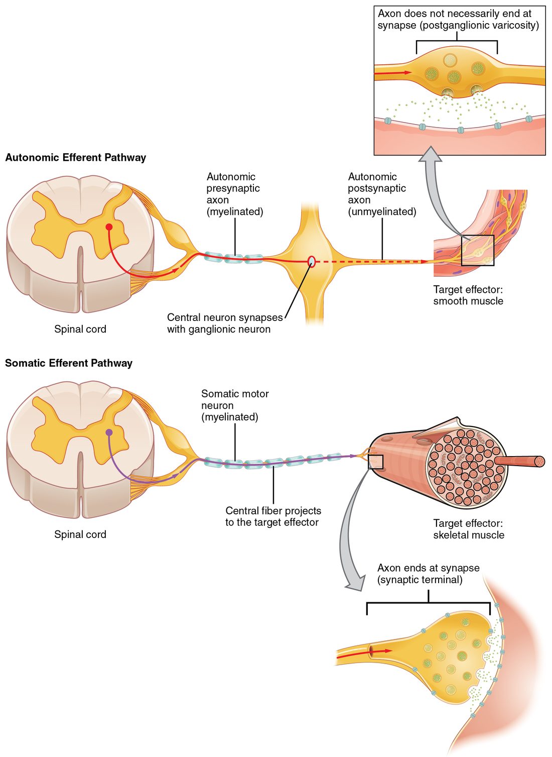
A visceral reflex sends its output through a chain of two motor neurons, which synapse in a ganglion outside the CNS, to smooth muscle, cardiac muscle or glands. A somatic reflex uses one motor neuron straight to skeletal muscle.
- Correct: It uses two neurons in series, synapsing in a ganglion outside the CNS: Correct. Two neurons and a ganglion on the way.
- It uses one motor neuron that runs all the way from the CNS to a skeletal muscle: That describes the somatic reflex.
- It has no sensory neuron, so the effector responds directly: Both kinds have a sensory receptor and sensory neuron. They differ on the motor side.
- Its motor axons are all heavily myelinated, so it is faster: The second axon of a visceral chain is unmyelinated, so visceral reflexes are slower than somatic ones.
6. Six weeks after an injury, a woman's knee and ankle jerks are 4+ on the right and 2+ on the left. Her right big toe goes up when the sole is stroked, and her right leg is weak and stiff. Where is the damage?
- In the dorsal roots of the right lumbar segments
- In the right sciatic nerve
- In the ventral horn of the right lumbar cord
- In the upper motor neuron pathway to the right leg
Show the answer
Hyperreflexia with clonus, a Babinski sign and a stiff, weak leg are upper motor neuron signs. The reflex arcs are intact but have lost the descending control that normally damps them.
- In the dorsal roots of the right lumbar segments: A broken sensory side of the arc would weaken or abolish the reflexes, not exaggerate them.
- In the right sciatic nerve: A damaged peripheral nerve breaks the arc and makes the reflexes weaker, with a floppy leg.
- In the ventral horn of the right lumbar cord: Losing ventral horn motor neurons removes the output of the arc, so reflexes fall and the muscle wastes.
- Correct: In the upper motor neuron pathway to the right leg: Correct. The arcs are released from descending control.
7. A person holds a very heavy box, and the tension in his biceps brachii rises steeply. Golgi tendon organs in the biceps fire. What is the effect on the biceps motor neurons?
- They are excited directly, so the biceps pulls harder
- They are inhibited through interneurons, so the biceps' force drops
- They are unaffected by tendon organ signals
- They are excited through interneurons in the opposite arm
Show the answer
Golgi tendon organs sense tension. Their sensory neurons excite inhibitory interneurons that inhibit the motor neurons of the same muscle: the tendon reflex. The biceps' force drops, and the triceps is excited.
- They are excited directly, so the biceps pulls harder: Direct excitation of the same muscle is what muscle spindles do in the stretch reflex. Tendon organs have the opposite effect.
- Correct: They are inhibited through interneurons, so the biceps' force drops: Correct. The tendon reflex damps the tension in the muscle.
- They are unaffected by tendon organ signals: Tendon organ signals do reach the brain, but they also drive the spinal tendon reflex.
- They are excited through interneurons in the opposite arm: Crossing to the opposite limb is a feature of the crossed-extensor reflex, not of the tendon reflex.
8. During the knee jerk, what stops the hamstrings from contracting against the kick?
- Spindle axons excite inhibitory interneurons that silence the hamstring motor neurons
- The hamstrings are stretched too, so their own spindles relax them
- Golgi tendon organs in the quadriceps fire first and switch off the hamstring motor neurons
- The cortex sends a signal to relax the hamstrings before the tap
Show the answer
Branches of the quadriceps spindle axons excite inhibitory interneurons, which release glycine onto the hamstring motor neurons and stop them firing. This is reciprocal inhibition.
- Correct: Spindle axons excite inhibitory interneurons that silence the hamstring motor neurons: Correct. Reciprocal inhibition through an inhibitory interneuron.
- The hamstrings are stretched too, so their own spindles relax them: Straightening the knee does stretch the hamstrings, but stretching a muscle excites its own motor neurons. It does not relax it.
- Golgi tendon organs in the quadriceps fire first and switch off the hamstring motor neurons: Tendon organs respond to tension, which rises only after the quadriceps contracts, and their main effect is on the quadriceps itself.
- The cortex sends a signal to relax the hamstrings before the tap: The reflex needs no signal from the brain, and it works when the tap comes as a surprise.
9Summary
A reflex is a rapid, involuntary, predictable response to a stimulus. Every reflex runs on a reflex arc: sensory receptor, sensory neuron, integration center, motor neuron and effector, and damage to any part can weaken or abolish it. Somatic reflexes act on skeletal muscle through one motor neuron; visceral (autonomic) reflexes act on smooth muscle, cardiac muscle and glands through two. The stretch reflex is monosynaptic: stretch fires muscle spindles, which excite the motor neurons of the same muscle, while interneurons inhibit the antagonist (reciprocal inhibition). The knee jerk and every other deep tendon reflex are stretch reflexes, graded 0 to 4+ with 2+ normal; reduced reflexes point to a broken arc, and brisk reflexes with clonus or a Babinski sign point to upper motor neuron damage. Golgi tendon organs sense tension and start the polysynaptic tendon reflex, which damps the muscle's force. The polysynaptic withdrawal reflex bends a limb away from harm, and the crossed-extensor reflex straightens the other limb to bear weight.