The spinal cord
1Why this matters
Mr. Adeyemi, 41, lifts a heavy box and feels severe low back pain. By evening both legs are weak, the skin between his thighs is numb, and he cannot pass urine. His spinal cord itself ended well above the injury, but a burst disc between the fourth and fifth lumbar vertebrae is crushing the bundle of nerve roots that hangs below it, and he needs surgery within hours.
2What this builds on
3Quick check before you start
1. Which region of the vertebral column lies just above the sacrum?
- Thoracic
- Lumbar
- Cervical
Show the answer
From top to bottom the column has 7 cervical, 12 thoracic and 5 lumbar vertebrae, then the sacrum and coccyx. The lumbar region sits just above the sacrum.
- Thoracic:
- Correct: Lumbar:
- Cervical:
2. Which division of the nervous system carries signals from sensory receptors toward the CNS?
- The motor (efferent) division
- The sensory (afferent) division
- The parasympathetic division
Show the answer
The sensory, or afferent, division carries signals in to the CNS. The motor, or efferent, division carries commands out; the parasympathetic division is part of the motor side.
- The motor (efferent) division:
- Correct: The sensory (afferent) division:
- The parasympathetic division:
3. Around the spinal cord, which space is a real space filled with fat and veins?
- The subdural space
- The epidural space
- The subarachnoid space
Show the answer
In the vertebral canal the dura is not attached to the bone, so the epidural space is real and holds fat and veins. The subarachnoid space holds CSF; the subdural space is only potential.
- The subdural space:
- Correct: The epidural space:
- The subarachnoid space:
4Anatomy
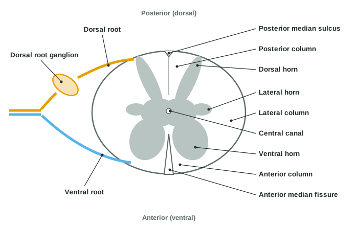
With labels hidden, select a box to reveal its label.
5How it works, step by step
- You step on a tack, and a sensory neuron in the sole of your foot fires.Its signal travels along its axon toward the spinal cord, running past its cell body, which sits to one side in a dorsal root ganglion.
- The same neuron's central process runs in through the dorsal root.It enters the back of the cord and ends in the dorsal horn.
- In the dorsal horn it excites interneurons and neurons whose axons turn up the cord.The signal climbs the white columns in an ascending tract toward the brain, and local interneurons pass it to the ventral horn.
- Motor neurons in the ventral horn are excited.Their axons leave through the ventral root and the joined nerve and reach the leg muscles, which contract and lift the foot.
6Core concepts
7A common mistake
The wrong idea: The spinal cord runs the full length of the vertebral column, so a needle in the lower back risks piercing it.
What actually happens: In an adult the cord ends at the conus medullaris, at about the first or second lumbar vertebra, because the vertebral column grew longer than the cord. Below that, the dural sac holds only the cauda equina, roots floating in CSF, down to about the second sacral vertebra. A lumbar puncture between L3 and L4 enters this fluid, and the roots slide away from the needle.
8Check yourself
Anything you miss goes into your review queue.
1. In this spinal cord cross section, back side at the top, which structure does pointer 7 mark?
- Dorsal horn
- Lateral horn
- Anterior column
- Ventral horn
Show the answer
Pointer 7 marks the broad gray horn reaching toward the front: the ventral (anterior) horn, which holds the cell bodies of motor neurons to skeletal muscle.
- Dorsal horn: The dorsal horn is the narrow gray horn reaching toward the back, at pointer 3.
- Lateral horn: The lateral horn is the small gray bump at the side, at pointer 4.
- Anterior column: The anterior column is white matter at the front beside the midline, at pointer 8, not gray matter.
- Correct: Ventral horn: Correct. The broad front horn of gray matter is the ventral horn.
2. Which numbered structure holds the cell bodies of sympathetic motor neurons, and at what levels of the cord is it found?
- Pointer 3, at every level
- Pointer 7, only in the enlargements
- Pointer 4, only from T1 to L2
- Pointer 11, only in the lumbar region
Show the answer
The lateral horn, the small gray bump at the side (pointer 4), holds sympathetic motor neurons. It is present only in the thoracic and upper lumbar segments, T1 to L2.
- Pointer 3, at every level: Pointer 3 is the dorsal horn, which is sensory and present at every level.
- Pointer 7, only in the enlargements: Pointer 7 is the ventral horn, which holds somatic motor neurons to skeletal muscle; it is largest in the enlargements but present at every level.
- Correct: Pointer 4, only from T1 to L2: Correct. The lateral horn holds sympathetic neurons, from T1 to L2.
- Pointer 11, only in the lumbar region: Pointer 11 is the dorsal root ganglion, which holds sensory cell bodies at every level.
3. Mr. Adeyemi, 41, has a burst disc between his fourth and fifth lumbar vertebrae. His legs are weak, the skin between his thighs is numb, and he cannot pass urine. Which structure is being compressed?
- The conus medullaris
- The cervical enlargement
- The lumbar enlargement of the cord
- The cauda equina
Show the answer
At L4–L5 the vertebral canal holds no spinal cord, only the cauda equina: the lumbar and sacral roots on their way down. Crushing them causes leg weakness, numbness of the saddle area and loss of bladder control, a surgical emergency.
- The conus medullaris: The conus medullaris lies at about L1–L2, above the burst disc.
- The cervical enlargement: The cervical enlargement is in the neck and supplies the upper limbs.
- The lumbar enlargement of the cord: The lumbar enlargement is part of the cord and lies behind the lower thoracic vertebrae, well above L4–L5.
- Correct: The cauda equina: Correct. At L4–L5 the canal holds only the cauda equina.
4. To treat severe pain, a surgeon cuts the dorsal roots of three neighboring segments on the right side. What would you expect in the band of skin and muscles supplied by the middle segment?
- Paralysis of the muscles, with normal sensation
- Loss of sensation, with normal muscle strength
- Loss of both sensation and strength
- No change, because the ventral roots carry the sensory signals
Show the answer
Dorsal roots carry sensory axons into the cord; ventral roots carry motor axons out. With the middle root and both its neighbors cut, no sensory route from that band reaches the cord, while the motor axons in the intact ventral roots still drive the muscles. Cutting one dorsal root alone would only dull the feeling, because neighboring roots overlap.
- Paralysis of the muscles, with normal sensation: Paralysis with normal sensation would follow cutting the ventral roots, which carry the motor axons.
- Correct: Loss of sensation, with normal muscle strength: Correct. Cutting the dorsal roots removes sensation and spares strength.
- Loss of both sensation and strength: Losing both would need both roots cut, or the nerve beyond where they join.
- No change, because the ventral roots carry the sensory signals: The ventral roots carry motor axons out; sensory signals enter through the dorsal roots.
5. Which statements about the dorsal root are true? Select all that apply.
- It carries sensory axons into the spinal cord
- Its ganglion holds the cell bodies of sensory neurons
- It carries motor axons out to skeletal muscles
- The neurons whose cell bodies sit in its ganglion are pseudounipolar
- Its cell bodies lie in the ventral horn
Show the answer
The dorsal root brings sensory axons into the back of the cord. Their cell bodies sit outside the cord in the dorsal root ganglion, and the neurons are pseudounipolar: one process runs to the body, the other into the cord.
- Correct: It carries sensory axons into the spinal cord: Yes. The dorsal root is the sensory way in.
- Correct: Its ganglion holds the cell bodies of sensory neurons: Yes. The dorsal root ganglion holds the sensory cell bodies.
- It carries motor axons out to skeletal muscles: No. Motor axons leave through the ventral root.
- Correct: The neurons whose cell bodies sit in its ganglion are pseudounipolar: Yes. Dorsal root ganglion neurons are pseudounipolar.
- Its cell bodies lie in the ventral horn: No. Ventral horn cell bodies send their axons out through the ventral root; dorsal root cell bodies sit in the ganglion.
6. A cross section of spinal cord has only a thin rim of white matter around a relatively large area of gray matter, and it has no lateral horns. Which level is it most likely from?
- Upper cervical
- Middle thoracic
- Upper lumbar
- Sacral
Show the answer
White matter thins going down the cord: fewer ascending axons have joined yet, and most descending axons have already left for their target segments. The thinnest white matter is at the sacral end, and lateral horns are absent below L2.
- Upper cervical: The upper cervical cord has the most white matter, since it carries every ascending and descending axon for the whole body.
- Middle thoracic: Middle thoracic segments have lateral horns, which this section lacks.
- Upper lumbar: Upper lumbar segments (to L2) still have lateral horns and more white matter than the sacral cord.
- Correct: Sacral: Correct. A thin white rim with no lateral horns fits the sacral cord.
7. This sequence traces a motor command from the brain to a leg muscle. Find the wrong step.
- The command leaves the motor areas of the cerebral cortex in a descending tract
- It runs down the white columns of the spinal cord
- It reaches motor neurons in the dorsal horn of a lumbar segment
- Their axons leave through the ventral root toward the muscle
Show the answer
Motor neurons to skeletal muscle sit in the ventral (anterior) horn, not the dorsal horn. The dorsal horn is the sensory side, where many incoming sensory axons end.
- The command leaves the motor areas of the cerebral cortex in a descending tract: Right. Motor commands travel down in descending tracts.
- It runs down the white columns of the spinal cord: Right. Tracts run in the white columns.
- Correct: It reaches motor neurons in the dorsal horn of a lumbar segment: This is the error. The motor neurons are in the ventral horn.
- Their axons leave through the ventral root toward the muscle: Right. Motor axons leave through the ventral root.
9Summary
The spinal cord runs from the foramen magnum to the conus medullaris at about L1–L2 in adults, inside the meninges. The cervical and lumbar enlargements hold the extra neurons for the limbs, and below the conus the cauda equina hangs in the dural sac, which reaches S2. In cross section, gray matter inside forms dorsal (sensory), ventral (somatic motor) and, from T1 to L2, lateral (sympathetic) horns around the central canal; white matter outside forms posterior, lateral and anterior columns, with the anterior median fissure in front and the posterior median sulcus behind. Sensory axons enter through the dorsal root, whose ganglion holds their cell bodies, and motor axons leave through the ventral root. Ascending tracts carry sensation up and descending tracts carry commands down; most cross the midline.