Nervous tissue is built from two kinds of cells: neurons, which carry signals, and glial cells, which make it possible for them to do so. This page covers neuron structure in detail and the types of glial cells: the parts of a neuron from the dendrites to the axon terminals, how neurons are classified by shape and by job, the four kinds of glia in the central nervous system and the two in the peripheral nervous system, how Schwann cells and oligodendrocytes build the myelin sheath, and why a cut nerve in your arm can grow back while damage to your spinal cord usually cannot.
What you already know
From the nervous tissue topic: a neuron receives signals on its dendrites and cell body, and sends its own signal away along a single axon to axon terminals, which release chemicals onto the next cell. Glial cells support and protect neurons, and unlike mature neurons they can divide. From the electrical signaling topics: when a stimulus pushes a neuron's membrane to threshold, it fires an all-or-none action potential that propagates along the axon. From the last topic: the brain and spinal cord form the CNS, and everything else forms the PNS. This page zooms in on the cells themselves.
The cell body in detail
The soma (soma = body) is another name for the cell body. It holds the nucleus and nearly all of the neuron's protein-making machinery. Stain a neuron and its soma is speckled with dark clumps, called Nissl bodies after the scientist who described them. They are stacks of rough ER covered with ribosomes. A neuron needs that much rough ER because its cell body must build proteins for an axon that may hold hundreds of times more cytoplasm than the soma itself.
Those proteins, and organelles such as mitochondria, reach the far end of the axon by axonal transport: motor proteins walk along microtubules, carrying cargo out from the soma and bringing worn-out material and chemical messages back. Fast transport moves vesicles hundreds of millimeters a day; slow transport moves the cytoskeleton and many enzymes only about a millimeter or a few millimeters a day. That slow rate will matter when you get to nerve regrowth.
From dendrite to axon terminal
Figure 1 shows a typical neuron from the CNS. Follow the path a signal takes through it.
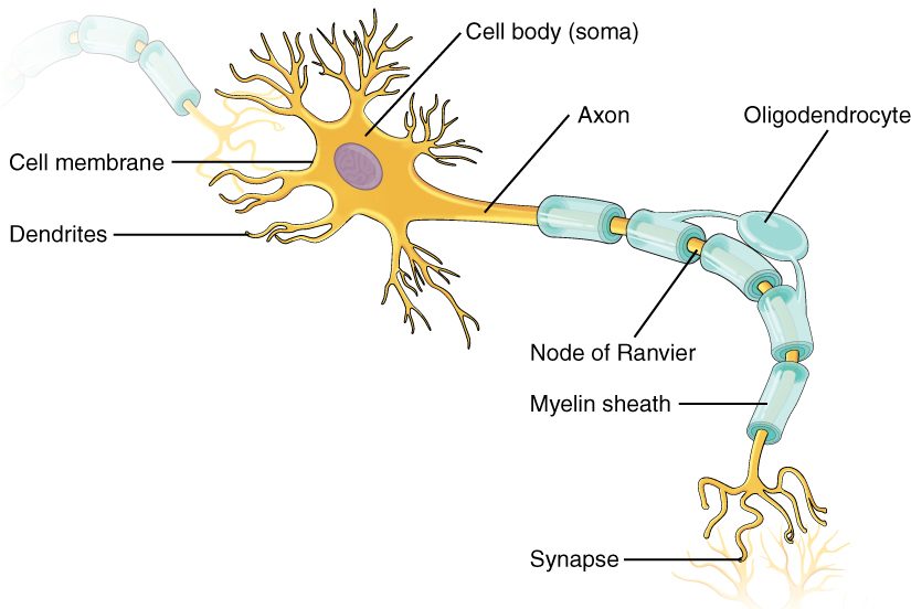
- Dendrites receive incoming signals. Many dendrites are studded with tiny knobs called dendritic spines, where axon terminals of other neurons make contact. A single large neuron can receive thousands of contacts.
- The axon hillock (hillock = small hill) is the cone-shaped region where the axon leaves the soma. It holds no Nissl bodies, which is how you spot it under a microscope.
- The initial segment is the first stretch of the axon beyond the hillock, before any insulation begins. Its membrane is packed with voltage-gated sodium channels, many times more densely than the soma's. So it reaches threshold first, and this is where the neuron's action potentials begin. That makes it the neuron's trigger zone: all the small graded potentials arriving at the dendrites and soma spread to it, and whether it reaches threshold decides whether the neuron fires.
- The axon itself carries the action potential away from the soma. Its plasma membrane is sometimes called the axolemma, and its cytoplasm is called axoplasm (-plasm = formed substance). Axoplasm holds microtubules and neurofilaments (the neuron's intermediate filaments) running along its length, the tracks and scaffolding for axonal transport.
- Axon collaterals (col- = together, later- = side) are side branches of the axon. They let one neuron send the same signal to several targets, sometimes including a branch that curls back to the region near its own soma.
- Axon terminals, the swollen tips of the axon's final branches, hold synaptic vesicles and release neurotransmitter onto the next cell.
| Dendrites | Axon | |
|---|---|---|
| Number per neuron | Usually many | One (it may branch into collaterals) |
| Direction of signal | Toward the soma | Away from the soma |
| Kind of electrical signal | Graded potentials | Action potentials |
| Length | Short, tapering, heavily branched | From under a millimeter to over a meter; uniform width |
| Nissl bodies (rough ER) | Present near the soma | Absent, from the hillock on |
| Myelin | None | Often present |
Classifying neurons by shape
Neurons come in many shapes, but anatomists sort them by one simple feature: how many processes (extensions) leave the soma. Figure 2 shows the three main types.
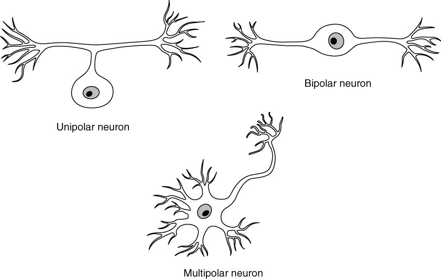
- Multipolar neurons (multi- = many, pol- = pole) have one axon and two or more dendrites. They are by far the most common type. Every motor neuron and nearly every interneuron in the CNS is multipolar.
- Bipolar neurons (bi- = two) have one axon and one dendrite, leaving opposite ends of the soma. They are rare, found only in a few sense organs: the light-sensing layer at the back of the eye, the patch of lining in the nose that detects smells, and the inner parts of the ear that sense sound and balance.
- Unipolar neurons (uni- = one) have a single short process that leaves the soma and then splits like a T into two long branches. One branch runs out to the sensory receptor ending in the skin or an organ; the other runs into the CNS. Most sensory neurons in your body are this type. Because the single process starts as two separate processes in the embryo and then fuses, they are also called pseudounipolar neurons (pseudo- = false).
In a unipolar neuron the signal does not pass through the soma at all. It starts near the sensory receptor ending, runs up the peripheral branch, past the T-junction and on into the CNS, with the soma sitting off to one side like a station beside the track. Both branches carry action potentials and are built like an axon. Some books still call the peripheral branch a dendrite because it carries signals toward the soma's side.
| Multipolar | Bipolar | Unipolar (pseudounipolar) | |
|---|---|---|---|
| Processes leaving the soma | Three or more: one axon, many dendrites | Two: one axon, one dendrite | One, which splits into two branches |
| How common | The vast majority of neurons | Rare | Common in the PNS |
| Usual job | Motor neurons and interneurons | Sensory, in a few sense organs | Sensory neurons for touch, pain, temperature, body position and organ stretch |
| Where the soma sits | Mostly CNS gray matter; also ganglia of the autonomic nervous system | In the sense organ itself | In a ganglion beside the brain or spinal cord |
Classifying neurons by job
You can also sort neurons by which way they carry information, which matches the divisions you met in the last topic.
- Sensory neurons (also called afferent neurons) carry signals from sensory receptors toward the CNS. Their somas sit in ganglia, outside the CNS.
- Interneurons (inter- = between) lie entirely inside the CNS and connect other neurons: sensory neurons to motor neurons, and neurons to each other in circuits of every size. Integration is their job. They make up the vast majority of your neurons.
- Motor neurons (also called efferent neurons), which you met at the neuromuscular junction, carry commands from the CNS out to effectors. Somatic motor neurons have their somas in the gray matter of the brain or spinal cord.
Put the two classifications together and a pattern appears: a neuron's shape follows its job and its position. Figure 3 traces a signal through all three functional types.
| Sensory neuron | Interneuron | Motor neuron | |
|---|---|---|---|
| Carries signals | From sensory receptors to the CNS | Between neurons within the CNS | From the CNS to effectors |
| Also called | Afferent neuron | Association neuron | Efferent neuron |
| Usual shape | Unipolar (bipolar in some sense organs) | Multipolar | Multipolar |
| Where the soma sits | A ganglion outside the CNS | CNS gray matter | CNS gray matter (somatic); a ganglion outside the CNS for the last neuron of an autonomic chain |
| Share of all neurons | A small fraction | The vast majority | A small fraction |
Glial cells: six kinds
You met glial cells in the nervous tissue topic as the support cells of nervous tissue. There are six kinds: four in the CNS and two in the PNS. Each one has a distinct job, and several of them have a partner on the other side of the CNS–PNS border that does a similar job in a different way.
How many glial cells are there? Direct counts of whole human brains find about 86 billion neurons and a similar number of other cells, most of them glia, so the overall ratio is close to one to one. It varies a great deal by region: glia far outnumber neurons in the white matter, and neurons outnumber glia in some densely packed gray matter.
Glia of the CNS
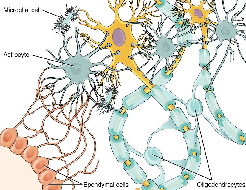
Figure 4 shows the four kinds of glial cells in the CNS, gathered around two neurons.
Astrocytes
Astrocytes (astr- = star, -cyte = cell) are star-shaped cells with many branching processes. They are the most varied glial cells in what they do:
- Controlling the fluid around neurons. When neurons fire, potassium ions leak out into the narrow spaces between cells. Astrocytes take up the excess potassium, and they take up neurotransmitter released at synapses, keeping the chemistry around neurons within its normal range.
- Wrapping the capillaries. The swollen ends of astrocyte processes, called end-feet, cover nearly the whole outer surface of the brain's capillaries. Signals from astrocytes help the capillary wall form an unusually tight seal between blood and brain tissue; you'll meet that seal in the topic on the brain's protection. Astrocytes also help widen local blood vessels when neurons nearby become active.
- Storing fuel. Astrocytes hold the brain's small store of glycogen and can pass fuel, such as lactate, to neurons.
- Building a scar. After an injury, astrocytes nearby enlarge, multiply and wall off the damaged area with a glial scar. It seals the wound, but it also blocks axons trying to grow back, as you'll see below.
Oligodendrocytes
Oligodendrocytes (oligo- = few, dendr- = branch, -cyte = cell) have a handful of arms. Each arm flattens into a sheet and wraps itself many times around a segment of an axon, forming the insulating layers of myelin described below. One oligodendrocyte can myelinate segments of dozens of different axons.
Microglia
Microglia (micro- = small) are small cells with fine, constantly moving processes. They are the resident immune cells of the CNS: they patrol the tissue, and when they detect damage or infection they multiply, move to the spot and engulf dead cells, debris and microbes by phagocytosis, acting like the macrophages of connective tissue. They also prune unneeded connections between neurons as the brain develops. Unlike the other glia, which arise from the same embryonic tissue as neurons, microglia come from immune precursor cells that settle in the brain early in development.
Ependymal cells
Ependymal cells (ependyma = upper garment) form a single layer of cube- to column-shaped cells lining the fluid-filled cavities inside the brain and the central canal that runs down the middle of the spinal cord. Many carry cilia, whose beating helps keep the fluid moving. Specialized ependymal cells, together with capillaries, produce that fluid, which cushions the brain and spinal cord; the topic on the brain's protection covers it.
Glia of the PNS
The PNS has only two kinds of glial cells. Figure 5 shows both around a sensory neuron.
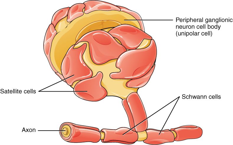
- Schwann cells (named after the physiologist Theodor Schwann) line up along PNS axons. Each one wraps a single segment of a single axon in myelin, or cradles several thin axons that it does not myelinate. They are also the key to nerve repair.
- Satellite cells (satelles = attendant) are flattened cells that cover the somas of neurons in ganglia, much as astrocytes surround neurons in the CNS. They support those neurons and regulate the fluid around them. Do not confuse them with the satellite cells of skeletal muscle, the stem cells that repair muscle fibers: same name, different cells.
| CNS glia | PNS glia | |
|---|---|---|
| Kinds | Four: astrocytes, oligodendrocytes, microglia, ependymal cells | Two: Schwann cells, satellite cells |
| Make myelin | Oligodendrocytes | Schwann cells |
| Surround neuron cell bodies and regulate their fluid | Astrocytes | Satellite cells |
| Clear debris and fight infection | Microglia, the resident immune cells | Schwann cells, with macrophages that arrive from the blood |
| Line fluid-filled spaces | Ependymal cells | No equivalent |
| Response to an axon injury | A glial scar forms; axons rarely regrow | Schwann cells guide the regrowing axon |
The myelin sheath
In the nervous tissue topic you read that some glial cells wrap axons in many layers of their own plasma membrane. That wrapping is the myelin sheath (myel- = marrow, from its fatty, marrow-like look), and the material itself is myelin. Because it is layer on layer of membrane with almost no cytoplasm between, myelin is mostly lipid, about 70 to 80% of its dry weight, with the rest protein. Lipid is a poor conductor, so myelin works as electrical insulation, and its fat gives white matter its color.
Myelin is not continuous. Each glial wrap covers one segment of axon, up to a millimeter or two long, called an internode. Between neighboring segments is a tiny gap, about a thousandth of a millimeter wide, where the axon's membrane is bare: a node of Ranvier (named after the anatomist Louis-Antoine Ranvier), also called a myelin sheath gap. Voltage-gated sodium channels are packed densely at the nodes and sparse under the myelin. Axons wrapped this way are myelinated; axons with no myelin are unmyelinated.
Myelin makes an action potential travel far faster along an axon, up to about 120 meters per second in the thickest myelinated axons compared with only about 0.5 to 2 meters per second in thin unmyelinated ones. How the insulation and the nodes produce that speed is the subject of the next topic.
How Schwann cells and oligodendrocytes build myelin
Both cells make myelin the same basic way. The glial cell flattens part of itself into a sheet, lays the sheet against the axon, and spirals around it, sometimes a hundred times, like a bandage wound around a finger. As it winds, the cytoplasm is squeezed out of the layers, leaving tightly packed membranes. Figure 6 compares the two cells.
The differences matter for repair:
- A Schwann cell devotes itself to one segment of one axon. After wrapping, its nucleus and remaining cytoplasm sit in a thin outer layer around the myelin, called the neurilemma (lemma = husk). That living outer layer is what lets Schwann cells help rebuild a damaged axon.
- An oligodendrocyte sends out several arms, and each arm myelinates a segment of a different axon. Its cell body sits among the axons, not on them, and no living outer layer surrounds the myelin.
| Schwann cell | Oligodendrocyte | |
|---|---|---|
| Location | PNS | CNS |
| Axons myelinated per cell | One segment of one axon | Segments of many axons (often dozens) |
| Where its nucleus sits | On the outside of its own wrap | In a cell body away from the axons |
| Outer living layer (neurilemma) | Yes | No |
| Handles unmyelinated axons by | Cradling several of them in grooves of one cell | Leaving them bare |
| Role after an axon is cut | Clears debris and forms a tube that guides regrowth | Leaves debris that contains growth-blocking proteins |
Myelination starts before birth and goes on for years. A newborn's nervous system is only partly myelinated, which is one reason babies' movements are slow and poorly coordinated; some regions of the brain keep adding myelin into your twenties.
Unmyelinated axons
Not every axon is myelinated. Thin axons, such as those carrying dull, aching pain and many autonomic axons, are unmyelinated. In the PNS they are still not bare: a single Schwann cell cradles several of them in grooves in its surface, without wrapping any of them in layers. In the CNS, unmyelinated axons simply run among the other cells. Signals travel along them much more slowly.
Why a cut PNS axon can grow back
A carpenter cuts a nerve at the base of his thumb. The surgeon sews the cut ends together and warns him that feeling will come back slowly, starting near the scar and moving out toward the fingertip over months. Figure 7 shows why.
Step by step:
- The far segment breaks down. The part of the axon beyond the cut has lost its supply of proteins from the soma. Within days it fragments, and its myelin falls apart with it. This is called Wallerian degeneration, after the physician Augustus Waller, who described it.
- Debris is cleared. Schwann cells in the far segment break down their own myelin and signal macrophages to come in from the blood. Together they clear the wreckage within a few weeks.
- The soma gears up. The neuron's cell body swells and its Nissl bodies disperse as it switches its protein production from signaling to growth.
- Schwann cells build a tube. The Schwann cells multiply and line up in a column inside the connective tissue sheath that once surrounded the axon, forming a regeneration tube. They release growth-promoting proteins and coat the tube with adhesive molecules that growing axons cling to.
- The axon regrows. Sprouts grow out from the cut end of the stump. One that finds the tube is guided along it at roughly 1 millimeter per day, about the speed of slow axonal transport.
- Reconnection. If the axon reaches a target, Schwann cells wrap it in new myelin and it can work again.
Recovery is often incomplete. If the cut ends are far apart, or scar tissue blocks the way, sprouts cannot find the tube and may form a painful tangle. Axons can also enter the wrong tube and reach the wrong target, for example a sensory axon regrowing to a different patch of skin. And if the journey takes too long, the muscle fibers without a nerve supply waste away before the axon arrives. That is why surgeons line up the cut ends precisely and repair nerves quickly.
Worked example: how long will regrowth take?
Problem. A nerve is cut and repaired 20 cm from the tip of the index finger. Axons regrow at about 1 mm per day once they cross the repair site. About how long until feeling can start to return to the fingertip?
- Put the distance in the same unit as the rate. 20 cm × 10 mm/cm = 200 mm.
- Write the relation. Time = distance ÷ rate.
- Substitute. Time = 200 mm ÷ 1 mm/day = 200 days.
- Convert to months. 200 days ÷ 30 days per month ≈ 6.7 months.
Answer. About 200 days, roughly six to seven months, and in practice somewhat longer: the sprouts need days to weeks to cross the repair site before steady growth begins. Feeling returns first near the scar and last at the fingertip.
Why CNS axons rarely grow back
Damage to the brain or spinal cord that cuts axons usually leaves permanent loss. It is not that CNS neurons cannot grow axons at all. In classic experiments, CNS axons grew for centimeters into a graft of peripheral nerve placed in the brain or spinal cord. The problem is mainly the surroundings the axon must grow through:
- No guiding tube. Oligodendrocytes do not multiply and line up to guide a regrowing axon the way Schwann cells do, and each one serves many axons, so it cannot devote itself to one.
- Growth-blocking myelin. CNS myelin contains proteins that make the tip of a growing axon stop and collapse. Microglia clear myelin debris slowly, over months to years rather than weeks, so those proteins linger.
- The glial scar. Astrocytes wall off the injury with a dense scar. It limits the spread of damage, but axons cannot cross it.
- A weaker growth program. Mature CNS neurons switch on fewer growth genes after injury than PNS neurons do.
Research on spinal cord repair targets each of these: blocking the myelin proteins, breaking down the scar, and transplanting cells that act like Schwann cells.
| PNS axon cut | CNS axon cut | |
|---|---|---|
| Myelinating glia | Schwann cells | Oligodendrocytes |
| Debris clearance | Fast (weeks): Schwann cells and macrophages from the blood | Slow (months or longer): microglia |
| Guidance for regrowth | Regeneration tube of Schwann cells | None |
| Growth-blocking signals | Few | Myelin proteins and the astrocyte scar |
| Usual outcome | Regrowth at about 1 mm/day, often with partial recovery | Little or no regrowth; loss is usually permanent |
Neurons don't divide; glia do
Regrowing an axon is not the same as replacing a neuron. Mature neurons do not divide, so a neuron whose soma dies is gone. Whether adult human brains make any new neurons at all, for example in a region important for memory, is still unsettled: studies of donated brains disagree, some finding signs of new neurons into old age and others finding almost none after childhood. Either way, adult neuron production is far too small to replace what an injury destroys.
Glial cells, by contrast, keep dividing: astrocytes to form scars, microglia to fight infection, oligodendrocyte precursors to replace lost myelin, Schwann cells to rebuild a nerve. That capacity has a downside. Most tumors that start in brain tissue itself arise from glial cells or their precursors, and are called gliomas; tumors made of neurons are rare.