Touch a hot pan and your hand jerks back before you even feel the pain. In that fraction of a second, electrical signals raced from your fingertip to your spinal cord and back out to the muscles of your arm. Every step ran through nervous tissue. This page introduces nervous tissue, its neurons and glia: the two kinds of cells it is built from, the parts of a neuron and which way signals travel through them, and what a nerve actually is.
What nervous tissue does
Nervous tissue is the fourth primary tissue type. It makes up your brain, your spinal cord and the nerves that branch from them to every part of your body. Its job is communication: it carries electrical signals quickly from one place to another and passes them from cell to cell.
Two features make that possible. Some of its cells are extremely long, so a single cell can connect your spinal cord to your big toe. And those cells produce rapid electrical changes across their plasma membranes and pass them along their length. How those electrical changes arise is the subject of the next chapter. Here you meet the cells that carry them.
Nervous tissue has two kinds of cells:
- Neurons carry the signals.
- Glial cells support, protect and feed the neurons, and they do not carry the long-distance signals themselves.
Neurons: the signaling cells
A neuron (neur- = nerve; also called a nerve cell) is a cell that receives signals, combines them, and sends a signal of its own to other cells: other neurons, muscle fibers or gland cells. Your brain alone holds about 86 billion of them.
Three features set neurons apart from most cells:
- Long extensions. Thin branches of the plasma membrane and cytoplasm reach out from the main part of the cell, some for a meter or more.
- Long life, little division. Mature neurons do not divide. Most of the neurons in your brain are as old as you are. So when neurons die, they are mostly not replaced.
- High demand for oxygen and glucose. Your brain is about 2% of your body weight but uses roughly 20% of the oxygen you consume at rest. Neurons store almost no fuel, so a few minutes without blood flow starts to kill them.
The parts of a neuron
Picture the neuron that runs from the base of your spine to the muscles of your foot. Its main body sits near your spinal cord, and one thread-thin extension runs the whole length of your leg. Figure 1 shows the parts of a typical neuron.
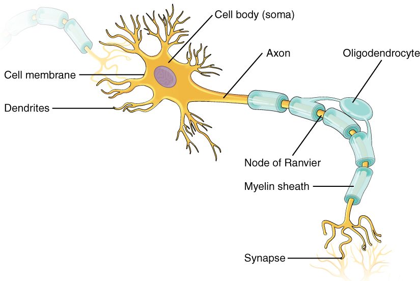
- The cell body is the part that holds the nucleus. It also holds most of the rough ER and Golgi, so it builds nearly all of the neuron's proteins. Those proteins are then carried out along the extensions.
- Dendrites (dendr- = tree) are short, heavily branched extensions of the cell body. They receive signals from other cells. A neuron can have many dendrites, and their branches can receive contacts from thousands of other neurons at once. The more a neuron branches, the more inputs it can collect.
- The axon (axon = axis) is a single long extension that carries the neuron's signal away from the cell body. Each neuron has only one, though it may branch near its end. Axons range from a fraction of a millimeter to over a meter long.
- At the end of the axon, its branches swell into small knobs called axon terminals (also called synaptic end bulbs). Each one sits very close to the next cell. When a signal arrives, the axon terminal releases chemicals by exocytosis, and those chemicals act on the next cell. The chemical-signaling topic explains how.
Which way signals travel
Signals move through a neuron in one direction:
- Dendrites (and the cell body itself) receive signals from other cells.
- The cell body combines those incoming signals.
- If they are strong enough, the axon carries a signal away from the cell body.
- The axon terminals release chemicals onto the next cell.
A memory aid: dendrites deliver in, axons send away. The next cell in line may be another neuron, whose dendrites then receive the signal, or a muscle fiber or gland cell that responds.
Why a cut axon dies beyond the cut
Because the cell body makes nearly all of a neuron's proteins, everything else depends on it. The axon is supplied by a steady stream of proteins and organelles carried along it from the cell body. Cut an axon, and the piece beyond the cut is separated from that supply. Within days that piece breaks down. The cell body and the stump still attached to it can survive. Whether the axon can grow back depends on where it is and which glial cells surround it; the nervous system chapter covers that.
Glial cells: the support cells
Glial cells (glia = glue; also called glia or neuroglia) are the other cells of nervous tissue. The name comes from an old belief that they were just the glue holding neurons together. They do far more than that. Depending on the kind, glial cells:
- hold neurons in place and give the tissue its structure
- wrap axons in many layers of their own plasma membrane, which insulates the axon and makes signals travel along it much faster
- control the makeup of the fluid around neurons, taking up excess ions and chemicals
- help supply neurons with fuel from the blood
- engulf dead cells and microbes
There are six kinds of glial cells, four in the brain and spinal cord and two in the rest of the body. You'll meet each one in the nervous system chapter.
Two facts about glia matter clinically. First, glial cells keep the ability to divide, unlike mature neurons. Second, as a result, most cancerous tumors that start in brain tissue itself grow from glial cells, not from neurons.
How many glial cells are there?
You may read that glial cells outnumber neurons ten to one. Careful counts of whole human brains, which dissolve the tissue and count the nuclei, do not support that figure. They find roughly equal numbers overall: about 86 billion neurons and a similar number of other cells, most of them glial cells. The ratio varies by region: glia far outnumber neurons in some parts, and neurons outnumber glia in others.
Nerves: cables of axons
Pull a nerve out of your arm and look at it closely, and you would find a white cord about as thick as a pencil. It is not one cell. A nerve is a bundle of many axons, running together outside the brain and spinal cord, wrapped and held together by connective tissue with its own blood vessels. It works like a telephone cable: many separate wires, each insulated, bundled inside one tough sheath. Figure 2 shows one in cross section.
A few points follow from that structure:
- A single nerve can hold thousands of axons from thousands of different neurons.
- One nerve usually carries signals in both directions. Some of its axons bring information in from your skin, joints and organs; others carry commands out to your muscles and glands.
- Cutting a nerve cuts every axon in it. That is why a deep cut at the wrist can leave several fingers both numb and weak.
- Bundles of axons inside the brain and spinal cord are not called nerves. They go by other names that you'll meet in the nervous system chapter.
A common mix-up: a nerve is not a neuron
Because the words sound alike, students often use "nerve" and "neuron" as if they meant the same thing. They are different levels of structure. A neuron is one cell. A nerve is an organ-level cable made of the axons of many neurons, plus connective tissue and blood vessels. A neuron's axon may run through a nerve, but its cell body is usually somewhere else.
| Neuron | Nerve | |
|---|---|---|
| What it is | One cell | A bundle of many axons with connective tissue |
| Level of organization | Cell | Organ |
| Contains cell bodies? | Yes, its own | No; the cell bodies lie elsewhere |
| Blood vessels inside? | No | Yes, in its connective tissue |
| Where found | Brain, spinal cord and throughout the body | Outside the brain and spinal cord |
| Direction of signals | One way: dendrites to cell body to axon | Both ways, in different axons |