Right now three kinds of muscle are working in you. The muscles holding your head up are skeletal muscle. Your heart is cardiac muscle. The walls of your stomach and your blood vessels are smooth muscle. This page is a skeletal, cardiac and smooth muscle comparison: what all muscle tissue shares, how to tell the three types apart under a microscope, and how their structure explains how each one behaves.
What makes a tissue muscle
Put a hand on your upper arm and bend your elbow. The muscle under your hand gets shorter and thicker. That is the defining act of muscle tissue: its cells contract, shortening with force.
The force comes from the two cytoskeleton proteins you met in the cell chapter. Muscle cells are packed with filaments of actin and myosin. Using energy from ATP, myosin grabs actin filaments and pulls them, and the cell shortens. The skeletal muscle chapter covers the details.
One rule follows directly: muscle can only pull. A contracting muscle cell shortens; it cannot push. When you straighten your elbow, a different muscle on the other side of your arm does the pulling.
Muscle fibers
A muscle cell is called a muscle fiber (also a myocyte; my-, myo- = muscle, -cyte = cell). The name "fiber" fits because skeletal muscle cells are long and thin, like threads. Don't confuse a muscle fiber, which is a whole living cell, with the protein fibers of connective tissue, which are nonliving matrix.
Four properties of muscle tissue
All three muscle types share four properties of muscle tissue:
- Responsiveness. A muscle fiber reacts to electrical and chemical signals by producing an electrical change across its plasma membrane. That change triggers contraction.
- Ability to contract. It shortens with force when triggered.
- Extensibility (ex- = out, tend- = stretch). It can be stretched beyond its resting length without damage. Your stomach wall stretches after a big meal; the muscles on the back of your thigh stretch when you touch your toes.
- Elasticity. After being stretched, it recoils to its resting length.
Striations: the stripes that sort muscle types
Under a microscope, skeletal and cardiac muscle fibers show fine, regular bands running across them, light and dark in turn, like the stripes on a barcode. These bands are striations (stria = streak), and muscle that has them is striated.
The stripes come from how the actin and myosin filaments are arranged. In striated muscle, they are stacked in short, repeating units lined up across the whole fiber. Where thick myosin filaments sit, the fiber looks dark; where only thin actin filaments sit, it looks light. Smooth muscle has the same filaments but arranged at angles in a crisscross lattice, with no lined-up units, so it shows no bands. It is nonstriated. That is where the name "smooth" comes from: its surface looks smooth under a microscope.
Figure 1 sets the three types side by side. Three questions identify any of them: Are there stripes? How many nuclei, and where? Are there dark discs joining the cells?
Skeletal muscle tissue
Skeletal muscle tissue makes up the muscles attached to your bones by tendons (and, in your face, to your skin). It moves your body, holds your posture, and releases heat as a by-product of its work.
- Long, cylindrical fibers. A single fiber is often several centimeters long, and some run nearly the length of their muscle.
- Many nuclei, at the edge. In the embryo, many small cells fuse end to end to form each fiber, so one fiber holds hundreds of nuclei, pressed against its plasma membrane.
- Striated. The bands are sharp and regular.
- Voluntary. A skeletal muscle contracts only when the nervous system signals it, and you can start those signals by choice. That is why skeletal muscle is also called voluntary muscle. Much of its work, such as keeping you upright and breathing, runs without conscious thought, but you can take over when you decide to.
Adult skeletal fibers don't divide. Small stem cells tucked against each fiber can multiply and fuse with damaged fibers, so skeletal muscle repairs modest injuries well.
Cardiac muscle tissue
Cardiac muscle tissue (cardi- = heart) is found only in the wall of your heart. Its job is to pump blood, about 100,000 contractions a day, without a break.
- Short, branched fibers. Each fiber branches and joins several neighbors, forming a connected network.
- One nucleus, in the center. Some fibers have two.
- Striated, like skeletal muscle.
- Involuntary. You cannot make your heart beat by deciding to. Some cardiac cells set their own rhythm, so the heart beats even with no signal from the brain. The nervous system and hormones only speed it up or slow it down.
Adult cardiac fibers almost never divide. About 1% or fewer of them are replaced each year, and fewer with age, so fibers that die are mostly lost for good, and the gap is filled with tough, noncontracting collagen.
Intercalated discs
Where one cardiac fiber meets the next, their membranes interlock in a zigzag joint called an intercalated disc (inter- = between, calat- = inserted). In a stained slide, the discs are dark lines across the fibers, and they are the quickest way to recognize cardiac muscle. Figure 2 zooms in on one.
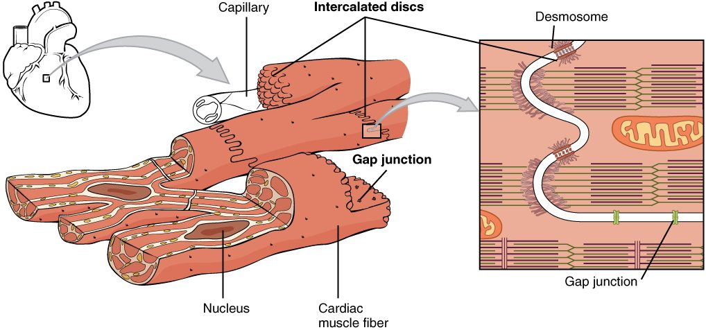
Each disc holds two kinds of cell junction from the cell chapter, and each does a specific job:
- Desmosomes rivet neighboring fibers together. When a fiber contracts, it pulls hard on its neighbors, and the desmosomes keep the fibers from being torn apart.
- Gap junctions are channels between the fibers. Ions flow through them, so an electrical change in one fiber spreads straight into the next.
Because the electrical change passes from fiber to fiber through gap junctions, a whole region of the heart contracts almost together, as one unit, instead of fiber by fiber.
Smooth muscle tissue
Smooth muscle tissue lines the walls of hollow organs: the stomach, intestines, urinary bladder, uterus, airways and blood vessels. Because it sits in the internal organs, it is also called visceral muscle (viscer- = internal organs).
- Spindle-shaped fibers. Each is thick in the middle and tapered at both ends, and much smaller than a skeletal fiber.
- One nucleus, in the center.
- Nonstriated.
- Involuntary. It responds to the nervous system, to hormones and to local conditions such as stretch, but never to a decision.
Smooth muscle contracts slowly and can stay contracted for a long time while using little ATP. In many hollow organs, gap junctions link its fibers, so a whole sheet contracts together in a slow wave that pushes contents along. Smooth muscle fibers can also divide, and they can grow larger. In pregnancy, the uterus wall thickens mainly because its fibers grow many times longer and wider, with some added by division.
Comparing the three muscle tissues
| Skeletal | Cardiac | Smooth | |
|---|---|---|---|
| Where | Attached to bones (and facial skin) | Heart wall only | Walls of hollow organs and blood vessels |
| Fiber shape | Long, cylindrical, unbranched | Short, branched | Spindle-shaped, tapered |
| Nuclei | Many, at the edge | One (sometimes two), central | One, central |
| Striations | Yes | Yes | No |
| Special junctions | None between fibers | Intercalated discs (desmosomes and gap junctions) | Gap junctions in many organs |
| Control | Voluntary | Involuntary; sets its own rhythm | Involuntary |
| Speed of contraction | Fast | Moderate | Slow, can be sustained |
| Replacing lost fibers | Good, from stem cells beside the fibers | Very poor | Good; fibers can divide |
Vasoconstriction and vasodilation
Step out into freezing air, and within a minute your fingers turn pale and cold. The smooth muscle in the walls of the small arteries feeding your fingers has contracted. Each of those arteries is wrapped in a ring of smooth muscle, so when the ring contracts, the vessel's radius shrinks. Narrowing of a vessel by its smooth muscle is vasoconstriction (vaso- = vessel, constrict- = squeeze). When the smooth muscle relaxes, blood pressure inside pushes the wall back out and the radius grows: vasodilation (dilat- = widen). A flushed face after exercise is vasodilation in the skin.
You already know from the chemistry and physics primer that resistance to flow depends on the fourth power of the radius. A small change in radius makes a large change in flow.
Worked example: halving an artery's radius.
- Resistance is proportional to 1 / radius4.
- The radius falls to 1/2 of its starting value.
- Radius4 becomes (1/2)4 = 1/16 of its starting value.
- Resistance is 1 divided by that, so it rises to 16 times its starting value.
- Flow equals the pressure gradient divided by resistance. With the same pressure gradient, flow falls to 1/16 of its starting value.
Halving the radius cuts flow through that vessel to about 6% of what it was.
Vasoconstriction and vasodilation are how your body shifts blood between organs from minute to minute. This is a short version. Blood vessel walls, and what signals their smooth muscle, return in full in the blood vessels topic of the cardiovascular chapter.
A common mix-up: striated does not mean voluntary
Because skeletal muscle is both striated and voluntary, students often treat the two words as meaning the same thing. They don't. Striation describes how the filaments are arranged. Voluntary describes how the muscle is controlled. Cardiac muscle is striated but involuntary. Keep the two ideas apart: stripes tell you the structure, not who is in charge.