Your nervous system is one connected network, but it is easier to learn when you divide it up. This page explains the organization of the nervous system: the central nervous system (CNS) and peripheral nervous system (PNS), the sensory and motor divisions that carry signals in and out, the somatic and autonomic parts of the motor side, and the names for clusters of cell bodies and bundles of axons, including gray matter and white matter. You met neurons, axons and nerves in the nervous tissue topic. Here you see how they are arranged into a system.
Three jobs: sensation, integration and response
You pick up a mug and find it is scalding hot. In well under a second, three things happen:
- Sensation. Sensory receptors in the skin of your fingers detect the heat and start signals.
- Integration. Neurons in your spinal cord and brain combine those signals with everything else they know (the mug is full, the table is right there) and settle on a response.
- Response. Signals go out to the muscles of your hand and arm, and you set the mug down fast.
Every task your nervous system performs follows that pattern:
- Sensory input (sens- = feel, perceive) is the information that sensory receptors collect about the world outside you and the state of your body: heat, pressure, stretch, light, blood chemistry.
- Integration (integr- = whole, make whole) is the processing step. Neurons combine many incoming signals, compare them with stored information, and determine what happens next. Most of your neurons do this job.
- Motor output (mot- = move) is the signal sent to an effector, a muscle or gland that responds. It is called "motor" even when the effector is a gland that secretes rather than a muscle that moves.
You have met this pattern before, in the feedback loop: a sensory receptor, an afferent pathway, a control center, an efferent pathway and an effector. The nervous system is built from exactly those parts.
Two parts by location: the CNS and the PNS
The first way to divide the nervous system is purely by where things are. Figure 1 shows the split.
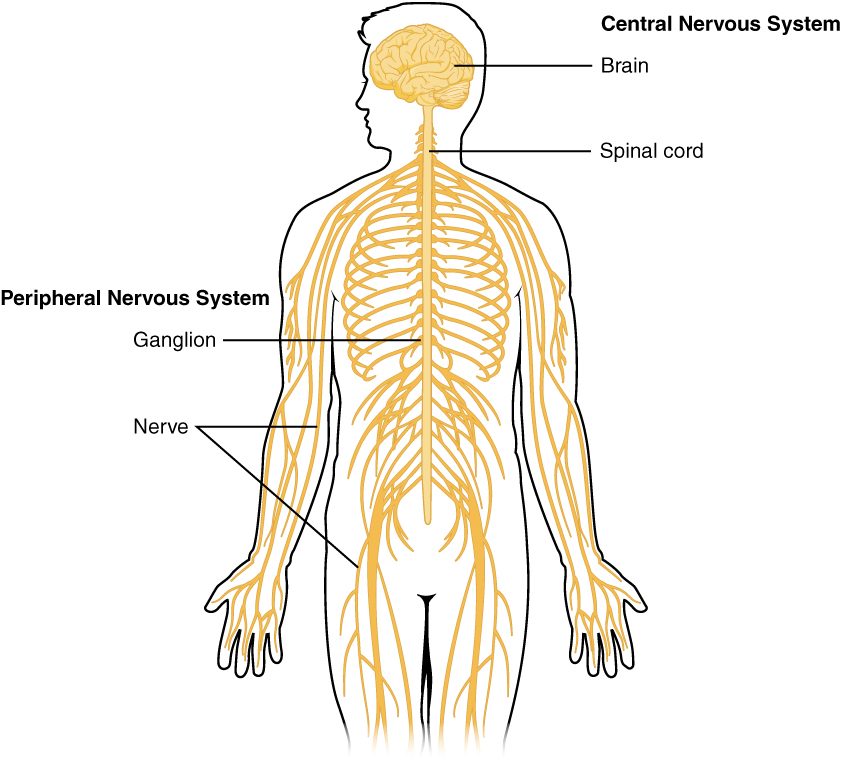
- The central nervous system (CNS; centr- = center) is your brain and spinal cord. Both sit inside bone: the brain in the cranial cavity and the spinal cord in the vertebral canal. The CNS is where integration happens.
- The peripheral nervous system (PNS; peri- = around, pher- = carry: the outer edge) is everything else: the nerves that run between the CNS and every part of your body, the clusters of neuron cell bodies that sit along those nerves, and the sensory receptors at their ends.
The PNS is the wiring and the CNS is the processor. A signal from your toe cannot be felt or acted on until PNS axons deliver it to the CNS, and a command from the CNS goes nowhere until PNS axons carry it out.
| Central nervous system (CNS) | Peripheral nervous system (PNS) | |
|---|---|---|
| What it includes | Brain and spinal cord | Nerves, clusters of cell bodies along them, sensory receptors |
| Where it lies | Inside the skull and the vertebral column | Throughout the body, outside the skull and vertebral column |
| Protection | Bone, plus extra wrappings and a cushion of fluid | Connective tissue wrappings only |
| Main job | Integration | Carrying signals in to the CNS and out from it |
| Cluster of cell bodies is called | A nucleus | A ganglion |
| Bundle of axons is called | A tract | A nerve |
The last two rows get their own section below.
Two directions: the sensory and motor divisions
The second way to divide the nervous system is by which way a signal travels relative to the CNS.
- The sensory division, also called the afferent division (af- = toward, fer- = carry), carries signals toward the CNS. Its axons bring input from sensory receptors in your skin, muscles, joints, eyes and ears, and from your internal organs.
- The motor division, also called the efferent division (ef- = away from, fer- = carry), carries signals away from the CNS to effectors: skeletal muscle, cardiac muscle, smooth muscle and glands.
These are the same afferent and efferent pathways you met in the feedback loop, now given names as divisions. A memory aid: afferent signals arrive at the CNS; efferent signals exit it.
Both divisions run mostly through the PNS, and they share the same nerves. A single nerve in your forearm holds afferent axons carrying touch and pain toward your spinal cord and efferent axons carrying commands to your hand muscles, side by side. That is why one deep cut can cause both numbness and weakness.
| Sensory (afferent) division | Motor (efferent) division | |
|---|---|---|
| Direction of signals | Toward the CNS | Away from the CNS |
| Starts at | Sensory receptors | The CNS |
| Ends at | The CNS | Effectors: muscles and glands |
| Step it serves | Sensory input | Motor output |
| Feedback loop part | Afferent pathway | Efferent pathway |
| If its axons are cut | Numbness: signals from the area never reach the CNS | Weakness or paralysis: commands never reach the effectors |
The motor division splits in two: somatic and autonomic
Think of two things your nervous system did this morning. You reached out and turned off your alarm. And, without any thought from you, your heart sped up as you stood, and your stomach started churning out digestive juice after breakfast. Different effectors, controlled in different ways.
- The somatic nervous system (somat- = body) controls skeletal muscle. Its commands travel from the CNS to the muscle along a single motor neuron, and they always excite the muscle, making it contract. Most somatic output is under conscious control: you decide to reach, kick or speak.
- The autonomic nervous system (ANS; auto- = self, nom- = law: self-governing) controls cardiac muscle, smooth muscle and glands. It sets your heart rate, the width of your blood vessels, the movements of your gut, your sweating and much more. You usually cannot control it by deciding to, and you are usually unaware of it.
"Voluntary" and "involuntary" are useful shorthand, but the real dividing line is the effector. Shivering and the steady rhythm of breathing both use skeletal muscle, so they are somatic output even though you do not decide to do them.
| Somatic nervous system | Autonomic nervous system | |
|---|---|---|
| Effectors | Skeletal muscle | Cardiac muscle, smooth muscle, glands |
| Usual control | Mostly conscious (voluntary) | Mostly unconscious (involuntary) |
| Neurons between the CNS and the effector | One motor neuron | A chain of two neurons, which meet in a ganglion outside the CNS |
| Effect on the effector | Always excites (the muscle contracts) | Can excite or inhibit, depending on the organ and the branch |
| Examples | Walking, writing, shivering | Heart rate, digestion, sweating, blood vessel width |
Both systems begin in the CNS and run out through the PNS. The somatic and autonomic systems are divisions of function, not of location.
The autonomic nervous system in brief
This is a short first look. The autonomic nervous system gets a full chapter of its own later in the course, where you'll see its anatomy, its chemical messengers and how it controls each organ.
The autonomic nervous system has two main branches, and most organs receive axons from both:
- The sympathetic division (sym- = together, path- = feeling) dominates during stress, exercise and danger. It is known as the fight-or-flight system. Picture being chased by a dog: your heart pounds, your airways widen, your palms sweat, and digestion is put on hold.
- The parasympathetic division (para- = beside) dominates during calm. It is known as the rest-and-digest system. After a meal on the couch, your heart rate slows, your gut moves food along and secretes digestive juices, and your bladder empties when you let it.
The two divisions are not an on switch and an off switch. Both send signals to most organs all the time. What changes is the balance: during a sprint the sympathetic side wins out; during a nap the parasympathetic side does. Where an organ gets both, their effects are usually opposite, so shifting the balance moves the organ one way or the other.
Your gut also has its own large nerve network in its wall, which can run many digestive movements by itself. You'll meet it in the digestive chapter.
The whole map
Figure 2 puts both ways of dividing the nervous system on one diagram.
Gray matter and white matter
Slice through a fresh brain or spinal cord and you see two shades of tissue: a pinkish-gray and a glossy white. The difference comes from which parts of the neurons are packed there.
- Gray matter is where neuron cell bodies, dendrites and axon terminals are concentrated, along with many glial cells and the ends of axons. This is where neurons contact one another, so gray matter is where integration happens.
- White matter is made mostly of axons running in bundles. Many of those axons are wrapped in layers of a fatty material made by glial cells. That fat gives white matter its pale, glistening color. The next topic, neurons and glial cells, names this wrapping and shows how it is built. White matter is where signals are carried from one region to another.
The two are arranged differently in the brain and the spinal cord, as Figure 3 shows. In most of the brain, gray matter forms a thin outer layer over deeper white matter, with extra islands of gray matter buried inside. In the spinal cord it is the other way round: gray matter forms a butterfly-shaped core, and white matter surrounds it.
| Gray matter | White matter | |
|---|---|---|
| Main contents | Cell bodies, dendrites, axon terminals, glial cells | Bundles of axons, most wrapped in fatty glial layers |
| Why that color | Little fatty wrapping, many cell bodies | The fatty wrapping around the axons |
| Main job | Integration: neurons connect and process | Carrying signals between regions |
| Position in the brain | Thin outer layer, plus deep islands | Deep, under the outer layer |
| Position in the spinal cord | Central butterfly-shaped core | Surrounding the core |
Clusters and bundles: nucleus, ganglion, tract and nerve
Neurons of the same kind tend to sit together, and their axons tend to travel together. So anatomists have names for a cluster of cell bodies and for a bundle of axons. The names change depending on whether the structure lies in the CNS or the PNS.
- A nucleus (nucle- = kernel; plural nuclei) is a cluster of neuron cell bodies inside the CNS. It is a small island of gray matter with a shared job, such as the clusters in the brain that help control breathing.
- A ganglion (ganglion = swelling or knot; plural ganglia) is a cluster of neuron cell bodies in the PNS. Ganglia show up as small swellings along nerves, often next to the spinal cord.
- A tract (tract- = drawn out, a stretch) is a bundle of axons inside the CNS. Tracts make up white matter and carry signals up and down the spinal cord and between brain regions.
- A nerve, as you learned in the nervous tissue topic, is a bundle of axons in the PNS, wrapped in connective tissue.
One axon can belong to both kinds of bundle. Figure 4 follows one sensory axon from your fingertip: it runs in a nerve up your arm, its cell body sits in a ganglion beside the spinal cord, and once it enters the spinal cord it runs on in a tract. The axon is the same; only the name of the bundle changes at the border of the CNS.
| In the CNS | In the PNS | |
|---|---|---|
| A cluster of neuron cell bodies | Nucleus | Ganglion |
| A bundle of axons | Tract | Nerve |
| Tissue the cluster belongs to | Gray matter | The ganglion's own tissue, with a connective tissue capsule |
| Tissue the bundle belongs to | White matter | The nerve, with connective tissue wrappings |
A common mix-up: autonomic does not mean peripheral
Because the PNS is "out in the body" and autonomic functions happen "automatically," students often merge the two, as if the CNS handled conscious actions and the PNS handled automatic ones. That is wrong. The two ways of dividing the nervous system cut across each other:
- CNS versus PNS is about location.
- Sensory versus motor, and somatic versus autonomic, are about function.
The somatic and autonomic systems each have parts in both locations. The neurons that decide your heart should speed up sit in the brain (CNS); the axons that carry that command to your heart run through nerves (PNS). Likewise, the neurons that plan a step sit in your brain, and the axons that carry the command to your leg muscles run in nerves. A nerve can carry somatic and autonomic axons side by side, which is why a cut at the wrist can stop the skin of a finger from sweating as well as making it numb and weak.