Every time you decide to move, a nerve cell tells a muscle fiber to fire, and it does so across a gap about 50 nanometers wide. That gap is where drugs used in surgery, poisons and some diseases do their work. This page walks through the neuromuscular junction steps in order: the motor neuron that carries the command, the junction itself, how calcium triggers the release of acetylcholine, how acetylcholine opens channels in the muscle fiber, the end plate potential that results, and how acetylcholinesterase ends the signal.
The motor neuron
You met the efferent pathway in the feedback loop: the path that carries a command from a control center out to an effector. For skeletal muscle, that path is a single kind of cell.
A motor neuron (motor = mover) is a neuron that carries commands from the brain or spinal cord out to an effector. The motor neurons that supply skeletal muscle have their cell bodies in the spinal cord or the brain. Each one sends a single long axon out through a nerve, sometimes a meter or more, to the muscle it controls. Most of these axons are wrapped in insulating layers made by glial cells, so their action potentials travel fast, up to about 120 m/s.
Near the muscle, the axon branches. Each branch ends on a different muscle fiber, so one motor neuron controls a group of fibers. Each fiber, in turn, normally receives just one branch, from one motor neuron, at a single junction near the middle of its length. That junction is the fiber's only source of commands: a skeletal muscle fiber contracts only when its motor neuron tells it to.
The neuromuscular junction
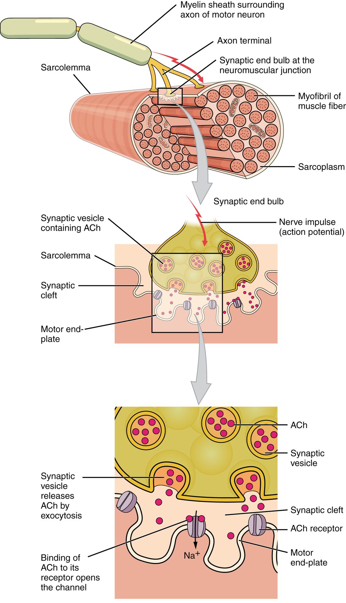
The neuromuscular junction (NMJ; neur/o = nerve, muscul/o = muscle) is the synapse between the axon terminal of a motor neuron and a skeletal muscle fiber (Figure 1). It has three parts:
- The axon terminal (synaptic end bulb) sits in a shallow dip on the fiber's surface. It is packed with synaptic vesicles, small membrane sacs each holding several thousand molecules of the neurotransmitter acetylcholine, and with mitochondria that supply the ATP it needs to make and package them.
- The synaptic cleft is the gap between the terminal and the fiber, about 50 nm wide here, more than twice the width of most synapses between neurons (about 20 nm). It is filled with extracellular fluid and a thin layer of basal lamina, and it holds the enzyme that will end the signal.
- The motor end plate is the patch of sarcolemma directly under the terminal. It is thrown into deep folds, which increase its surface area. The crests of the folds are packed with acetylcholine receptor proteins, about 10,000 in every square micrometer. The depths of the folds and the membrane around the end plate carry voltage-gated sodium channels.
Acetylcholine
Acetylcholine (ACh; say a-SEE-til-KOH-leen) is the neurotransmitter that motor neurons release onto skeletal muscle. It is a small molecule built from two parts, an acetyl group and choline, and its name says so.
- The axon terminal makes ACh itself, joining choline to an acetyl group with an enzyme.
- A transport protein in the vesicle membrane loads the ACh into synaptic vesicles.
- Many loaded vesicles sit docked at the terminal membrane that faces the cleft, ready to be released.
ACh is also used at many other synapses, by some autonomic neurons and in the brain. At the neuromuscular junction it always excites: there are no inhibitory synapses on skeletal muscle fibers. A muscle fiber relaxes only when its motor neuron stops firing.
Step by step: from nerve impulse to muscle action potential
Here is what happens each time an action potential reaches the end of a motor neuron's axon. You saw the general version of this sequence in chemical signaling; at the neuromuscular junction every step is fast and reliable.
- An action potential arrives at the axon terminal. It depolarizes the terminal membrane.
- Voltage-gated calcium channels open. Calcium ions (Ca2+) are about 10,000 times more concentrated in the extracellular fluid than in the cytosol, and the inside of the terminal is negative, so calcium rushes in down its electrochemical gradient.
- Calcium triggers exocytosis. Calcium binds sensor proteins on the docked vesicles. Within a fraction of a millisecond, dozens to a hundred or more vesicles fuse with the terminal membrane and empty their ACh into the cleft.
- ACh diffuses across the cleft. Fifty nanometers takes it only microseconds.
- ACh binds receptor proteins on the motor end plate. Each receptor protein must bind two ACh molecules before it opens.
- The channels open and positive charge flows in. The end plate depolarizes: this is the end plate potential.
- The end plate potential triggers an action potential. Charge spreads to the neighboring membrane, which reaches threshold, and an action potential sweeps away from the end plate toward both ends of the fiber.
- Acetylcholinesterase splits ACh, and the signal ends. Within a millisecond or two, the cleft is cleared and the channels close.
The rest of this page takes the key steps one at a time.
Calcium-triggered transmitter release
Calcium is the link between the electrical signal and the chemical one. The action potential itself releases nothing: it opens calcium channels, and the calcium that enters makes the vesicles fuse. This is calcium-triggered transmitter release.
Two consequences follow:
- Release needs calcium outside the cell. If the extracellular calcium around a junction is removed, action potentials still reach the terminal, but no ACh is released and the muscle does not respond.
- Release comes in packets. Each vesicle holds roughly the same amount of ACh, so ACh is released in fixed packets, one per vesicle. An action potential releases many packets at once. Even at rest, a single vesicle occasionally fuses by chance and causes a tiny depolarization of the end plate, far too small to reach threshold.
Acetylcholine receptors at the end plate
The receptor proteins on the motor end plate are nicotinic receptor proteins, named after a compound in tobacco leaves that also binds and opens them. (Another family of ACh receptor proteins, found on the heart and many glands, does not respond to that compound; you will meet both families in the autonomic chapter.)
A nicotinic receptor protein is a ligand-gated channel. Five protein subunits ring a central pore, and two of them carry a binding site for ACh.
- When both sites are occupied, the protein changes shape and the pore opens for about a millisecond.
- The open pore lets small positive ions through: sodium in and potassium out.
- At a resting potential of about −90 mV, sodium's inward push is far stronger than potassium's outward push. More positive charge enters than leaves, and the membrane depolarizes.
The muscle's receptor protein binds ACh only briefly, then lets it go. Whether it binds again depends on whether any ACh is still in the cleft, and that is set by the enzyme in the next section but one.
The end plate potential
The end plate potential (EPP) is the depolarization of the motor end plate caused by ACh opening the channels of its receptor proteins. It is a graded potential, the kind you met in the electrical signals topic:
- Its size depends on how much ACh is released and how many ACh-gated channels open.
- It is local: it spreads a short way along the sarcolemma and fades.
- The receptor-packed crests of the folds do not fire. The EPP triggers an action potential in the fold depths and the membrane around the end plate, which are rich in voltage-gated sodium channels.
What makes the neuromuscular junction special is the size of the EPP. At synapses between neurons in the brain, one input usually shifts the voltage by less than a millivolt, and a neuron fires only when many inputs add up. At the neuromuscular junction, one action potential in the motor neuron releases enough ACh to depolarize the end plate several times more than needed to reach threshold: a safety margin of about three to five times. So every action potential in a motor neuron produces an action potential in each muscle fiber it supplies, one for one, even when the motor neuron fires rapidly and each release gets a little smaller.
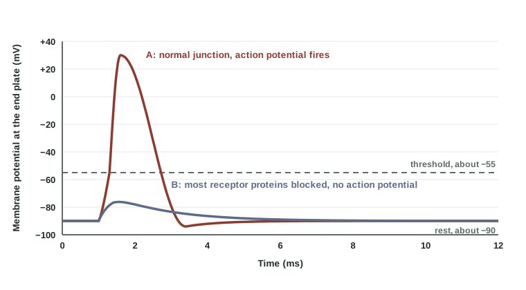
Figure 2 shows why the safety margin matters. A healthy junction can lose a large share of its receptor proteins, or release less ACh, and still reach threshold. Only when the EPP shrinks below threshold does the fiber stop responding. That is the point at which a drug that blocks receptor proteins, or a disease that destroys them, causes weakness.
| End plate potential | Action potential in the muscle fiber | |
|---|---|---|
| Where it happens | The motor end plate only | The sarcolemma next to the end plate, then the whole fiber |
| What opens the channels | ACh binding (ligand-gated channels) | Depolarization to threshold (voltage-gated sodium and potassium channels) |
| Size | Graded: varies with the ACh released and the receptor proteins available | All-or-none: the same full size every time |
| Over distance | Fades within a short distance | Regenerated along the whole fiber |
| Needs threshold? | No | Yes |
| Its job | Bring the neighboring membrane to threshold | Carry the signal along the fiber and into its T tubules |
Acetylcholinesterase: ending the signal
If ACh stayed in the cleft, it would keep binding receptor proteins, the end plate would stay depolarized, and the fiber could not respond cleanly to the next command. Acetylcholinesterase (AChE; -ase = enzyme) is the enzyme that prevents this. It is anchored in the basal lamina of the synaptic cleft, between the terminal and the end plate.
- AChE hydrolyzes ACh into acetate and choline. Neither one can open the ACh-gated channels.
- It works extremely fast: one enzyme molecule can split thousands of ACh molecules per second. Much of the released ACh is destroyed within about a millisecond, some of it before it ever reaches a receptor protein.
- A transport protein in the axon terminal takes the choline back into the terminal, where it is joined to a new acetyl group to make more ACh.
- With ACh gone, the ACh-gated channels close, the end plate returns to rest, and the junction is ready for the next action potential.
So one action potential in the motor neuron produces one brief pulse of ACh, one EPP and one action potential in the fiber. A steady contraction needs a steady stream of motor neuron action potentials.
Drugs, poisons and disease at the junction
Because the neuromuscular junction works in clear steps, you can predict what happens when a step is blocked. Each row below targets one step.
| Step affected | Example | What it does | Result |
|---|---|---|---|
| Release | Botulinum toxin, from the bacterium that causes botulism | Destroys proteins the vesicles need to fuse, so ACh is not released | Weakness that spreads and can stop breathing; in tiny local doses (Botox), relaxes chosen muscles for months |
| Binding | Curare-type drugs, such as rocuronium | Occupy the ACh binding sites on nicotinic receptor proteins without opening them | EPP too small to reach threshold; paralysis of skeletal muscle, used during surgery |
| Breakdown | Neostigmine; organophosphate insecticides and nerve agents | Block AChE, so ACh lingers in the cleft | Neostigmine reverses curare-type drugs. A large block causes repeated firing and quivering, then weakness as the end plate stays depolarized |
Blocking the receptor proteins
A curare-type drug competes with ACh for the binding sites. With enough drug present, too few ACh-gated channels open, and the EPP stays below threshold, as in trace B of Figure 2. The motor neuron still fires and still releases ACh, but the muscle fibers do not respond. During surgery this relaxes the muscles completely, including the diaphragm, so a ventilator must breathe for the patient.
Because the block is competitive, more ACh can overcome it. That is how neostigmine works at the end of surgery: by slowing AChE, it lets ACh build up in the cleft until it outcompetes the remaining drug.
Blocking acetylcholinesterase
When AChE is blocked, ACh keeps binding and rebinding receptor proteins after each release. At first the EPP lasts longer and can trigger several action potentials instead of one, so muscles quiver. If the block is large and lasting, as in organophosphate poisoning, the end plate stays depolarized. The voltage-gated sodium channels around it close and cannot reopen while the membrane stays depolarized, as you saw in the electrical signals topic, so the fiber stops firing. The result is weakness and, in severe cases, failure of the breathing muscles.