The neuromuscular junction
1Why this matters
Before Ms. Alvarez's abdominal surgery, the anesthesiologist puts her to sleep and then injects rocuronium. Within a minute every skeletal muscle in her body goes limp, including her diaphragm, so a ventilator breathes for her. Her nerves still fire and her muscles are healthy: the drug acts in the narrow gap where each nerve ending meets a muscle fiber. When surgery ends, a second drug, neostigmine, lets her breathe on her own again.
2What this builds on
3Quick check before you start
1. What opens a ligand-gated channel?
- A change in membrane voltage
- A specific molecule binding to it
- Stretching of the membrane
Show the answer
A ligand-gated channel opens when a specific molecule, its ligand, binds a site on the channel protein. Voltage opens voltage-gated channels, and stretch opens mechanically gated ones.
- A change in membrane voltage:
- Correct: A specific molecule binding to it:
- Stretching of the membrane:
2. A depolarization stops short of threshold. What happens?
- A smaller action potential fires
- A full action potential fires after a delay
- It fades away with no action potential
Show the answer
Action potentials are all-or-none. A depolarization that does not reach threshold is a graded potential that simply fades.
- A smaller action potential fires:
- A full action potential fires after a delay:
- Correct: It fades away with no action potential:
3. At a synapse between a neuron and its target cell, what carries the signal across the synaptic cleft?
- A neurotransmitter, by diffusion
- Ions flowing through gap junctions
- The action potential itself
Show the answer
The two cells do not touch. The axon terminal releases a neurotransmitter, which diffuses across the cleft and binds receptor proteins on the other cell.
- Correct: A neurotransmitter, by diffusion:
- Ions flowing through gap junctions:
- The action potential itself:
4Anatomy
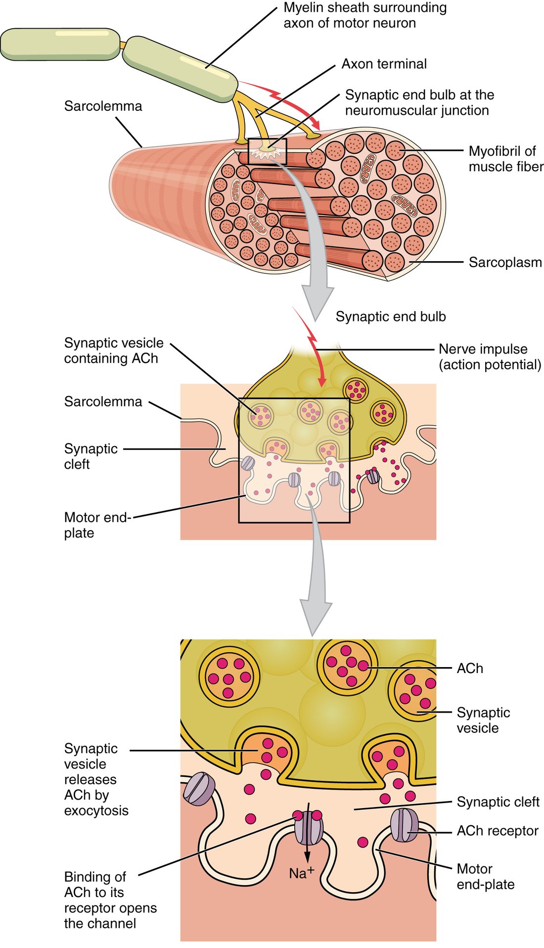
With labels hidden, select a box to reveal its label.
5How it works, step by step
- An action potential travels down the motor neuron's axon and depolarizes its axon terminal.Voltage-gated calcium channels in the terminal open, and calcium ions rush in.
- Calcium binds sensor proteins on the docked synaptic vesicles.The vesicles fuse with the terminal membrane and release acetylcholine into the synaptic cleft by exocytosis.
- Acetylcholine diffuses across the cleft and binds nicotinic receptor proteins on the motor end plate.Their channels open, sodium flows in, and the end plate depolarizes: the end plate potential.
- The end plate potential spreads to the neighboring sarcolemma and brings it to threshold.Voltage-gated sodium channels open, and an action potential spreads along the muscle fiber.
- Acetylcholinesterase in the cleft splits acetylcholine into acetate and choline.The channels of the receptor proteins close and the end plate returns to rest, ready for the next nerve impulse.
6Core concepts
7A common mistake
The wrong idea: The nerve's electrical impulse jumps straight across into the muscle fiber.
What actually happens: The axon terminal and the muscle fiber do not touch, and no current passes between them. The action potential in the terminal lets calcium in, calcium releases acetylcholine, and acetylcholine diffuses across the cleft and opens channels in the end plate. The muscle fiber then makes its own, new action potential. That chemical step is why drugs and toxins that change release, binding or breakdown of acetylcholine can weaken or paralyze muscles whose nerves are working normally.
8Check yourself
Anything you miss goes into your review queue.
1. Put the events of transmission at the neuromuscular junction in order.
- An action potential depolarizes the axon terminal
- Voltage-gated calcium channels open and calcium enters the terminal
- Synaptic vesicles fuse with the terminal membrane and release acetylcholine
- Acetylcholine diffuses across the synaptic cleft
- Acetylcholine binds nicotinic receptor proteins and their channels open
- The end plate potential brings the neighboring sarcolemma to threshold
Show the answer
The nerve impulse opens calcium channels; calcium triggers exocytosis of acetylcholine; acetylcholine crosses the cleft and opens ligand-gated channels on the end plate; the resulting end plate potential brings the nearby membrane to threshold, and the muscle fiber fires an action potential.
- Correct order: 1. An action potential depolarizes the axon terminal 2. Voltage-gated calcium channels open and calcium enters the terminal 3. Synaptic vesicles fuse with the terminal membrane and release acetylcholine 4. Acetylcholine diffuses across the synaptic cleft 5. Acetylcholine binds nicotinic receptor proteins and their channels open 6. The end plate potential brings the neighboring sarcolemma to threshold
2. A patient receives a drug that blocks most of the acetylcholine receptor proteins at the motor end plate. Her motor neurons keep firing normally. Predict the change in each variable.
| Variable | Change |
|---|---|
| Action potentials reaching the axon terminal | — |
| Acetylcholine released per nerve impulse | — |
| Size of the end plate potential | — |
| Action potentials in the muscle fibers | — |
| Strength of contraction | — |
Show the answer
Blocking the receptor proteins breaks the chain at the muscle side: acetylcholine is released normally but cannot open enough channels. The end plate potential shrinks below threshold, the fibers stop firing, and the muscle weakens, even though the nerve is working.
- Action potentials reaching the axon terminal: no change. The drug acts on the muscle side of the junction; the motor neuron and its axon are unaffected.
- Acetylcholine released per nerve impulse: no change. Calcium entry and exocytosis in the terminal are unchanged, so the same amount of acetylcholine is released.
- Size of the end plate potential: down. With most receptor proteins blocked, fewer ligand-gated channels open, so less sodium enters the end plate.
- Action potentials in the muscle fibers: down. Once the end plate potential falls below threshold, the neighboring sarcolemma no longer fires.
- Strength of contraction: down. Fibers that no longer fire action potentials do not contract, so the muscle weakens or goes limp.
3. In a lab, a nerve–muscle preparation is placed in a fluid with no calcium. Stimulating the nerve still produces action potentials that reach the axon terminals, but the muscle does not respond. What explains this?
- Without calcium outside, the muscle fibers' voltage-gated sodium channels stay closed
- The action potential cannot depolarize the terminal without calcium
- Calcium entering the terminal is what triggers the vesicles to release acetylcholine
- Acetylcholinesterase needs calcium to split acetylcholine
Show the answer
Release is calcium-triggered. The action potential opens voltage-gated calcium channels, and the calcium that flows in makes the vesicles fuse. With no calcium outside, nothing flows in and no acetylcholine is released.
- Without calcium outside, the muscle fibers' voltage-gated sodium channels stay closed: The fibers' sodium channels do not need outside calcium to open; the chain fails earlier, at release from the terminal.
- The action potential cannot depolarize the terminal without calcium: The question says the action potentials do reach and depolarize the terminals; it is the next step that fails.
- Correct: Calcium entering the terminal is what triggers the vesicles to release acetylcholine: Correct. No calcium entry means no exocytosis of acetylcholine.
- Acetylcholinesterase needs calcium to split acetylcholine: If the enzyme failed, acetylcholine would linger and the muscle would respond more, not less.
4. Trace B was recorded from the same end plate as trace A after a drug was given. Why does trace B show no action potential?
- The drug raised the threshold of the sarcolemma
- The drug stopped the axon terminal from releasing any acetylcholine
- The end plate potential was too small to reach threshold
- The drug held the membrane depolarized so the sodium channels could not reopen
Show the answer
In trace B the membrane depolarizes only to about −75 mV and fades, well below the threshold line at about −55 mV. With most receptor proteins blocked, too few channels open, and a graded potential that does not reach threshold triggers nothing.
- The drug raised the threshold of the sarcolemma: The threshold line is the same for both traces; what changed is how far the membrane depolarized.
- The drug stopped the axon terminal from releasing any acetylcholine: Trace B still shows a small depolarization, so some acetylcholine was released and bound. With no release at all there would be no bump.
- Correct: The end plate potential was too small to reach threshold: Correct. The end plate potential stayed below threshold.
- The drug held the membrane depolarized so the sodium channels could not reopen: Trace B returns to rest within a few milliseconds; it does not stay depolarized.
5. Select every statement that is true of the end plate potential.
- Its size depends on how much acetylcholine binds
- It is all-or-none
- It is caused by ligand-gated channels opening
- It spreads along the whole fiber without fading
- In a healthy junction, it is several times larger than needed to reach threshold
Show the answer
The end plate potential is a graded, local depolarization made by acetylcholine opening ligand-gated channels. In a healthy junction it is large enough to bring the neighboring membrane to threshold with room to spare. The action potential it triggers is what is all-or-none and travels the whole fiber.
- Correct: Its size depends on how much acetylcholine binds: Correct. More acetylcholine and more open channels make a bigger end plate potential.
- It is all-or-none: It is graded; the action potential it triggers is all-or-none.
- Correct: It is caused by ligand-gated channels opening: Correct. Acetylcholine opens the nicotinic receptor channels.
- It spreads along the whole fiber without fading: It fades within a short distance; the action potential carries the signal along the fiber.
- Correct: In a healthy junction, it is several times larger than needed to reach threshold: Correct. This safety margin makes transmission reliable.
6. A student wrote the steps at the neuromuscular junction. One step is wrong. Which one?
- The action potential opens voltage-gated calcium channels in the axon terminal
- Calcium triggers exocytosis of acetylcholine
- Acetylcholine opens nicotinic receptor channels, and sodium flows in
- The end plate potential triggers an action potential in the neighboring sarcolemma
- The axon terminal takes acetylcholine back up intact to end the signal
Show the answer
At the neuromuscular junction, acetylcholinesterase in the cleft ends the signal by splitting acetylcholine into acetate and choline. Only the choline is taken back into the terminal.
- The action potential opens voltage-gated calcium channels in the axon terminal: This step is right. Depolarization of the terminal opens its calcium channels.
- Calcium triggers exocytosis of acetylcholine: This step is right. Calcium entry makes the vesicles fuse and release acetylcholine.
- Acetylcholine opens nicotinic receptor channels, and sodium flows in: This step is right. The receptor protein is a ligand-gated channel, and sodium's inward push dominates.
- The end plate potential triggers an action potential in the neighboring sarcolemma: This step is right. The end plate potential brings the nearby membrane, rich in voltage-gated sodium channels, to threshold.
- Correct: The axon terminal takes acetylcholine back up intact to end the signal: This is the error. Acetylcholine is split in the cleft, not taken back intact.
7. A farmworker is splashed with an organophosphate insecticide, which blocks acetylcholinesterase. At first his muscles quiver; hours later they are weak, and he struggles to breathe. What explains the later weakness?
- His motor neurons have used up all their stored acetylcholine
- Acetylcholine has destroyed the receptor proteins
- The end plate stays depolarized and nearby sodium channels cannot reopen
- The insecticide also blocks voltage-gated calcium channels in his axon terminals
Show the answer
With the enzyme blocked, acetylcholine lingers and keeps the end plate depolarized. Early on, the prolonged end plate potential fires repeated action potentials, so muscles quiver. With lasting depolarization, voltage-gated sodium channels around the end plate close and stay closed, so the fiber can no longer fire.
- His motor neurons have used up all their stored acetylcholine: The terminals still make and release acetylcholine; the problem is that it is not cleared.
- Acetylcholine has destroyed the receptor proteins: Acetylcholine binds and releases its receptor proteins without destroying them.
- Correct: The end plate stays depolarized and nearby sodium channels cannot reopen: Correct. A persistently depolarized membrane cannot fire new action potentials.
- The insecticide also blocks voltage-gated calcium channels in his axon terminals: Organophosphates block acetylcholinesterase, not calcium channels.
9Summary
A motor neuron carries commands from the brain or spinal cord to skeletal muscle fibers; each fiber has one neuromuscular junction, where the motor neuron's axon terminal faces the folded motor end plate across a synaptic cleft about 50 nm wide. An action potential in the terminal opens voltage-gated calcium channels, and entering calcium triggers exocytosis of synaptic vesicles full of acetylcholine. Acetylcholine diffuses across the cleft and binds nicotinic receptor proteins, ligand-gated channels that let sodium in, producing the end plate potential: a graded depolarization several times larger than needed to reach threshold. It triggers an action potential in the neighboring sarcolemma, one for every motor neuron impulse. Acetylcholinesterase in the cleft splits acetylcholine into acetate and choline within about a millisecond, ending the signal; the choline is taken back into the terminal. Botulinum toxin blocks release, curare-type drugs block the receptor proteins, and AChE inhibitors make acetylcholine linger.