Excitation–contraction coupling
1Why this matters
Twenty minutes into a routine knee operation, 24-year-old Daniel's jaw muscles lock tight, his heart races and his temperature starts to climb fast. The anesthetist stops the anesthetic gas and gives dantrolene. Daniel was born with a faulty calcium release channel in his muscle fibers, harmless until the gas made it leak. To see why one leaky channel can lock every muscle in the body, you need the chain that links a muscle fiber's action potential to its contraction.
2What this builds on
3Quick check before you start
1. What does the end plate potential do at a healthy neuromuscular junction?
- Brings the neighboring sarcolemma to threshold, so the fiber fires an action potential
- Travels the whole length of the fiber without fading
- Releases calcium from the axon terminal
Show the answer
The end plate potential is a large, local, graded depolarization. It brings the sarcolemma next to the end plate to threshold, and that membrane fires the action potential that travels along the fiber.
- Correct: Brings the neighboring sarcolemma to threshold, so the fiber fires an action potential:
- Travels the whole length of the fiber without fading:
- Releases calcium from the axon terminal:
2. In a resting muscle fiber, what stops the myosin heads from binding actin?
- The Z discs hold the thin filaments away from the thick ones
- Tropomyosin covers the myosin-binding sites on actin
- The myosin heads carry no energy until they are signaled
Show the answer
At rest, tropomyosin lies over the myosin-binding sites on actin, held in place by troponin.
- The Z discs hold the thin filaments away from the thick ones:
- Correct: Tropomyosin covers the myosin-binding sites on actin:
- The myosin heads carry no energy until they are signaled:
3. The sodium–potassium pump uses ATP directly to move ions against their gradients. What kind of transport is that?
- Facilitated diffusion
- Secondary active transport
- Primary active transport
Show the answer
Primary active transport uses the energy of ATP directly, through a pump that is itself an ATPase.
- Facilitated diffusion:
- Secondary active transport:
- Correct: Primary active transport:
4Anatomy
With labels hidden, select a box to reveal its label.
5How it works, step by step
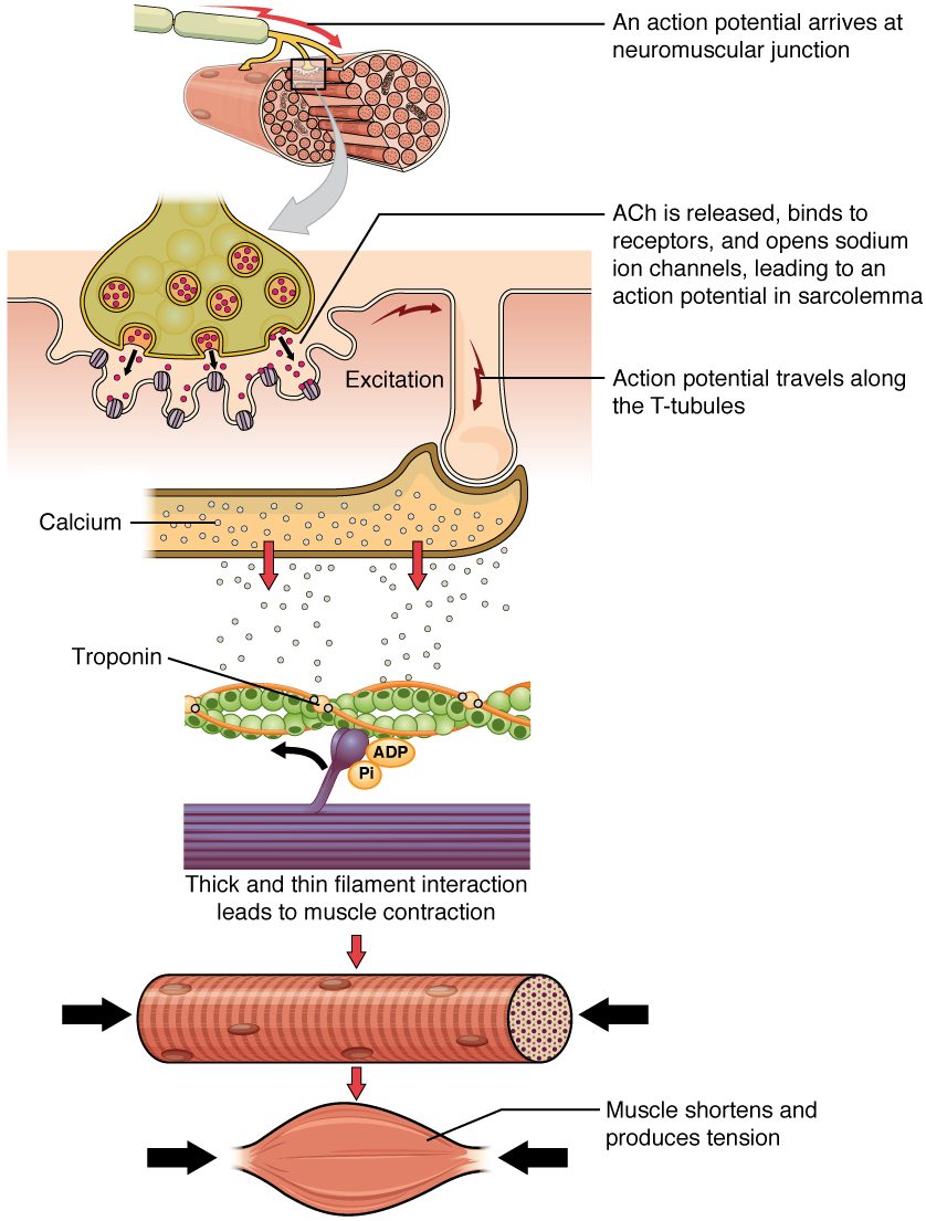
- The end plate potential brings the sarcolemma to threshold.An action potential spreads along the sarcolemma and down every T tubule into the fiber.
- The action potential depolarizes the T tubule membrane at each triad.DHP receptors, the voltage sensors, change shape and pull open the ryanodine receptors in the terminal cisternae.
- The ryanodine receptors are open.Calcium flows out of the sarcoplasmic reticulum, and its concentration in the sarcoplasm rises about a hundredfold.
- Calcium binds troponin.Troponin changes shape and tropomyosin rolls off the myosin-binding sites on actin.
- The binding sites are uncovered.Myosin heads bind actin and pull, and the sarcomeres shorten until calcium pumps return the calcium to the SR.
6Core concepts
7A common mistake
The wrong idea: The calcium that triggers skeletal muscle contraction flows in from outside the cell.
What actually happens: In skeletal muscle, the calcium comes from the sarcoplasmic reticulum inside the fiber. The action potential in the T tubule changes the shape of the DHP receptor, which is physically linked to the ryanodine receptor and pulls it open. Voltage alone is the trigger, so a skeletal muscle fiber stimulated directly (not through its nerve) keeps contracting for a while in a bath with no calcium. Heart muscle is the one that depends on calcium entering from outside.
8Check yourself
Anything you miss goes into your review queue.
1. Put the steps of excitation–contraction coupling in a skeletal muscle fiber in order.
- An action potential spreads along the sarcolemma
- The action potential travels down the T tubules
- DHP receptors change shape and pull open ryanodine receptors
- Calcium flows out of the sarcoplasmic reticulum
- Calcium binds troponin
- Tropomyosin uncovers the myosin-binding sites on actin
Show the answer
The action potential spreads over the fiber and into the T tubules. At the triads, the voltage-sensing DHP receptors pull open the ryanodine receptors, calcium leaves the SR, binds troponin, and tropomyosin moves off the binding sites so myosin can bind actin.
- Correct order: 1. An action potential spreads along the sarcolemma 2. The action potential travels down the T tubules 3. DHP receptors change shape and pull open ryanodine receptors 4. Calcium flows out of the sarcoplasmic reticulum 5. Calcium binds troponin 6. Tropomyosin uncovers the myosin-binding sites on actin
2. The action potential is over within about 5 ms, yet force keeps rising until about 35 ms. What keeps the filaments pulling after the action potential has ended?
- More action potentials fire in the T tubules after the first
- Calcium remains in the sarcoplasm until the pumps remove it
- Acetylcholine stays bound to the end plate
- The ryanodine receptors stay open for about 35 ms
Show the answer
Calcium stays bound to troponin until the SR pumps bring the sarcoplasmic calcium back down, which takes tens of milliseconds. Force builds while calcium is present, even though the release channels closed when the action potential ended.
- More action potentials fire in the T tubules after the first: The top trace shows a single action potential; nothing else fires.
- Correct: Calcium remains in the sarcoplasm until the pumps remove it: Correct. The calcium trace falls slowly because pumping back takes time.
- Acetylcholine stays bound to the end plate: Acetylcholinesterase clears acetylcholine within a millisecond or two.
- The ryanodine receptors stay open for about 35 ms: The release channels close when the membrane repolarizes; the calcium trace starts falling within 10 ms.
3. In skeletal muscle, what is the job of the DHP receptor in the T tubule membrane?
- It pumps calcium back into the sarcoplasmic reticulum
- It senses the voltage change and pulls open the SR release channel
- It binds acetylcholine released by the motor neuron
- It lets in the calcium from outside the cell that then binds troponin
Show the answer
The DHP receptor is the voltage sensor. When the T tubule depolarizes it changes shape, and because it is linked to the ryanodine receptor, it pulls that release channel open.
- It pumps calcium back into the sarcoplasmic reticulum: Pumping calcium back is the job of the SR calcium ATPase.
- Correct: It senses the voltage change and pulls open the SR release channel: Correct. It is the voltage sensor linked to the release channel.
- It binds acetylcholine released by the motor neuron: Acetylcholine binds nicotinic receptor proteins on the motor end plate, not in the T tubules.
- It lets in the calcium from outside the cell that then binds troponin: In skeletal muscle, the calcium that binds troponin comes from the SR, not from outside; the DHP receptor's job is to sense voltage.
4. An experimental drug blocks the calcium pumps of the sarcoplasmic reticulum. A single action potential is triggered immediately afterward. Predict the change in each variable, compared with a normal fiber.
| Variable | Change |
|---|---|
| Calcium released from the SR by this action potential | — |
| Time calcium stays high in the sarcoplasm | — |
| Time until the fiber relaxes | — |
| Calcium stored in the SR afterward | — |
Show the answer
The SR calcium pumps end contraction by clearing calcium. Blocking them leaves release unchanged but makes calcium, and therefore contraction, last much longer, and it empties the store.
- Calcium released from the SR by this action potential: no change. Release through the ryanodine receptors does not depend on the pumps, so this first release is normal.
- Time calcium stays high in the sarcoplasm: up. The pumps are what remove calcium from the sarcoplasm; without them it lingers.
- Time until the fiber relaxes: up. Calcium stays bound to troponin, so tropomyosin keeps the binding sites uncovered and the filaments keep pulling.
- Calcium stored in the SR afterward: down. Released calcium is not pumped back, so the store is not refilled for the next action potential.
5. A skeletal muscle fiber is placed in a bath with no calcium and stimulated directly with an electrode. It keeps contracting normally for a while. What does this show?
- Its calcium comes from the SR, released by voltage
- Skeletal muscle contracts without any calcium
- The electrode supplies the calcium
- Troponin does not need calcium in skeletal muscle
Show the answer
Direct stimulation bypasses the neuromuscular junction, whose transmitter release does need outside calcium. The fiber's action potential then opens the SR release channels through the linked DHP receptors, so contraction uses calcium stored inside the fiber.
- Correct: Its calcium comes from the SR, released by voltage: Correct. Voltage opens the SR store; outside calcium is not needed.
- Skeletal muscle contracts without any calcium: Calcium is still the switch; it simply comes from inside the fiber.
- The electrode supplies the calcium: An electrode delivers current, not calcium ions.
- Troponin does not need calcium in skeletal muscle: Troponin must bind calcium before tropomyosin moves; the calcium comes from the SR.
6. Daniel has an inherited defect that makes his ryanodine receptors leak calcium when he breathes certain anesthetic gases. During surgery his muscles go rigid and his temperature climbs fast. What best explains both signs?
- The gas blocks acetylcholinesterase and the muscles fire nonstop
- Nonstop calcium release keeps filaments pulling and burns ATP, releasing heat
- The gas stops the SR calcium pumps and no ATP is used
- The defect blocks action potentials in the T tubules
Show the answer
Leaking release channels keep calcium high, so troponin stays on and the muscles stay contracted. The calcium pumps work nonstop to clear calcium and the myosin heads keep splitting ATP. All that ATP use releases heat, so body temperature climbs.
- The gas blocks acetylcholinesterase and the muscles fire nonstop: The problem is in the SR release channel, not at the neuromuscular junction.
- Correct: Nonstop calcium release keeps filaments pulling and burns ATP, releasing heat: Correct. Uncontrolled calcium release drives both the rigidity and the heat.
- The gas stops the SR calcium pumps and no ATP is used: The pumps keep working hard and using ATP; that is part of where the heat comes from.
- The defect blocks action potentials in the T tubules: The electrical steps are normal; the defect is in the channel the voltage sensor opens.
7. Why is muscle relaxation described as an active process?
- The muscle fiber pushes itself back to its resting length
- The motor neuron sends a separate signal that makes the fiber relax
- ATP-driven pumps return calcium to the SR
- Acetylcholine is pumped back into the axon terminal
Show the answer
Relaxation requires the sarcoplasmic calcium to fall, and that means pumping it back into the SR against a gradient of thousands to one, using ATP. (The myosin heads also need ATP to let go of actin.)
- The muscle fiber pushes itself back to its resting length: Muscle can only pull. Elastic proteins, opposing muscles and gravity lengthen it.
- The motor neuron sends a separate signal that makes the fiber relax: There are no inhibitory signals to skeletal muscle; the motor neuron simply stops firing.
- Correct: ATP-driven pumps return calcium to the SR: Correct. Calcium ATPases use ATP to clear the sarcoplasm.
- Acetylcholine is pumped back into the axon terminal: Acetylcholine is split by acetylcholinesterase in the cleft; only its choline is taken back.
9Summary
Excitation–contraction coupling links the muscle fiber action potential to contraction, and calcium is the link. The action potential, lasting only 2 to 5 ms, spreads from the end plate along the sarcolemma and down the T tubules. At each triad, DHP receptors in the T tubule membrane sense the voltage change and, being physically linked to them, pull open the ryanodine receptors of the sarcoplasmic reticulum. Calcium flows out of the SR and binds troponin, which moves tropomyosin off the myosin-binding sites on actin, and myosin heads bind and pull. Relaxation reverses the chain: the motor neuron stops firing, acetylcholinesterase clears the cleft, the release channels close, and SR calcium pumps (calcium ATPases) use ATP to pump calcium back. Calcium leaves troponin and tropomyosin covers actin again. Because pumping takes tens of milliseconds, force outlasts the brief action potential.