Every thought, movement and sensation depends on neurons passing signals to one another at synapses, and most of the drugs that act on your nervous system act there too. This page walks through the synaptic transmission steps in order, from the action potential arriving at an axon terminal to the neurotransmitter being cleared away. It then explains electrical synapses, excitatory and inhibitory postsynaptic potentials, how a neuron adds up its inputs, the two classes of receptor protein, the major neurotransmitters, and how drugs and toxins change each step.
The synapse in detail
You met the basic synapse in chemical signaling: a junction where a neuron passes a signal to another cell, with a narrow synaptic cleft between them. Now name the two neurons. The presynaptic neuron (pre- = before) sends the signal from its axon terminal. The postsynaptic neuron (post- = after) receives it, usually on a dendrite or the cell body. The same neuron is postsynaptic at the thousands of synapses it receives and presynaptic at the synapses its own axon makes.
A single motor neuron in your spinal cord receives around 10,000 synapses. Some excite it, some inhibit it, and whether it fires depends on the sum. The rest of this page builds up to that sum.
Electrical synapses
At an electrical synapse, the two neurons are joined by gap junctions, the protein tunnels between neighboring cells that you met in cell junctions. Ions flow straight through them from one cell's cytosol into the other's, so current passes directly, as it does between patches of one axon.
- Almost no delay. There is no messenger to release, so the signal crosses in a tiny fraction of a millisecond.
- Usually two-way. Current can flow in either direction.
- Synchronizing. Groups of neurons joined this way tend to fire together, in step. You find electrical synapses in some brain regions where that matters, and in the developing nervous system.
- Little control. A gap junction passes whatever current arrives. It cannot amplify or invert the signal, although chemical messengers can slowly turn the coupling up or down.
The same kind of gap junction joins the cells of cardiac muscle, which is how an action potential spreads from cell to cell across the heart.
Chemical synapses
Most synapses in your body are chemical. At a chemical synapse, the two cells are separated by a synaptic cleft about 20 nm wide, far too wide for current to jump. The signal crosses as a neurotransmitter. That extra step costs about half a millisecond, the synaptic delay, but it gives the synapse a great deal of control: the same arriving action potential can excite one cell and inhibit another, be amplified, or be changed by drugs.
| Electrical synapse | Chemical synapse | |
|---|---|---|
| How the cells connect | Gap junctions join their cytosols | A synaptic cleft about 20 nm wide separates them |
| What crosses | Ions (current) directly | A neurotransmitter, by diffusion |
| Delay | Almost none | About 0.5 ms or more |
| Direction | Usually both ways | One way: presynaptic to postsynaptic |
| Effect on the next cell | Follows the first cell's voltage | Excitatory or inhibitory, depending on the receptor protein |
| Can drugs and learning adjust it? | Very little | Yes, at many steps |
| Where found | Some brain regions; between the cells of cardiac muscle and of single-unit smooth muscle | Most synapses, including the neuromuscular junction |
Chemical synaptic transmission, step by step
You saw the core of this at the neuromuscular junction. The same sequence runs at every chemical synapse (Figure 1):
- An action potential reaches the axon terminal. It depolarizes the terminal membrane.
- Voltage-gated calcium channels open. Calcium is about 10,000 times more concentrated in the extracellular fluid than in the cytosol, and the inside is negative, so calcium rushes in down its electrochemical gradient.
- Calcium triggers exocytosis. Synaptic vesicles filled with neurotransmitter are docked at the membrane. Calcium binds sensor proteins on them, and within a fraction of a millisecond the vesicles fuse with the membrane and release their contents into the cleft.
- The neurotransmitter diffuses across the cleft. Twenty nanometers takes well under a millisecond.
- It binds receptor proteins on the postsynaptic membrane. Binding is specific and reversible, like any ligand on its receptor protein.
- Ion channels open or close, and the postsynaptic membrane potential changes. Some receptor proteins are channels themselves; others act through a G protein. The result is a graded potential in the postsynaptic cell.
- The neurotransmitter is removed and the signal ends, within milliseconds for most fast synapses. (The ways it is removed have their own section below.)
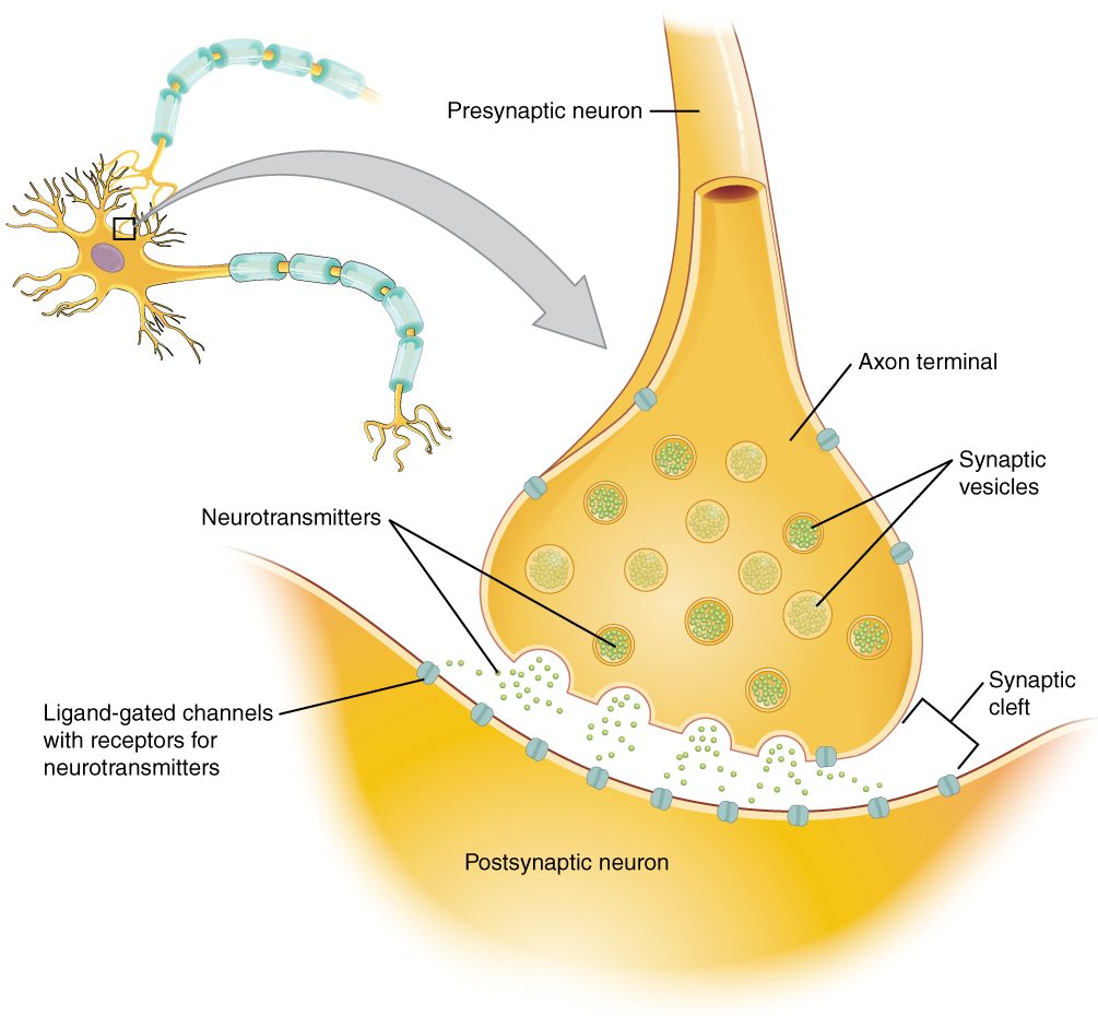
Notice what sets the amount released: calcium. More action potentials per second let more calcium in, and more vesicles fuse. Remove calcium from the fluid around a synapse and the action potential still arrives, but nothing is released.
Two classes of receptor protein
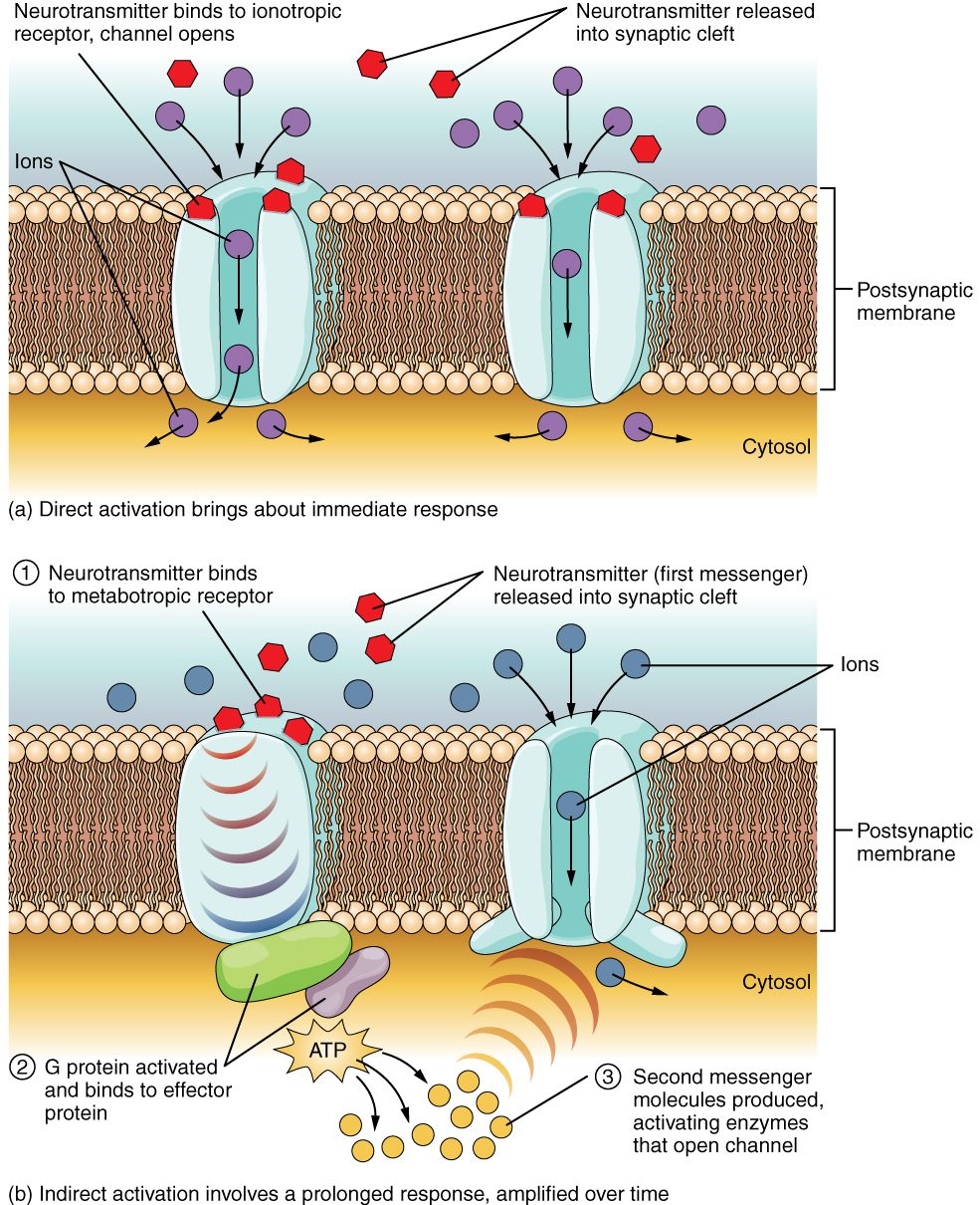
What the neurotransmitter does depends on the receptor protein it binds. There are two classes (Figure 2):
- An ionotropic receptor protein is itself a ligand-gated ion channel (iono- = ion, trop- = turning or acting on). Binding opens the pore at once. The effect starts within a millisecond and ends as soon as the neurotransmitter leaves. The nicotinic receptor proteins at the neuromuscular junction are ionotropic.
- A metabotropic receptor protein is a G-protein-coupled receptor (metabo- = change, as in metabolism). It has no pore. Binding switches on a G protein inside the cell, which either acts on a nearby ion channel directly or switches on an enzyme that makes a second messenger. The effect starts after tens of milliseconds or more, lasts from hundreds of milliseconds to minutes, and is amplified, because each step activates many copies of the next. It can also change enzymes and even which genes the cell uses.
| Ionotropic receptor protein | Metabotropic receptor protein | |
|---|---|---|
| What it is | A ligand-gated ion channel | A G-protein-coupled receptor protein |
| How it changes the membrane potential | Opens its own pore | Opens or closes separate channels through a G protein or second messenger |
| Speed of onset | Under a millisecond | Tens of milliseconds or more |
| Duration of effect | Milliseconds | Hundreds of milliseconds to minutes |
| Amplification | None: one binding, one channel | Yes: one binding, many second messenger molecules |
| Other effects | None beyond ion flow | Can change enzymes and gene expression |
| Example | Nicotinic receptor proteins for acetylcholine at the neuromuscular junction | Receptor proteins for acetylcholine on the heart cells that set its rate, which slow the heart |
Look at the two examples in the last row. Acetylcholine excites skeletal muscle through an ionotropic receptor protein, and slows the heart through a metabotropic one that opens potassium channels. The receptor protein, not the neurotransmitter, decides what happens.
Excitatory and inhibitory postsynaptic potentials
The graded potential a neurotransmitter causes in a postsynaptic neuron is a postsynaptic potential (PSP). It comes in two kinds.
An excitatory postsynaptic potential (EPSP) is a small depolarization that moves the membrane toward threshold. Example: glutamate binds ionotropic receptor proteins that are channels for sodium and potassium. Sodium's inward push is far stronger at −70 mV than potassium's outward push, so the net effect is positive charge in, and the membrane depolarizes.
An inhibitory postsynaptic potential (IPSP) is a change that moves the membrane away from threshold or holds it there. Example: GABA binds ionotropic receptor proteins that are chloride channels. Chloride's equilibrium potential in most adult neurons lies at or slightly below the resting potential, so chloride flowing in hyperpolarizes the membrane a little, or at least holds it near rest while other inputs try to depolarize it. Opening potassium channels, as some metabotropic receptor proteins do, also inhibits: potassium leaves and the membrane heads toward −90 mV.
| EPSP | IPSP | |
|---|---|---|
| Change in membrane potential | Depolarization | Hyperpolarization, or held near rest |
| Effect on reaching threshold | Brings it closer | Pushes it away |
| Typical channels opened | Channels for sodium and potassium together (net sodium in) | Chloride channels (chloride in) or potassium channels (potassium out) |
| Typical neurotransmitters | Glutamate; acetylcholine at some synapses | GABA, glycine |
| Size at one synapse | Usually under 1 mV in the brain | Usually under 1 mV |
| Graded or all-or-none? | Graded; fades with distance | Graded; fades with distance |
A single EPSP in the brain is tiny, far less than the 15 mV needed to go from −70 to −55 mV. Compare the neuromuscular junction, where one action potential in a motor neuron releases enough acetylcholine to bring the muscle fiber to threshold every time. Neuron-to-neuron synapses are built to vote, not to command.
Summation: how a neuron adds up its inputs
PSPs are graded potentials, so they add together. The place where the sum counts is the trigger zone, the initial segment just beyond the axon hillock, which has the most voltage-gated sodium channels and the lowest threshold. PSPs spread there from the dendrites and cell body, shrinking as they go. If the sum at the trigger zone reaches threshold, an action potential fires. This adding-up is summation, and it comes in two forms (Figure 3):
- Temporal summation (tempus = time): one presynaptic neuron fires several times in quick succession. Each EPSP begins before the last has faded, so they stack.
- Spatial summation (spatium = space): several presynaptic neurons, at different places on the cell, fire at about the same time. Their EPSPs arrive together and add.
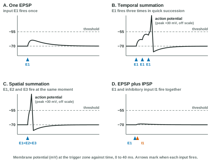
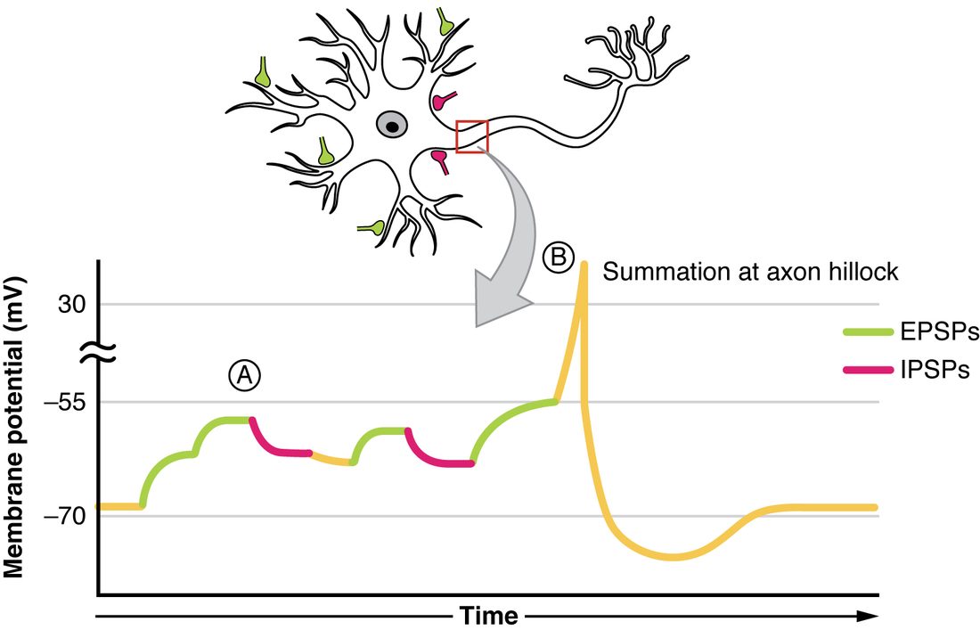
EPSPs and IPSPs add algebraically: an IPSP subtracts from whatever depolarization the EPSPs build. Figure 4 shows a real sequence: EPSPs climb toward threshold, IPSPs knock the potential back, and only when enough EPSPs arrive together does the trigger zone fire.
Worked example: will the neuron fire?
Problem. A neuron rests at −70 mV and its threshold is −55 mV. At its trigger zone, each EPSP from its excitatory inputs is worth +4 mV and each IPSP from its inhibitory input is worth −3 mV. Three excitatory inputs and one inhibitory input fire at the same moment. Does it fire? How many excitatory inputs would it need, with the inhibitory one still active?
- Find the depolarization needed. −55 − (−70) = 15 mV.
- Add the inputs. 3 × (+4) + 1 × (−3) = 12 − 3 = +9 mV.
- Compare. The membrane reaches −70 + 9 = −61 mV, below threshold. No action potential.
- Find the inputs needed. The EPSPs must supply 15 + 3 = 18 mV. 18 ÷ 4 = 4.5, so 5 excitatory inputs are needed: 5 × 4 − 3 = 17 mV, reaching −53 mV.
Answer. No; with the IPSP active it needs 5 simultaneous EPSPs. (Real PSPs do not add perfectly and fade as they spread, but the logic of the vote is the same.)
If the sum stays above threshold, the trigger zone fires again as soon as each refractory period allows. Stronger combined input means a higher firing rate. A neuron turns thousands of graded inputs into one all-or-none output whose rate carries the message.
The major neurotransmitters
More than a hundred substances act as neurotransmitters. A handful do most of the work. They fall into a few chemical groups:
| Group | Neurotransmitter | Where and what it does | Main way it is removed |
|---|---|---|---|
| Acetylcholine (its own group) | Acetylcholine | Excites skeletal muscle at the neuromuscular junction; used by many autonomic neurons and in the brain for attention and arousal | Broken down in the cleft by acetylcholinesterase |
| Amino acids | Glutamate | The main excitatory neurotransmitter of the brain and spinal cord | Reuptake into neurons and astrocytes |
| Amino acids | GABA (gamma-aminobutyric acid) | The main inhibitory neurotransmitter of the brain | Reuptake |
| Amino acids | Glycine | A major inhibitory neurotransmitter of the spinal cord | Reuptake |
| Biogenic amines | Norepinephrine | Alertness and arousal in the brain; released by most sympathetic neurons onto their targets | Reuptake, then enzymes |
| Biogenic amines | Dopamine | Movement control, motivation and reward | Reuptake, then enzymes |
| Biogenic amines | Serotonin | Mood, sleep, appetite; also in the gut | Reuptake, then enzymes |
| Neuropeptides | Endorphins and related peptides | Damp down pain signals; released during stress and hard exercise | Broken down by enzymes |
| Gases | Nitric oxide | Made on demand and diffuses out; not stored in vesicles | Breaks down within seconds |
A few terms and roots:
- Biogenic amines (bio- = life, -genic = produced by; an amine is a nitrogen-containing group) are made in neurons from single amino acids. Dopamine, norepinephrine and serotonin are the main ones; histamine, which you met in inflammation, is also one. Most act through metabotropic receptor proteins, so their effects are slower and longer.
- Norepinephrine is also called noradrenaline. Both names point to the glands on top of the kidneys (epi- = upon, nephr- = kidney; ad- = at, ren- = kidney).
- Serotonin was named for being found in blood serum (sero-) and changing the tone of blood vessels (-tonin).
- Neuropeptides are short chains of amino acids made in the cell body and shipped down the axon in large vesicles. They are released mainly during rapid firing, often alongside a smaller neurotransmitter, and act through metabotropic receptor proteins. Endorphins (endo- = within + morphine: the body's own morphine) are neuropeptides that act on the same receptor proteins as opioid drugs.
Two points hold for all of them. First, whether a neurotransmitter excites or inhibits depends on the receptor proteins it meets; glutamate and GABA are called excitatory and inhibitory because nearly all their receptor proteins in the adult brain work that way. Second, many neurons release more than one neurotransmitter, often a small molecule plus a neuropeptide.
Ending the synaptic signal
A synapse must be cleared quickly, or the next action potential could not be told apart from the last. Three routes remove a neurotransmitter from the cleft:
- Diffusion away from the cleft. It always happens, and it is the main route for some neuropeptides.
- Degradation by enzymes. Acetylcholinesterase in the cleft splits acetylcholine into acetate and choline in about a millisecond; the terminal takes the choline back to make more. Enzymes in and around neurons break down neuropeptides and, after reuptake, the biogenic amines.
- Reuptake: transporter proteins in the presynaptic membrane, or in nearby astrocytes, pump the neurotransmitter back in, powered by the sodium gradient (secondary active transport). The terminal repackages much of it into vesicles. Glutamate, GABA, glycine, dopamine, norepinephrine and serotonin are all removed mainly this way.
Block removal and the neurotransmitter lingers, binding again and again. The postsynaptic response grows larger and lasts longer.
Drugs and toxins at the synapse
Each step of synaptic transmission is a target. Working out a drug's effect is a matter of finding its step and following the chain.
| Step | Example | What it does | Result |
|---|---|---|---|
| Release | Botulinum toxin | Destroys proteins vesicles need to fuse, so acetylcholine is not released | Weakness and paralysis; in tiny doses (Botox), relaxes chosen muscles |
| Release | Tetanus toxin (from the bacterium that causes the disease tetanus) | Blocks release from inhibitory interneurons in the spinal cord | Motor neurons lose their brakes: rigid muscles and spasms, such as lockjaw |
| Binding | Curare-type drugs | Block nicotinic receptor proteins at the neuromuscular junction | Paralysis; used in surgery |
| Binding | Opioids (morphine, fentanyl) | Bind the metabotropic receptor proteins endorphins use | Pain relief; in overdose, slowed breathing |
| Binding | Naloxone | Occupies the same receptor proteins without activating them, pushing opioids off | Reverses an opioid overdose |
| Binding | Benzodiazepines, alcohol | Make GABA's chloride channels open more readily | More inhibition: calm, sleepiness, slowed breathing in excess |
| Degradation | Nerve agents, organophosphate insecticides | Block acetylcholinesterase | Acetylcholine builds up: twitching, then weakness and failed breathing |
| Reuptake | SSRIs (selective serotonin reuptake inhibitors) | Block serotonin transporters | More serotonin, for longer, at its synapses |
| Reuptake | Cocaine | Blocks dopamine, norepinephrine and serotonin transporters | Stronger, longer signaling by all three; its rewarding effect comes mainly from dopamine |
A note on SSRIs: they raise serotonin at synapses within hours, but their effect on depression takes weeks, and the old idea that depression is simply "low serotonin" is not supported by current evidence. How they help is still being worked out.
Neural circuits
Neurons are wired into neural circuits, patterns of connection that shape where a signal goes. Two patterns are basic (Figure 5):
- Divergence (di- = apart, vergere = to turn): one neuron's axon branches to synapse on many postsynaptic neurons. One signal spreads to many. Example: a single neuron in the brain that starts a movement can reach many motor neurons, which together work a whole group of muscles.
- Convergence (con- = together): many presynaptic neurons synapse on one postsynaptic neuron. Many signals feed one. Example: a motor neuron in the spinal cord receives input from the brain, from sensory neurons and from interneurons, and its firing reflects the sum. Convergence is what spatial summation works on.