Synapses and neurotransmitters
1Why this matters
Mr. Ruiz, 45, sprayed an organophosphate insecticide on a hot afternoon without gloves. An hour later he is drenched in sweat and drooling, the muscles of his arms are twitching in small ripples, and his breathing is getting weak. Nothing in the poison excites his nerves directly. It jams the enzyme that clears acetylcholine from his synapses, so every acetylcholine signal his neurons send keeps acting long after it should have stopped.
2What this builds on
3Quick check before you start
1. What can pass directly from one cell's cytosol to its neighbor's through a gap junction?
- Ions and small molecules
- Whole organelles
- Nothing: gap junctions seal the space between cells
Show the answer
Gap junctions are protein tunnels that link two cells' cytosols, so ions and small molecules pass straight through. Sealing the space between cells is the job of tight junctions.
- Correct: Ions and small molecules:
- Whole organelles:
- Nothing: gap junctions seal the space between cells:
2. At the neuromuscular junction, what directly triggers the synaptic vesicles to release acetylcholine?
- Sodium entering the axon terminal
- Calcium entering the axon terminal
- Acetylcholinesterase in the cleft
Show the answer
The action potential opens voltage-gated calcium channels in the axon terminal. Calcium entering triggers the vesicles to fuse with the membrane and release acetylcholine.
- Sodium entering the axon terminal:
- Correct: Calcium entering the axon terminal:
- Acetylcholinesterase in the cleft:
3. A messenger binds a membrane receptor protein that has no pore. The receptor protein switches on a G protein inside the cell. What usually happens next?
- The messenger enters the nucleus
- The receptor protein opens and lets ions through
- An enzyme makes a second messenger such as cAMP
Show the answer
A G protein switches on a membrane enzyme that makes a second messenger, such as cyclic AMP, which carries the signal on inside the cell.
- The messenger enters the nucleus:
- The receptor protein opens and lets ions through:
- Correct: An enzyme makes a second messenger such as cAMP:
4Anatomy
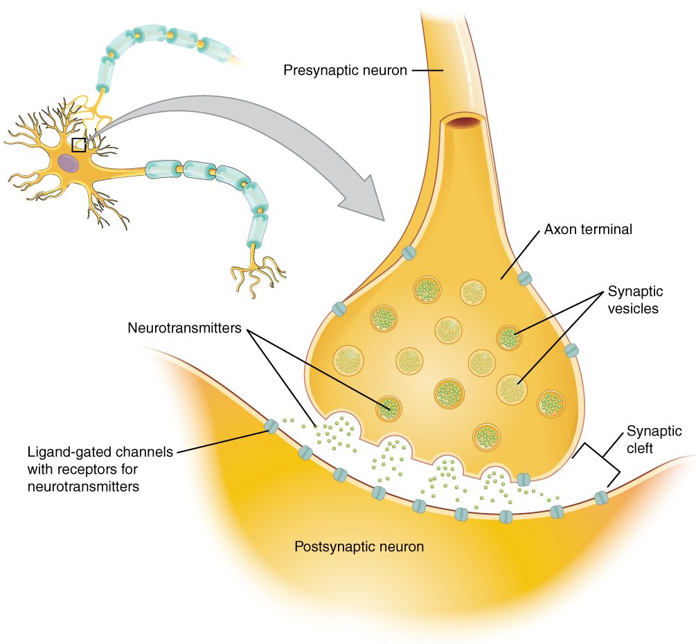
With labels hidden, select a box to reveal its label.
5How it works, step by step
- An action potential reaches the axon terminal of the presynaptic neuron.The depolarization opens voltage-gated calcium channels, and calcium rushes into the terminal.
- Calcium binds sensor proteins on docked synaptic vesicles.The vesicles fuse with the membrane and release neurotransmitter into the synaptic cleft by exocytosis.
- The neurotransmitter diffuses across the cleft and binds receptor proteins on the postsynaptic membrane.Ionotropic receptor proteins open at once; metabotropic ones act more slowly through G proteins. Either way, ion channels open or close.
- Ions flow through the opened channels.The postsynaptic membrane depolarizes (an EPSP) or hyperpolarizes or is held near rest (an IPSP).
- EPSPs and IPSPs from many synapses spread to the trigger zone and add together (spatial and temporal summation).If the sum reaches threshold, the postsynaptic neuron fires an action potential.
- Diffusion, enzymes and reuptake transporters clear the neurotransmitter from the cleft.The receptor proteins empty, the channels close, and the synapse is ready for the next signal.
6Core concepts
7A common mistake
The wrong idea: Each neurotransmitter is either excitatory or inhibitory in itself.
What actually happens: The receptor protein, not the neurotransmitter, decides the effect. Acetylcholine excites skeletal muscle through ionotropic receptor proteins that let sodium in, and slows the heart through metabotropic receptor proteins that open potassium channels. Glutamate and GABA are called excitatory and inhibitory only because nearly all of their receptor proteins in the adult brain work that way.
8Check yourself
Anything you miss goes into your review queue.
1. Put the steps of chemical synaptic transmission in order.
- An action potential reaches the axon terminal
- Voltage-gated calcium channels open and calcium enters
- Synaptic vesicles fuse with the membrane and release neurotransmitter
- Neurotransmitter diffuses across the synaptic cleft
- Neurotransmitter binds receptor proteins and ion channels open
- A postsynaptic potential changes the membrane potential of the next cell
Show the answer
Depolarization of the terminal opens calcium channels; calcium triggers exocytosis; the neurotransmitter crosses the cleft and binds receptor proteins; channels open or close, and the ion flow produces an EPSP or IPSP in the postsynaptic cell. Removal of the neurotransmitter then ends the signal.
- Correct order: 1. An action potential reaches the axon terminal 2. Voltage-gated calcium channels open and calcium enters 3. Synaptic vesicles fuse with the membrane and release neurotransmitter 4. Neurotransmitter diffuses across the synaptic cleft 5. Neurotransmitter binds receptor proteins and ion channels open 6. A postsynaptic potential changes the membrane potential of the next cell
2. A researcher injects current into one neuron. The neighboring neuron depolarizes with almost no delay, and when the experiment is reversed the first neuron follows the second just as fast. What connects them?
- A chemical synapse using an ionotropic receptor protein
- A chemical synapse using a metabotropic receptor protein
- Gap junctions forming an electrical synapse
- A neuromuscular junction
Show the answer
Near-instant, two-way transfer of current is the signature of an electrical synapse: gap junctions let ions flow straight from one cytosol into the other.
- A chemical synapse using an ionotropic receptor protein: Even a fast chemical synapse has a synaptic delay of about half a millisecond, and it works only from presynaptic to postsynaptic.
- A chemical synapse using a metabotropic receptor protein: Metabotropic effects take tens of milliseconds or more, and they are one-way.
- Correct: Gap junctions forming an electrical synapse: Correct. Ions pass directly through gap junctions, in either direction.
- A neuromuscular junction: A neuromuscular junction links a neuron to a skeletal muscle fiber, not two neurons, and it is a one-way chemical synapse.
3. A patient starts an SSRI, a drug that blocks the transporters that carry serotonin back into the presynaptic terminal. Predict each variable at the patient's serotonin synapses in the first hours, compared with before the drug.
| Variable | Change |
|---|---|
| Serotonin taken back into the presynaptic terminal | — |
| Time serotonin stays in the synaptic cleft after each release | — |
| Activation of postsynaptic serotonin receptor proteins | — |
| Calcium entry into the terminal per action potential | — |
Show the answer
Blocking reuptake removes the main exit route for serotonin. Each release lasts longer and activates postsynaptic receptor proteins more, while the steps that cause release are unchanged.
- Serotonin taken back into the presynaptic terminal: down. The transporters that perform reuptake are blocked.
- Time serotonin stays in the synaptic cleft after each release: up. With reuptake, the main removal route, blocked, only slow diffusion clears serotonin from the cleft.
- Activation of postsynaptic serotonin receptor proteins: up. Serotonin that lingers binds the receptor proteins again and again, so they are active for longer.
- Calcium entry into the terminal per action potential: no change. The drug acts on the reuptake transporters, not on the voltage-gated calcium channels that open when the action potential arrives.
4. At point A on this graph, several EPSPs have added together, yet the neuron has not fired an action potential. Why not?
- The neuron is in its absolute refractory period
- The summed depolarization is still below threshold
- EPSPs cannot add together; only the latest one counts
- The trigger zone lacks voltage-gated sodium channels
Show the answer
Point A sits above rest but below the −55 mV line. The EPSPs have summed, but not by enough to reach threshold, so no action potential fires.
- The neuron is in its absolute refractory period: The refractory period follows an action potential, and none has fired yet.
- Correct: The summed depolarization is still below threshold: Correct. Summation has moved the membrane toward threshold but not to it.
- EPSPs cannot add together; only the latest one counts: The stepwise climb to A shows the EPSPs adding; that is summation.
- The trigger zone lacks voltage-gated sodium channels: The trigger zone, the initial segment just beyond the hillock, has the densest voltage-gated sodium channels in the neuron. The problem at A is simply that the sum is below threshold.
5. In panel D, an excitatory input and an inhibitory input fire together and the membrane barely moves. What would make this neuron reach threshold?
- Firing the inhibitory input I1 more often
- Blocking the reuptake of the inhibitory neurotransmitter at the I1 synapse
- Removing the inhibitory input and firing three excitatory inputs together
- Lowering the calcium in the fluid around the excitatory axon terminal
Show the answer
EPSPs and IPSPs add algebraically. Removing the IPSP and adding more simultaneous EPSPs, as in panel C, gives enough depolarization to reach threshold.
- Firing the inhibitory input I1 more often: More IPSPs subtract more from the EPSP and push the membrane further from threshold.
- Blocking the reuptake of the inhibitory neurotransmitter at the I1 synapse: Blocking reuptake of an inhibitory neurotransmitter prolongs its effect, making the IPSP larger and longer.
- Correct: Removing the inhibitory input and firing three excitatory inputs together: Correct. More excitation and no inhibition is what panel C shows reaching threshold.
- Lowering the calcium in the fluid around the excitatory axon terminal: Less calcium outside means less neurotransmitter released from the excitatory terminal, so a smaller EPSP.
6. Acetylcholine makes skeletal muscle fibers contract but slows the heart. What explains the opposite effects?
- The heart receives a chemically different form of acetylcholine from its nerves
- The two tissues carry different receptor proteins for acetylcholine
- Acetylcholine is broken down faster in skeletal muscle
- Heart cells lack voltage-gated channels
Show the answer
The receptor protein decides the effect. Skeletal muscle has ionotropic nicotinic receptor proteins, which let sodium in and depolarize the fiber. The heart cells that set its rate have metabotropic receptor proteins, which through a G protein open potassium channels and slow them.
- The heart receives a chemically different form of acetylcholine from its nerves: Acetylcholine is the same molecule wherever it is released.
- Correct: The two tissues carry different receptor proteins for acetylcholine: Correct. Same neurotransmitter, different receptor proteins, different results.
- Acetylcholine is broken down faster in skeletal muscle: Faster breakdown would shorten an effect, not reverse it.
- Heart cells lack voltage-gated channels: Heart cells rely on voltage-gated channels for their action potentials.
7. Select every statement that is true of metabotropic receptor proteins.
- They act through G proteins
- Their pore opens the instant the neurotransmitter binds
- Their effects can last seconds to minutes
- They can amplify a signal through a second messenger
- They include the nicotinic receptor proteins at the neuromuscular junction
- They can change which genes a cell uses
Show the answer
Metabotropic receptor proteins are G-protein-coupled receptor proteins. They work more slowly and for longer than channels, amplify signals through second messengers, and can change enzymes and gene expression.
- Correct: They act through G proteins: Correct. The G protein is the link to everything they do.
- Their pore opens the instant the neurotransmitter binds: They have no pore. An instant-opening pore describes an ionotropic receptor protein.
- Correct: Their effects can last seconds to minutes: Correct. G protein and second messenger effects outlast the neurotransmitter's binding.
- Correct: They can amplify a signal through a second messenger: Correct. One bound receptor protein can lead to many second messenger molecules.
- They include the nicotinic receptor proteins at the neuromuscular junction: Nicotinic receptor proteins at the neuromuscular junction are ionotropic: they are ligand-gated channels.
- Correct: They can change which genes a cell uses: Correct. Second messengers can switch on enzymes that change gene expression.
8. An infant develops botulism. Botulinum toxin destroys the proteins synaptic vesicles need to fuse with the membrane. Her cry is weak, she feeds poorly and her limbs are floppy. At which step of transmission does the toxin act?
- Conduction of the action potential along the motor axon
- Binding of acetylcholine to nicotinic receptor proteins
- Breakdown of acetylcholine by acetylcholinesterase
- Release of acetylcholine from the axon terminal
Show the answer
Action potentials still reach the terminal and calcium still enters, but the vesicles cannot fuse, so acetylcholine is not released. Muscle fibers get no signal, and they are weak and floppy.
- Conduction of the action potential along the motor axon: The toxin acts at the terminal, after conduction. The action potential still arrives.
- Binding of acetylcholine to nicotinic receptor proteins: Receptor proteins are unaffected; there is simply no acetylcholine to bind them.
- Breakdown of acetylcholine by acetylcholinesterase: Blocking breakdown would leave extra acetylcholine and cause twitching, the opposite of floppy weakness.
- Correct: Release of acetylcholine from the axon terminal: Correct. No vesicle fusion means no release.
9Summary
At an electrical synapse, gap junctions pass current directly, almost instantly and usually both ways. At a chemical synapse, an action potential in the presynaptic neuron's axon terminal opens voltage-gated calcium channels; calcium triggers exocytosis of neurotransmitter, which crosses the cleft and binds receptor proteins on the postsynaptic neuron. Ionotropic receptor proteins are ligand-gated channels (fast, brief); metabotropic ones work through G proteins (slower, longer, amplified). The result is an EPSP, a depolarization toward threshold, or an IPSP, which moves the membrane away from it. The trigger zone adds these by temporal and spatial summation and fires if the sum reaches threshold. The major neurotransmitters are acetylcholine, the amino acids glutamate, GABA and glycine, the biogenic amines norepinephrine, dopamine and serotonin, neuropeptides such as endorphins, and nitric oxide. The signal ends by diffusion, degradation and reuptake, and drugs and toxins act by changing release, binding, degradation or reuptake. Neurons are wired in circuits with divergence and convergence.