The functions of the skin come straight from its layers. The stratum corneum makes it a barrier; nerve endings in the epidermis and dermis make it a sense organ; its blood vessels and sweat glands make it the main place your body trades heat with the world, the core of thermoregulation; and its keratinocytes use sunlight to make vitamin D. This page takes those functions in turn, and ends with the body temperature loop that ties the skin's heat-handling parts together.
The skin as a barrier
Sit in a bath for an hour and you do not swell up with water. Stand in dry desert air for a day and you do not dry out through your skin. Spill water on your arm and nothing soaks in; rub on a greasy ointment and some of it does. All of that comes from the layers you have already met.
The skin as a barrier means the ways the intact skin keeps water in and keeps microbes, chemicals and UV light out. It works on several levels at once.
A physical barrier
- Dead, keratin-filled cells. The stratum corneum is 15 to 30 or more layers of flat, dead cells packed with cross-linked keratin. They have no living machinery for a microbe to hijack.
- Cells joined tightly. Desmosomes bind the cells, so the sheet does not tear easily.
- Lipid sheets between the cells. The lipids released by lamellar granules fill every gap, like mortar between bricks. Water and water-soluble molecules are hydrophilic and cannot cross the lipid easily, so water stays in and waterborne chemicals stay out.
- Dry and constantly shed. The surface holds little water for microbes to grow in, and as surface cells are shed, the microbes stuck to them go too.
A chemical barrier
- Slightly acidic. Sweat and sebum make the skin surface slightly acidic, about pH 5. Many harmful microbes grow poorly at that pH, and the enzymes that build the lipid sheets work best there.
- Germ-killing molecules. Fatty acids in sebum and small germ-killing peptides in sweat and made by keratinocytes slow or kill some bacteria and fungi.
Cellular and UV defenses
- Langerhans cells in the epidermis and macrophages in the dermis take up microbes that get past the surface.
- Melanin caps the nuclei of keratinocytes and absorbs UV light before it reaches their DNA.
What gets through
The barrier is a lipid barrier, so lipid-soluble (hydrophobic) substances can dissolve through it slowly. That is how medicated skin patches deliver some drugs through intact skin, and why organic solvents and some pesticides are absorbed through the skin. Water-soluble substances, including most drugs in water-based creams, barely cross unless the stratum corneum is damaged.
Break the barrier and each of those protections fails at once: water leaks out, microbes get in, and chemicals reach living cells. The next topic shows what that means after a large burn.
The skin as a sense organ
Close your eyes and run a fingertip over a coin. You can feel its raised edge, the texture of its face, and, if you press harder, the pressure against your finger. A hand resting on a buzzing phone feels the vibration, and a hot pan or a pinprick feels like pain. Different nerve endings handle each of these.
The skin's touch receptors, also called tactile receptors, are sensory receptors in the epidermis and dermis that respond to touch, pressure, vibration, stretch, temperature and pain. Each is the ending of a sensory nerve fiber. There are two broad kinds:
- A free nerve ending is the bare, branching tip of a sensory nerve fiber, with no covering. Free nerve endings reach into the lower epidermis and throughout the dermis. They respond to pain, to warmth or cold, and to itch, and some to light touch.
- An encapsulated ending is a nerve ending wrapped in a capsule of connective tissue or specialized cells. The capsule shapes how the ending responds to mechanical forces.
The main touch receptors of the skin:
| Free nerve endings | Tactile (Merkel) cells | Tactile (Meissner) corpuscles | Lamellated (Pacinian) corpuscles | Hair follicle endings | |
|---|---|---|---|---|---|
| Structure | Bare branching nerve tips | A tactile cell pressed against a flattened nerve ending | An oval capsule around coiled nerve endings | A large capsule of many layers, like an onion, around one nerve ending | Nerve endings wrapped around the base of a follicle |
| Where | Lower epidermis and all of the dermis | Stratum basale | Dermal papillae, mainly of hairless skin | Deep dermis and hypodermis | Around hair follicles in the dermis |
| Responds to | Pain, heat, cold, itch, some touch | Steady light pressure, shape and texture | Light touch and slow vibration | Deep pressure and fast vibration | Bending of a hair |
| Response to steady pressure | Keeps signaling | Keeps signaling | Signals when pressure changes, then falls quiet | Signals when pressure changes, then falls quiet | Signals when the hair moves, then falls quiet |
| Encapsulated? | No | No | Yes | Yes | No |
The tactile corpuscle (Meissner corpuscle) and lamellated corpuscle (Pacinian corpuscle) each have two names: the descriptive name used here, and the name of the anatomist who first described it. You will see both in exams. You can find a lamellated corpuscle, deep in the dermis, in Figure 1.
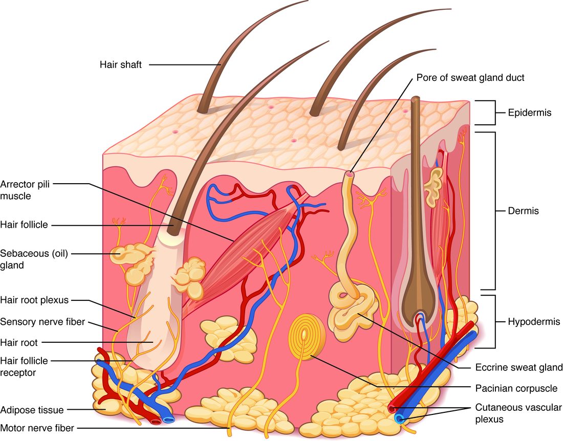
The endings that signal only when pressure changes explain why you feel your socks when you put them on and then stop noticing them. The endings that keep signaling let you feel how hard you are gripping a cup for as long as you hold it.
How finely you can feel depends on how densely the endings are packed. On a fingertip, two pencil points about 2 to 3 mm apart feel like two touches. On your back they must be several centimeters apart before they stop feeling like one.
Heat exchange with the environment
Your cells make heat all the time: the chemical reactions of metabolism, taken together, release heat. That heat must leave through your skin, and a little through your breath, at the same rate it is made, or your internal temperature will climb. Heat always moves down a temperature gradient, from warmer to cooler. Heat exchange with the environment happens by four routes:
- Radiation (thermal radiation): your skin gives off infrared rays, invisible light that carries heat, to any cooler surface nearby, without touching it. Standing near a cold window, you lose heat to the glass even when the room air is warm. The sun and a fire heat you the same way.
- Conduction: heat passes directly to an object you touch. Sitting on cold stone draws heat from you. Water conducts heat about 25 times faster than air, which is why cold water chills you so much faster than air at the same temperature.
- Convection: moving air or water carries away the warmed layer next to your skin and replaces it with cooler fluid. Wind and a fan speed up heat loss this way.
- Evaporation: water on your skin turns to vapor, and the energy that change takes comes from your skin. Sweat evaporating is the main example; some water also evaporates from your airways with every breath.
| Radiation | Conduction | Convection | Evaporation | |
|---|---|---|---|---|
| How heat moves | Infrared rays to surfaces not touching you | Directly into an object you touch | Carried off by air or water flowing past | Used up turning water on the skin into vapor |
| Needs | Surroundings cooler than your skin | Contact with a cooler object | Cooler air or water, moving | Air not already full of water vapor |
| Everyday example | Losing heat to cold walls | Sitting on cold stone | Wind; swimming in cool water | Sweat drying on your skin |
| Share of heat loss at rest in a cool room | About 60% | About 3% | About 15% | About 20% |
| Can it add heat to you? | Yes, from the sun or a fire | Yes, from a hot surface | Yes, in air hotter than your skin | No: it only removes heat |
The last row matters on a hot day. Your skin sits at about 33 to 35 °C. Once the air and surroundings are hotter than that, radiation, conduction and convection all run backward and add heat. Evaporation is then the only way left to lose heat.
How much heat sweat removes
Turning water into vapor takes a lot of energy. Each liter of sweat that evaporates from your skin removes about 580 kcal of heat. Sweat that drips off removes almost none, because it carries the heat away without changing into vapor. In humid air, where the air already holds a lot of water vapor, sweat evaporates slowly, and more of it drips.
Worked example 1: heat removed by sweating
Problem. During an hour of yard work, 0.6 L of sweat evaporates from Carlos's skin. How much heat does that remove?
- Write the rule. Heat removed = volume evaporated × about 580 kcal per liter.
- Substitute. Heat removed = 0.6 L × 580 kcal/L.
- Calculate. 0.6 × 580 = 348 kcal.
Answer. About 350 kcal removed in the hour.
Worked example 2: how much sweat must evaporate
Problem. On a day when the air is hotter than her skin, a cyclist makes 870 kcal of heat per hour. Radiation, conduction and convection remove none. How much sweat must evaporate each hour just to remove the heat she makes?
- Find the only route. The air is hotter than her skin, so evaporation is the only way to lose heat.
- Rearrange the rule. Volume evaporated = heat to remove ÷ 580 kcal per liter.
- Substitute. Volume = 870 kcal ÷ 580 kcal/L.
- Calculate. 870 ÷ 580 = 1.5 L.
Answer. At least 1.5 L of sweat must evaporate every hour. If some drips off unevaporated, she must sweat more than that, and she loses all of it as water from her body.
The body temperature loop (basic)
You met body temperature as the model negative feedback loop in Homeostasis and feedback loops. The skin supplies the sensors and most of the effectors.
Dermal blood flow: the skin's thermostat valve
Dermal blood flow is the flow of blood through the vessels of the dermis. The heat your cells make is carried by the blood; how much of that blood passes through the skin decides how much heat reaches the surface to be lost.
- Vasodilation of the small arteries feeding the dermis sends more warm blood to the skin. The skin surface warms, its gradient to the surroundings steepens, and more heat is lost by radiation, conduction and convection. Skin blood flow can rise from about 0.3 L/min at rest in comfortable air to 6 to 8 L/min in severe heat.
- Vasoconstriction of those vessels keeps warm blood deeper in the body. The skin surface cools, the gradient to the surroundings shrinks, and less heat is lost. The skin and the fat of the hypodermis then act as a layer of insulation.
In most of your skin, the widening in heat is driven by nerve signals that actively relax the vessel walls. In the hairless skin of the palms, soles and lips, it comes mainly from nerves easing the steady signal that normally keeps those vessels narrowed.
Sweating and shivering
- Sweating by the eccrine sweat glands puts water on the skin to evaporate. It starts when the body warms and can reach 1 to 2 L an hour in hard exercise in the heat.
- Shivering is rapid, involuntary contraction of skeletal muscles in small bursts. The contractions move nothing outside the body, so nearly all their energy is released as heat. Shivering can raise heat production three- to fivefold for a time.
The two columns of the loop:
| Loop part | Body too warm | Body too cold |
|---|---|---|
| Stimulus | Exercise or hot surroundings warm the body | Cold air or water cools the skin and blood |
| Sensory receptor (sensor) | Warm-sensitive nerve endings in the skin; temperature-sensitive neurons in the brain | Cold-sensitive nerve endings in the skin; temperature-sensitive neurons in the brain |
| Afferent pathway | Sensory nerves to the brain | Sensory nerves to the brain |
| Control center | The brain's temperature control center | The brain's temperature control center |
| Efferent pathway | Nerves to dermal vessels and eccrine sweat glands | Nerves to dermal vessels, skeletal muscles and arrector pili muscles |
| Effectors | Smooth muscle of dermal vessels; eccrine sweat glands | Smooth muscle of dermal vessels; skeletal muscles; arrector pili |
| Response | Vasodilation raises dermal blood flow; sweating adds evaporation; more heat lost | Vasoconstriction lowers dermal blood flow; shivering makes heat; less heat lost, more made |
| Feedback type | Negative | Negative |
Figure 2 draws the "too warm" half. Two practical points follow from it:
- Humidity weakens the loop. Sweating still happens in humid heat, but little of the sweat evaporates, so the response removes less heat and body temperature keeps rising.
- Heavy sweating costs water and salt. Every liter of sweat comes out of your body's fluid, so long work in the heat needs drinking to match.
Behavior, such as moving into shade or putting on a coat, is often the most powerful response of all. The full picture of body temperature control returns in the metabolism chapter.
Vitamin D synthesis in the skin
In the 1800s, children in the smoky, sunless industrial cities of northern Europe grew up with soft, bowed leg bones. Those living in sunny countryside rarely did. The difference was a molecule their skin made in sunlight.
Vitamin D synthesis in the skin is the conversion of a close relative of cholesterol in keratinocytes into vitamin D by ultraviolet light:
- Keratinocytes in the stratum basale and stratum spinosum hold a steroid called 7-dehydrocholesterol in their plasma membranes. It is the molecule your cells turn into cholesterol in the last step of making it, so it is a close relative of cholesterol.
- Ultraviolet B (UVB) light, the shorter-wavelength part of sunlight's UV, reaches those layers. Its energy breaks one ring of the 7-dehydrocholesterol molecule open, making previtamin D3.
- Over the next few hours to days, body warmth rearranges previtamin D3 into vitamin D3, also called cholecalciferol (chole- = bile, calci- = calcium, -fer = carrying).
- Vitamin D3 moves into the dermal capillaries, riding in the blood bound to a carrier protein.
Your skin cannot overdose you on vitamin D. Extra UV light converts previtamin D3 and vitamin D3 into inactive products, so after a certain amount of sun, making more stops. Too much vitamin D comes only from supplements.
Several things reduce how much vitamin D your skin makes, and each works through the same step: less UVB reaching 7-dehydrocholesterol.
- More melanin. Melanin absorbs UVB. Darker skin needs a longer exposure to make the same amount.
- Sunscreen and clothing block UVB.
- Winter at high latitudes. When the sun is low, UVB travels a long path through the atmosphere and is absorbed before it reaches the ground. Farther than about 40 degrees from the equator, north or south, winter sun makes little or no vitamin D.
- Window glass blocks nearly all UVB, so sun through a window makes almost none.
- Age. Older skin holds less 7-dehydrocholesterol.
You can also take vitamin D in from food, such as oily fish, egg yolks and fortified milk, or supplements. Plant and fungal sources supply a slightly different form, vitamin D2, which the body handles the same way. Because the skin can make all you need with enough sun, vitamin D is not strictly a vitamin; it acts more like the raw material for a hormone.
Activating vitamin D
Vitamin D from the skin or from food is inactive. Activating vitamin D takes two more steps, each adding a hydroxyl group (–OH) in a different organ (Figure 3):
- The liver adds the first –OH, making 25-hydroxyvitamin D. This is the main form in the blood, and its level is what a vitamin D blood test measures.
- The kidneys add a second –OH, making the active hormone.
Without enough of the active hormone, the intestine absorbs only a small share of the calcium in food. Growing bone needs a steady supply of calcium and phosphate to harden its new tissue. In a child, too little vitamin D leaves new bone soft and poorly mineralized, and the legs bow under the body's weight. This is rickets. It also causes slow growth and bone pain. Adults with too little vitamin D get a similar softening of existing bone, which the bone chapter covers.
Because the last step happens in the kidneys, a person with badly damaged kidneys can have plenty of 25-hydroxyvitamin D and still make too little of the active hormone. Sun and diet cannot fix that; the active hormone itself must be given as a drug.
Putting it together
Each function traces back to a structure. The dead, keratin-filled cells and lipid sheets of the stratum corneum, with an acidic surface, make the barrier. Free nerve endings, tactile cells and the two kinds of corpuscle make the skin a sense organ. Dermal blood flow and eccrine sweat glands, run by the brain's temperature control center in a negative feedback loop, set how much heat you lose by radiation, conduction, convection and evaporation. And keratinocytes, given UVB, start the pathway that ends with the kidneys making the hormone that lets your intestine absorb calcium.