Two glands in your neck, one wrapped around the front of your trachea and four tiny ones tucked behind it, set two very different things. Thyroid hormone function is to set how fast nearly every cell in your body uses oxygen and fuel and makes heat, and to allow normal growth and brain development. Parathyroid hormone function is to hold your blood calcium in a narrow range. This page covers the structure of the thyroid gland, how it builds its hormones from iodine, how the thyroid axis controls them, what goes wrong when there is too much or too little, and then how parathyroid hormone, calcitriol and calcitonin together control blood calcium.
Two patients with neck surgery
Ms. Duarte, 38, has lost 6 kg in two months while eating more than usual. She feels hot all the time, her hands shake, her heart races at rest, and her eyes look as if they are staring. Her thyroid gland is making far too much hormone. After treatment fails, a surgeon removes her whole thyroid gland.
The next evening her lips tingle, and her hands cramp into claws when a blood pressure cuff is inflated on her arm. Her blood calcium has fallen. The surgery did not remove the calcium-controlling part of the thyroid gland; it bruised the four tiny glands on its back. By the end of this page you will be able to explain both problems.
Structure of the thyroid gland
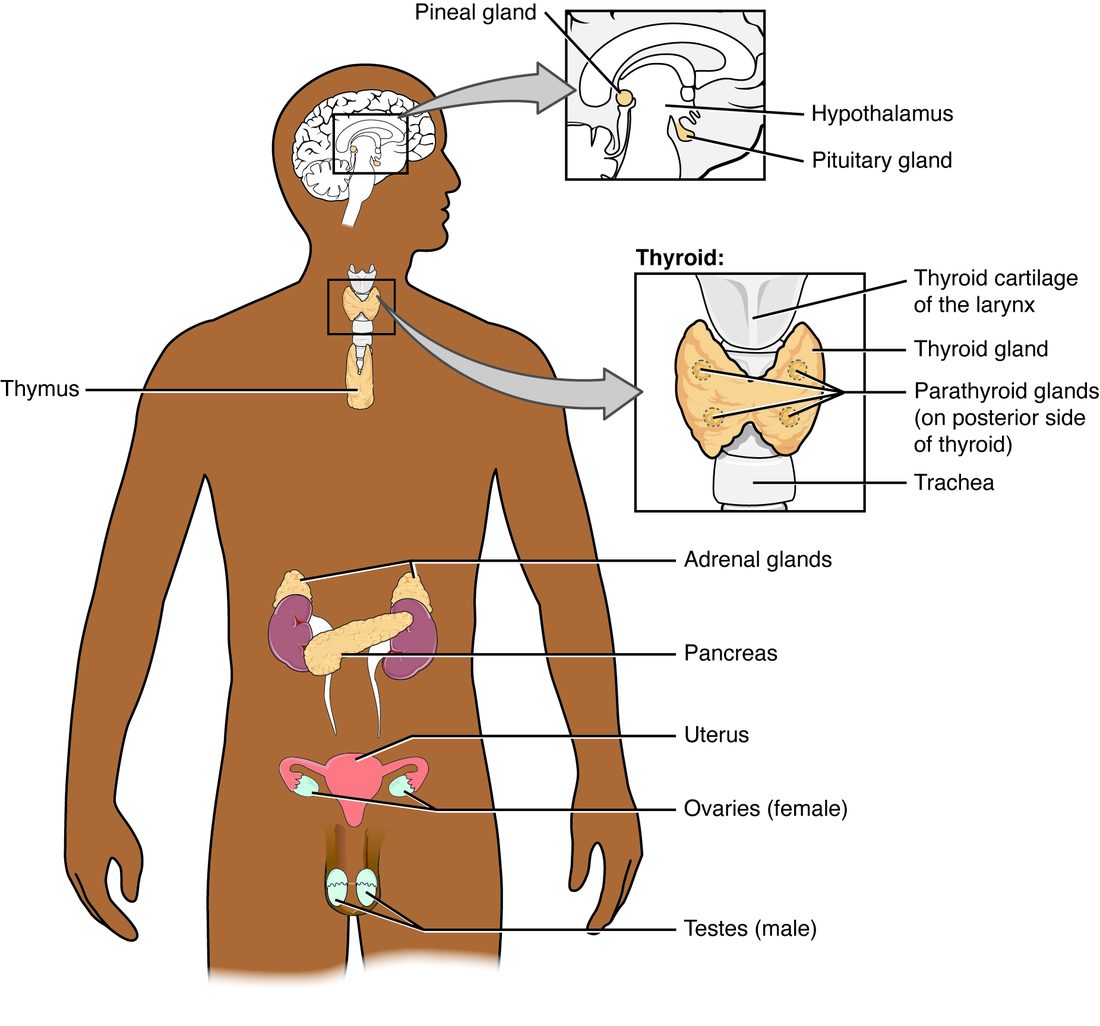
The thyroid gland (Greek thyreos, a door-shaped shield) is a butterfly-shaped gland in the front of the neck, just below the Adam's apple (Figure 1). Its two lobes lie on either side of the trachea, joined across the front by a narrow bridge of tissue, the isthmus. It weighs about 20 g and has one of the richest blood supplies of any organ for its size.
Under the microscope, the gland is packed with thousands of thyroid follicles, which you met as an example of simple cuboidal epithelium (Figure 2):
- Each follicle is a hollow sphere whose wall is a single layer of follicular cells, which make the thyroid gland's main hormones.
- The hollow is filled with thyroid colloid, a sticky, protein-rich fluid. Its main protein is thyroglobulin (-globulin = a kind of rounded protein), a large protein made by the follicular cells, on which the hormones are built and stored.
- Between the follicles lie scattered parafollicular cells (para- = beside), also called C cells (C for the hormone they make, calcitonin).
How tall the follicular cells are tells you how hard the gland is working. Under strong stimulation by TSH they become tall columns, and the colloid shrinks as it is used up. In a resting gland they are flat and the follicles are swollen with colloid.
Thyroid hormones
The thyroid hormones are two amine hormones built from the amino acid tyrosine and the element iodine:
- Thyroxine (T4) carries four iodine atoms. It is about 90% of what the gland releases.
- Triiodothyronine (T3; tri- = three) carries three. It is the active form: it binds the thyroid receptor protein about ten times more tightly than T4.
Most T3 is not made in the thyroid gland at all. Enzymes in the liver, kidneys and other target cells snip one iodine off T4 to make T3. So T4 is best thought of as a long-lasting reserve that tissues activate as they need it.
How the gland makes them
Your body cannot make iodine; you get it from food, mainly iodized salt, dairy foods and seafood, and you need about 150 micrograms a day. The follicular cells build the hormones in steps (Figure 2):
- Trapping. A cotransporter in the follicular cell's membrane pulls iodide ions (I−) in from the blood along with sodium, concentrating iodide in the gland 20 to 40 times above the blood. This is secondary active transport, driven by the sodium gradient.
- Making thyroglobulin. The cell makes thyroglobulin and releases it into the colloid by exocytosis.
- Attaching iodine. At the cell's inner surface, an enzyme converts iodide to a reactive form of iodine and attaches it to tyrosines in thyroglobulin: one or two iodines per tyrosine.
- Coupling. Pairs of iodinated tyrosines, still part of thyroglobulin, join: two with two iodines each make T4; one with one and one with two make T3.
- Storage. The hormones stay attached to thyroglobulin in the colloid. The gland stores enough for two to three months, far more than any other endocrine gland.
- Release. When TSH arrives, the follicular cells take colloid back in by endocytosis. Lysosomes digest the thyroglobulin, freeing T4 and T3, which leave the cell into the capillaries.
Travel and action
The thyroid hormones are amines, but they are hydrophobic. So, as you saw in Hormones and how they act, they travel almost entirely bound to carrier proteins in the plasma: more than 99% is bound, which gives T4 a half-life of about a week. Free hormone enters target cells through membrane transporters, T4 is converted to T3, and T3 binds a receptor protein in the nucleus. The hormone–receptor complex binds hormone response elements and changes the transcription of many genes. The effects therefore start slowly, over hours to days, and last.
What they do
Almost every cell in your body has thyroid receptor protein. The main effects:
- Oxygen use and heat. Cells make more sodium–potassium pumps and more mitochondria, so they use more ATP and more oxygen, and more of the energy in their fuel ends up as heat. This is the calorigenic effect (calor- = heat).
- Fuel use. Cells take up and burn more glucose, break down more stored fat, and turn over more protein.
- The heart and the sympathetic system. Heart cells make more beta-1 receptor proteins and faster contractile proteins, so the heart beats faster and more forcefully. This is the permissive effect on epinephrine you met in Hormones and how they act.
- Growth and the brain. Thyroid hormones are needed, together with growth hormone, for normal bone growth, and they are essential for the growth of neurons and the formation of myelin in the fetus and during the first two to three years after birth.
Regulation of thyroid hormone
The thyroid gland is the target gland of the thyroid axis you met in the last topic (Figure 3):
- The hypothalamus releases TRH into the hypophyseal portal system.
- TRH makes the anterior pituitary release TSH.
- TSH binds receptor proteins on follicular cells. Through cAMP, it speeds every step: iodide trapping, thyroglobulin making, iodination and the endocytosis of colloid that releases T4 and T3. Over weeks, it also makes the gland grow.
- T4 and T3 rise in the blood and act on target cells.
- Long-loop negative feedback: the thyroid hormones inhibit TSH release from the anterior pituitary, their main site of feedback, and TRH release from the hypothalamus. TSH falls, and so does thyroid output.
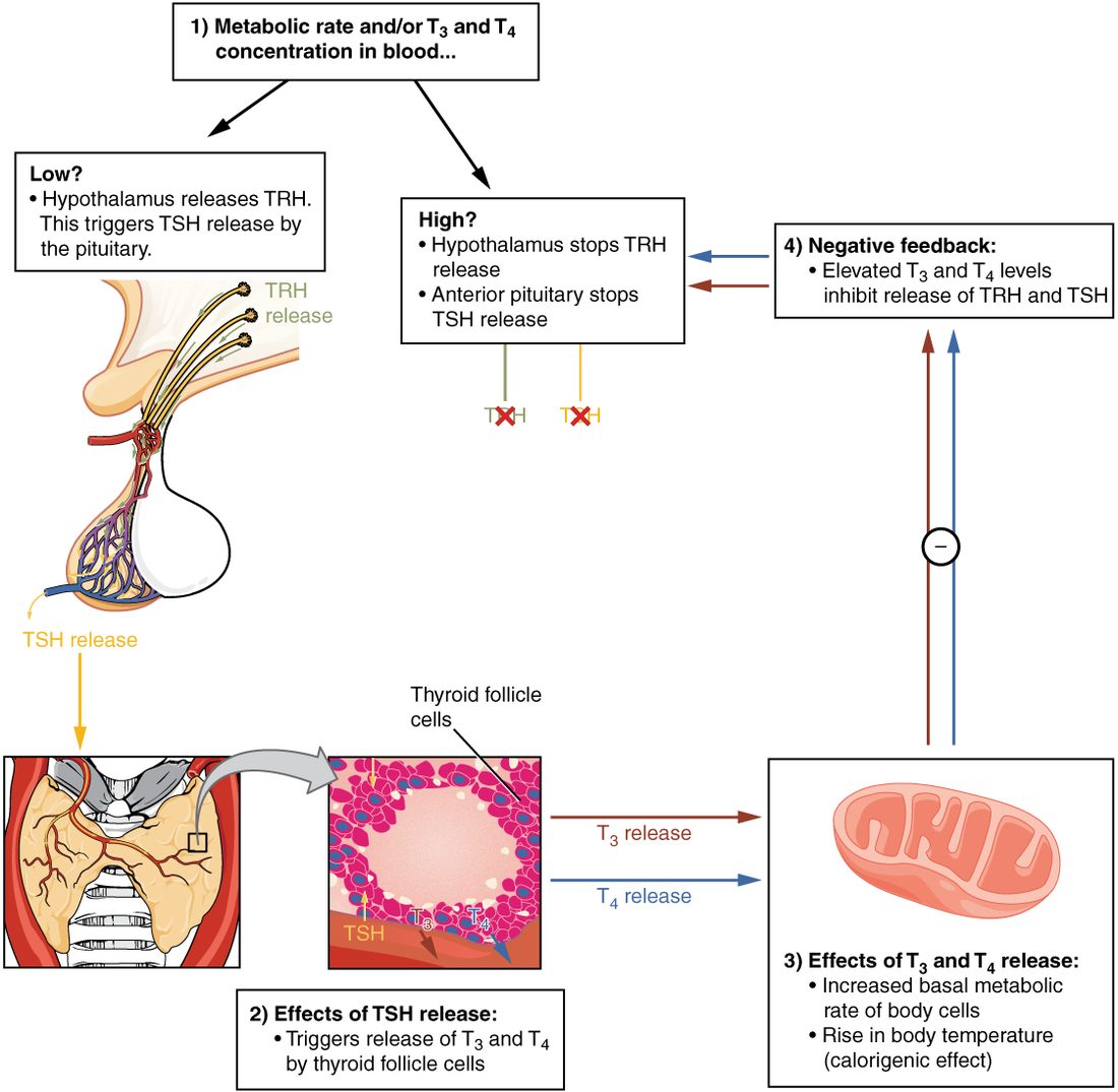
This loop keeps free T4 remarkably steady in each person. Because the pituitary is so sensitive to feedback, a small fall in free T4 produces a large rise in TSH. That is why a TSH blood test is usually the first test for a thyroid problem: it moves a lot for a small change in the thyroid gland's output.
Thyroid disorders
Almost every sign of too much or too little thyroid hormone follows from the effects above: more or less oxygen use, heat, sympathetic sensitivity and turnover in every tissue.
| Hypothyroidism | Hyperthyroidism | |
|---|---|---|
| Thyroid hormone level | Low | High |
| Most common cause in adults | The immune system destroying the gland (Hashimoto thyroiditis); worldwide, iodine deficiency | Graves disease: an immune protein that switches on TSH receptor proteins |
| TSH level | High, when the gland itself fails | Very low, suppressed by feedback |
| Heat and temperature | Feels cold; less heat made | Feels hot, sweats; more heat made |
| Body weight | Gains a little, despite a poor appetite | Loses weight, despite a big appetite |
| Heart | Slow heart rate | Fast heart rate at rest, pounding heartbeat, sometimes an irregular rhythm |
| Nervous system | Slowed thinking, tiredness, low mood, slow reflexes | Anxiety, restlessness, fine tremor of the hands, brisk reflexes |
| Skin and hair | Dry, thick, puffy skin; coarse hair | Warm, moist skin; fine hair |
| Gut | Slow bowel movements | Frequent bowel movements |
| Goiter? | Often, when TSH is high | Often, in Graves disease |
Hypothyroidism
Hypothyroidism (hypo- = under) is too little thyroid hormone. In countries with iodized salt, the usual cause is Hashimoto thyroiditis, in which the immune system gradually destroys the follicular cells. Worldwide, the most common cause is too little iodine in the diet. A daily tablet of synthetic T4 (levothyroxine) replaces the missing hormone, and the dose is adjusted until TSH is back in the normal range.
Hypothyroidism in newborns
Congenital hypothyroidism (congenital = present at birth), also called neonatal hypothyroidism (neo- = new, nat- = born), is a missing, underdeveloped or poorly working thyroid gland in a newborn. It affects roughly 1 in 2,000 to 4,000 babies. Because thyroid hormone is essential for brain growth after birth, untreated babies develop permanent intellectual disability and short stature. Babies usually look normal at first, partly because some of the mother's hormone crossed to them before birth. That is why most countries screen every newborn with a heel-prick blood test in the first days of life: started within the first two weeks, T4 tablets allow normal development.
Hyperthyroidism and Graves disease
Hyperthyroidism (hyper- = over) is too much thyroid hormone. The most common cause is Graves disease. In Graves disease the immune system makes a protein that binds the TSH receptor protein on follicular cells.
This immune protein does not block the receptor protein; it switches it on, like TSH. The pituitary's feedback cannot stop it, so the gland grows and releases hormone nonstop, while TSH is driven almost to zero. The same immune attack inflames the tissue behind the eyes, which can push the eyes forward. Treatment slows the gland with drugs that block iodination, destroys part of it with radioactive iodine (the gland takes it up just like ordinary iodide), or removes it surgically; beta blockers ease the racing heart and tremor in the meantime.
Goiter
A goiter is any enlargement of the thyroid gland, seen as a swelling at the front of the neck. Two classic drivers act through the TSH receptor protein:
- Iodine deficiency. Without iodine, the gland cannot make T4 and T3. Their levels fall, feedback is lost, and TSH rises. TSH keeps driving the follicular cells to grow and make thyroglobulin, and the gland swells, sometimes enormously, even though it cannot make hormone. This was common in mountain and inland regions until salt was iodized.
- Graves disease. The stimulating immune protein drives growth just as TSH does.
So a goiter alone does not tell you whether the gland is making too much or too little hormone. Nodules and inflammation can also enlarge it.
Calcitonin
The parafollicular (C) cells of the thyroid gland release calcitonin, a peptide hormone, when blood calcium rises. Calcitonin binds receptor proteins on osteoclasts and quickly stops them breaking down bone. With less calcium coming out of bone, blood calcium falls. Calcitonin also makes the kidneys excrete a little more calcium.
How much does this matter? In adults, not much. People whose thyroid glands have been removed, like Ms. Duarte, have no calcitonin, yet their blood calcium stays normal as long as their parathyroid glands are intact. People with tumors of the C cells have very high calcitonin, and their blood calcium is also normal. Calcitonin may matter more in childhood, when bone is being built fast, and during pregnancy and breastfeeding, when the body is protecting the mother's skeleton. It is also given as a drug to lower very high calcium quickly.
The parathyroid glands
The parathyroid glands (para- = beside) are usually four small, yellow-brown glands, each about the size of a grain of rice, on the back of the thyroid gland: an upper and a lower pair (Figure 4). Some people have three, five or more, and one can sit lower in the neck or even in the chest.
Most of their cells are chief cells, which make parathyroid hormone. Chief cells carry a calcium-sensing receptor protein on their surface. When blood calcium falls, fewer calcium ions bind it, and the cells release more hormone within seconds; when calcium rises, release falls. This is a humoral stimulus, and the chief cells are both the sensor and the control center of the loop.
Parathyroid hormone
Parathyroid hormone (PTH) is a peptide hormone and the main hormone that raises blood calcium. You met it briefly with bone; here is the full picture. PTH acts on three places (Figure 4):
- Bone. PTH binds receptor proteins on osteoblasts, not osteoclasts. The osteoblasts respond by releasing a signal that makes more osteoclasts form and makes existing ones work harder. The osteoclasts dissolve bone matrix, and calcium and phosphate enter the blood. (A steady high level of PTH breaks bone down, but short daily pulses of it, given as a drug, build bone and are used to treat severe osteoporosis.)
- Kidneys. PTH makes the cells of the kidney's tubules pull more calcium back from the urine into the blood, so less calcium is lost. It also makes them keep less phosphate, so more phosphate is lost in the urine. That matters: the phosphate released from bone would otherwise combine with calcium and lower free calcium.
- Kidneys, making calcitriol. PTH switches on the kidney enzyme that makes calcitriol, the active form of vitamin D. Calcitriol then acts on the intestine.
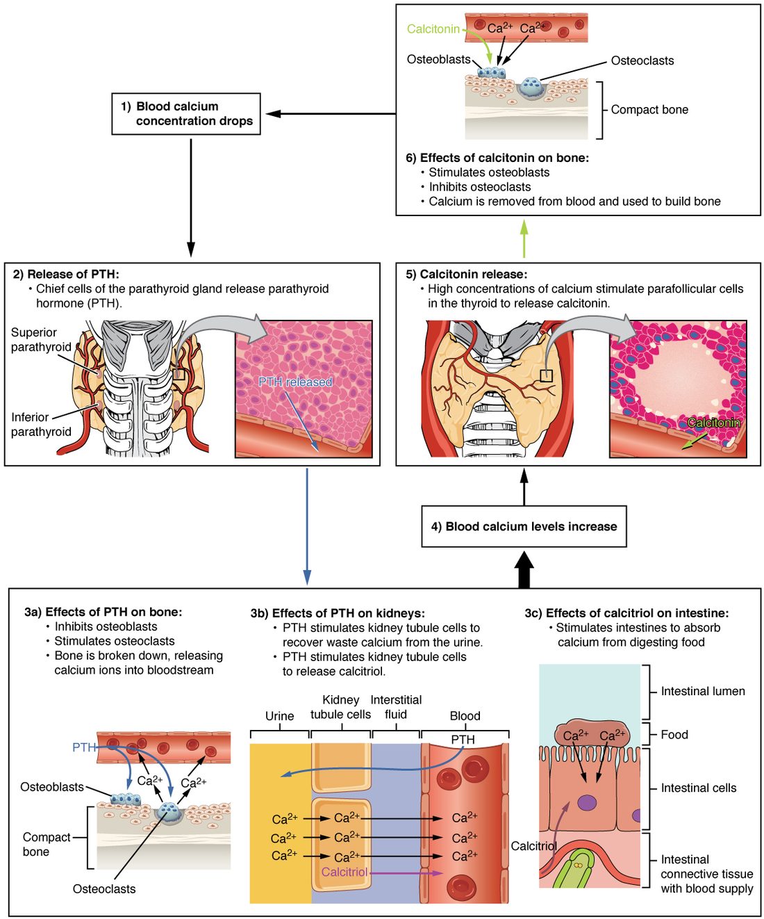
The net effect is higher blood calcium and lower blood phosphate. As calcium rises back into the normal range, about 8.5 to 10.5 mg/dL, the chief cells slow their release: negative feedback.
Calcitriol
Calcitriol is the active form of vitamin D, and it is a hormone. You met vitamin D in Functions of the skin: ultraviolet light turns a cholesterol derivative in your skin into vitamin D, and you also get it from food. That vitamin D is inactive. Two steps activate it (Figure 5):
- The liver adds a hydroxyl group (OH), making 25-hydroxyvitamin D, the main circulating form. Its blood level is what doctors measure to judge your vitamin D stores.
- The kidneys add a second hydroxyl group, making 1,25-dihydroxyvitamin D: calcitriol (calci- = calcium, -triol = three OH groups). PTH switches this step on, and a low blood phosphate also speeds it; calcitriol itself holds it back.
Calcitriol is lipid-soluble. It acts like a steroid: it enters target cells, binds a receptor protein in the nucleus, and changes transcription. Its main effects:
- Intestine: the lining cells make more calcium channels, calcium-binding proteins and pumps, so they absorb much more calcium, and more phosphate, from food. Without calcitriol you absorb only about 10–15% of the calcium you eat; with it, 30–40% or more.
- Bone: by keeping calcium and phosphate high enough in the blood, it lets new bone matrix mineralize. It also helps PTH release calcium from bone when calcium is low.
- Kidneys: it helps the tubules keep calcium.
When vitamin D runs short
With too little vitamin D, from too little sunlight, a poor diet or kidney disease that blocks the last step, the intestine absorbs too little calcium and phosphate. Blood calcium dips, PTH rises to hold it up, and PTH also drives phosphate into the urine. Too little calcium phosphate reaches the osteoid, so new bone matrix stays soft:
- In children, whose epiphyseal plates are still growing, this is rickets, which you met with the skin: soft, bowed leg bones and wide, knobbly growth plates.
- In adults, it is osteomalacia (osteo- = bone, malac- = softening): bones that ache, fracture easily and do not show the bowing of rickets because they are no longer growing.
| Osteomalacia | Osteoporosis | |
|---|---|---|
| What is wrong with the bone | Matrix is laid down but not mineralized: soft bone | Too little bone, but what is there is normally mineralized |
| Usual cause | Too little vitamin D, or too little calcitriol | Bone breakdown outpacing bone building, with age, especially in women after their periods stop |
| Blood calcium and phosphate | Often low or low-normal; PTH high | Usually normal |
| Treatment | Vitamin D and calcium | Drugs that slow osteoclasts or build bone, with calcium and vitamin D |
Parathyroid disorders
Hyperparathyroidism
Hyperparathyroidism is too much PTH, usually from a benign tumor of one parathyroid gland. Blood calcium runs high (hypercalcemia) and phosphate low. Bones lose mineral and ache; extra calcium filtered into the urine forms stones in the kidneys; high calcium makes nerve and muscle cells less excitable, so patients feel tired, weak, low in mood and foggy, and their gut slows. Medical students remember it as "bones, stones, groans and moans". Many cases today are found early, from a routine blood test, before any of these develop. Removing the overactive gland cures it.
Hypoparathyroidism
Hypoparathyroidism is too little PTH. The most common cause is damage to or removal of the parathyroid glands during thyroid surgery, as happened to Ms. Duarte. Blood calcium falls (hypocalcemia). You saw in Bone and blood calcium why that is dangerous: low extracellular calcium makes voltage-gated sodium channels open more easily, so nerves and muscles become overexcitable. The result is tingling around the mouth and in the fingers, muscle cramps and spasms (tetany), and, if severe, spasm that narrows the airway at the voice box, and seizures. The clawed hand when a blood pressure cuff is inflated is a classic bedside sign. Calcium and calcitriol by mouth, or calcium through a vein in an emergency, treat it.
| Hyperparathyroidism | Hypoparathyroidism | |
|---|---|---|
| PTH | High | Low |
| Blood calcium | High | Low |
| Blood phosphate | Low | High |
| Nerves and muscles | Less excitable: weakness, tiredness, slow gut | Overexcitable: tingling, cramps, tetany, seizures |
| Bone | Mineral lost | Dense, little turnover |
| Usual cause | A benign tumor of one gland | Damage during neck surgery |
The blood calcium loop in full
Putting the three hormones together, here is the feedback loop for blood calcium:
- Calcium falls. Chief cells of the parathyroid glands sense it and release PTH. PTH releases calcium from bone, makes the kidneys keep calcium and lose phosphate, and switches on calcitriol, which makes the intestine absorb more calcium. Calcium rises back to normal, and PTH release falls. This is the main loop.
- Calcium rises. PTH release falls, so bone breakdown slows, the kidneys let more calcium go, and less calcitriol is made. The C cells release calcitonin, which briefly inhibits osteoclasts; in adults this is a minor extra brake.
Notice the time scales: PTH on the kidneys and bone acts within minutes to hours, while calcitriol on the intestine, working through new proteins, takes a day or more. Together they hold blood calcium within about 10% of its set point.