A reflex is a fast, automatic response that the same stimulus sets off the same way every time, and every reflex runs on the same five-part circuit, the reflex arc. This page walks through the reflex arc part by part, the stretch reflex behind the knee jerk (and why the "deep tendon reflex" is really a stretch reflex), how clinicians grade tendon reflexes, the tendon reflex that responds to muscle tension, the withdrawal and crossed-extensor reflexes that pull your foot off a tack and keep you standing, and the difference between somatic and visceral reflexes.
What makes a response a reflex
A doctor taps just below your kneecap and your lower leg kicks forward. You did not decide to kick, you could not easily stop it, and it happens the same way every time the tap lands in the same place. A moment later you notice the tap, but the kick has already happened.
That is a reflex (re- = back, flex- = bend: a signal "bent back" out of the CNS): a rapid, involuntary response to a particular stimulus, produced by a fixed circuit of neurons, so that the same stimulus produces the same response each time. Three features set a reflex apart from a voluntary movement:
- Fast. The knee jerk starts about 20 milliseconds after the tap, long before you could react on purpose.
- Involuntary. It does not need a decision, and it runs even in someone who is unconscious. It does not need the brain at all if the circuit lies in the spinal cord.
- Stereotyped (predictable). A given stimulus gives the same response, which is exactly what makes reflexes useful to test.
Some reflexes are built in from birth, such as the knee jerk and a newborn's grasp. Others are learned by repetition, such as braking when a light turns red. This page is about the built-in ones.
The reflex arc
Every reflex runs on a reflex arc: the chain of structures a signal passes through from stimulus to response. It has five parts, always in this order (Figure 1):
- Sensory receptor. It detects the stimulus and produces a graded receptor potential. In the knee jerk, it is a sensory receptor inside the thigh muscle that responds to stretch.
- Sensory neuron. It carries action potentials into the CNS. For a spinal reflex, its cell body sits in a dorsal root ganglion and its axon enters through the dorsal root.
- Integration center. The place in the CNS where the incoming signal is passed on to outgoing neurons: one or more synapses in the gray matter of the spinal cord or brainstem. It may be a single synapse or a chain of interneurons.
- Motor neuron. It carries action potentials out of the CNS. For a spinal reflex to a skeletal muscle, this is the lower motor neuron, whose axon leaves through the ventral root.
- Effector. The muscle or gland that responds: in the knee jerk, the quadriceps femoris, which contracts and straightens the knee.
A useful way to use the arc: if a reflex is missing, the fault can be in any of the five parts. A cut dorsal root, a damaged segment of the cord, a cut ventral root or a diseased muscle can each abolish the same knee jerk. The rest of the exam tells you which part failed.
One synapse or many
- A monosynaptic reflex (mono- = one) has a single synapse in its integration center: the sensory neuron synapses directly on the motor neuron. The stretch reflex is the classic example, and it is the fastest reflex you have.
- A polysynaptic reflex (poly- = many) has one or more interneurons between the sensory and motor neurons. Each extra synapse adds a little delay, about half a millisecond to a millisecond, but interneurons let one input reach many motor neurons, excite some and inhibit others, and spread across several segments or to the other side of the cord. Most reflexes are polysynaptic.
Somatic and visceral reflexes
Reflexes are sorted by their effector, which matches the two parts of the motor division you met in the organization of the nervous system:
- A somatic reflex (somat- = body) acts on skeletal muscle. The knee jerk and pulling your hand off a hot pan are somatic reflexes.
- A visceral reflex (viscer- = internal organs), also called an autonomic reflex, acts on smooth muscle, cardiac muscle or glands, through the autonomic nervous system. Your pupils narrowing in bright light, saliva flowing when food touches your tongue, and your heart slowing when the pressure in your large arteries rises are visceral reflexes.
The sensory half of the two kinds is alike. The motor half differs, as Figure 2 shows: a somatic reflex uses one motor neuron that runs all the way from the CNS to the muscle, while a visceral reflex uses a chain of two, which synapse in a ganglion outside the CNS.
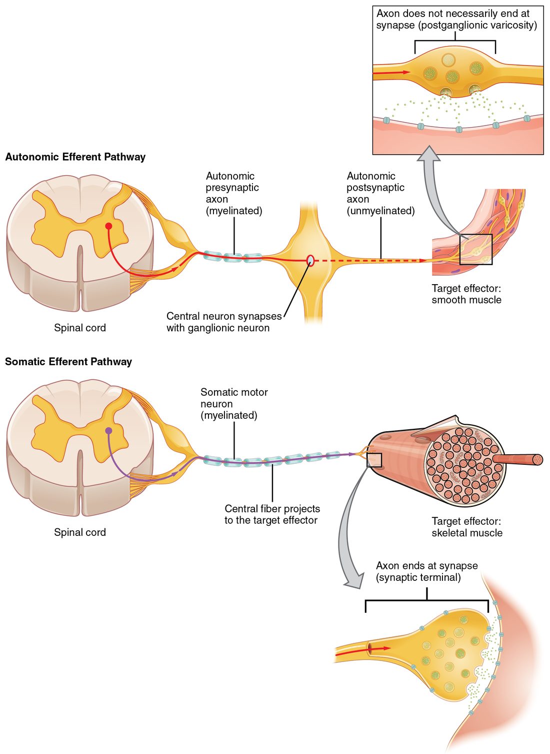
| Somatic reflex | Visceral (autonomic) reflex | |
|---|---|---|
| Effector | Skeletal muscle | Smooth muscle, cardiac muscle or glands |
| Motor neurons from CNS to effector | One | Two, synapsing in a ganglion outside the CNS |
| Motor axons | Myelinated all the way, fast | First myelinated, second unmyelinated, slower |
| Effect on the target | Always excites (contraction) | Can excite or inhibit |
| Sensory side | Sensory receptors mostly in skin, muscles and joints | Sensory receptors mostly in internal organs and vessels |
| Examples | Knee jerk; pulling your foot off a tack | Pupils narrowing in bright light; saliva flowing when you eat |
The rest of this page is about somatic spinal reflexes, the ones tested in every physical exam. The visceral reflexes come back with the autonomic nervous system and with the organs they control.
The stretch reflex
The sensor: the muscle spindle
Scattered through almost every skeletal muscle are small sensory organs called muscle spindles, named for their shape: fat in the middle and tapering at both ends, like the spindle of a spinning wheel. Each is a capsule a few millimeters long holding a handful of thin, modified muscle fibers. Sensory nerve endings wrap around the middle of those fibers. Muscle spindles are proprioceptors: they lie parallel to the ordinary muscle fibers, so when the whole muscle is stretched, they are stretched too, and stretch opens mechanically gated channels in the wrapped endings. The more and the faster the muscle stretches, the faster the spindle's sensory neuron fires.
The circuit
The stretch reflex is the contraction of a muscle in response to its own stretch. Follow it for the knee jerk, which clinicians call the patellar reflex:
- The tap lands on the patellar ligament, just below the kneecap (clinicians usually call it the patellar tendon). The ligament is pushed in, which pulls on the quadriceps femoris and stretches it for a split second.
- Muscle spindles in the quadriceps stretch, and their sensory neurons fire a burst of action potentials.
- The sensory axons enter the spinal cord through the dorsal roots of about L2 to L4.
- In the ventral horn, they synapse directly on the lower motor neurons of the same muscle and release glutamate. One synapse: the stretch reflex is monosynaptic.
- Those motor neurons fire, and acetylcholine at their neuromuscular junctions makes the quadriceps contract.
- The knee straightens, and the lower leg kicks forward.
A stretch reflex resists a change in muscle length: stretch a muscle and it contracts back. That is why the same circuit helps keep you upright. When you start to sway forward, your calf muscles are stretched, their spindles fire, and the calf muscles contract and pull you back.
The antagonist relaxes: reciprocal inhibition
The quadriceps straightens the knee, and the hamstrings bend it. If the hamstrings contracted at the same moment, they would fight the kick. They do not, because branches of the same spindle sensory axons also excite inhibitory interneurons in the cord. These release glycine onto the motor neurons of the hamstrings, which produces IPSPs and stops them firing. Exciting one muscle while inhibiting its antagonist is called reciprocal inhibition. So a stretch reflex has a monosynaptic part (to the stretched muscle) and a polysynaptic part (to its antagonist).
Deep tendon reflexes and how they are graded
When a clinician taps a tendon with a reflex hammer, the test is called a deep tendon reflex. The name is misleading: the sensor is not in the tendon. The tap stretches the muscle through its tendon, and the muscle spindles respond. Every deep tendon reflex is a stretch reflex. Each one checks a particular set of spinal cord segments:
| Reflex | Where you tap | What happens | Main segments |
|---|---|---|---|
| Biceps | Biceps brachii tendon at the front of the elbow | Elbow bends | C5–C6 |
| Brachioradialis | Brachioradialis tendon above the wrist, thumb side | Forearm bends and turns | C5–C6 |
| Triceps | Triceps brachii tendon above the back of the elbow | Elbow straightens | C7 |
| Patellar (knee jerk) | Patellar ligament below the kneecap | Knee straightens | L3–L4 |
| Ankle jerk | Calcaneal (Achilles) tendon at the back of the ankle | Foot points down (plantar flexion) | S1 |
A rhyme many students use counts up from the ankle: "one, two, buckle my shoe" (S1–S2, ankle), "three, four, kick the door" (L3–L4, knee), "five, six, pick up sticks" (C5–C6, biceps), "seven, eight, lay them straight" (C7–C8, triceps). The table lists the main segment of each pair.
The grading scale
The briskness of each response is graded on a scale of 0 to 4+ and always compared with the same reflex on the other side:
| Grade | Meaning |
|---|---|
| 0 | Absent, even with reinforcement |
| 1+ | Present but weak (diminished); may need reinforcement to appear |
| 2+ | Normal |
| 3+ | Brisker than average; may or may not be abnormal |
| 4+ | Very brisk, with clonus: a run of rhythmic jerks after a single stretch |
Reinforcement means asking the person to clench their teeth or hook their fingers together and pull just as you tap. It makes a weak reflex easier to see, so a reflex is only called absent if it stays absent with reinforcement.
What the grades point to follows from the arc and from what you learned about motor pathways:
- Reduced or absent (0 or 1+) in one reflex: a break somewhere in that arc, such as a compressed nerve root, a damaged peripheral nerve or damaged ventral horn neurons. Absent reflexes everywhere suggest a disease of peripheral nerves. Some healthy people have weak reflexes everywhere, which is why symmetry matters more than the number.
- Increased (3+ or 4+), with clonus or a Babinski sign: upper motor neuron damage. The arc is intact but has lost the descending control that normally damps it.
Worked example: how long does a knee jerk take?
Problem. In an adult, the sensory path from the quadriceps to the spinal cord segments of the knee jerk is about 0.6 m long, and the motor path back is also about 0.6 m. Spindle sensory axons conduct at about 70 m/s and the motor axons at about 60 m/s. The central synapse adds about 1 ms, and the neuromuscular junction plus the start of contraction add about 3 ms. Estimate the time from tap to muscle response.
- Time in the sensory axon. time = distance ÷ speed = 0.6 m ÷ 70 m/s = 0.0086 s = 8.6 ms.
- Time in the motor axon. 0.6 m ÷ 60 m/s = 0.010 s = 10 ms.
- Add the synapse. One central synapse: 1 ms.
- Add the muscle. Neuromuscular junction and start of contraction: 3 ms.
- Total. 8.6 + 10 + 1 + 3 = 22.6 ms.
Answer. About 23 ms, close to what is measured. Notice that almost all of it is spent in the axons. A disease that strips myelin from peripheral axons, and so slows conduction, lengthens the delay and weakens the reflex, while one extra synapse would add only about a millisecond.
The tendon reflex
Muscles have a second kind of proprioceptor. The Golgi tendon organ sits where muscle fibers join their tendon. It is a capsule of collagen strands woven through with sensory nerve endings. Because it lies in series with the muscle fibers, it is squeezed when the muscle pulls on its tendon, so it reports muscle tension (force), mostly from active contraction.
The tendon reflex runs from it: rising tension fires the tendon organ's sensory neuron, which excites inhibitory interneurons in the cord. These inhibit the motor neurons of the same muscle, and other interneurons excite the motor neurons of the antagonist. The contracting muscle's force is damped. Because interneurons are involved, the tendon reflex is polysynaptic.
| Muscle spindle | Golgi tendon organ | |
|---|---|---|
| Location | Among the muscle fibers, parallel to them | At the junction of muscle and tendon, in series with the fibers |
| Detects | Muscle length and how fast it changes (stretch) | Muscle tension (force) |
| Reflex it starts | Stretch reflex | Tendon reflex |
| Synapses in the arc | One to the same muscle (monosynaptic) | Two or more (polysynaptic) |
| Effect on the same muscle | Excites: it contracts | Inhibits: its force drops |
| Effect on the antagonist | Inhibits it | Excites it |
The withdrawal and crossed-extensor reflexes
You step on a tack with your left foot. Before you feel any pain, your left knee and hip bend and lift the foot off the tack, and your right leg stiffens to take your whole weight. Two reflexes did this (Figure 3):
- The withdrawal reflex, also called the flexor reflex: nociceptors in the skin fire, and their sensory neurons excite interneurons that excite the motor neurons of the flexor muscles of the same limb and inhibit those of its extensors. The limb bends away from the stimulus.
- The crossed-extensor reflex: other interneurons cross the midline of the cord and do the opposite on the other side, exciting the extensors and inhibiting the flexors of the opposite limb, which straightens and supports the body.
These reflexes show what interneurons add:
- Spread across segments. Lifting a leg needs hip, knee and ankle flexors, whose motor neurons lie in several lumbar and sacral segments. Interneurons carry the signal up and down the cord to all of them (divergence).
- Grading. A light prick lifts the foot a little; a deep stab pulls the whole limb up hard, because a stronger stimulus fires more sensory neurons faster and recruits more interneurons and motor units.
- Afterdischarge. Chains of interneurons keep firing for a moment after the stimulus stops, so the limb stays withdrawn briefly.
- Delay. With several synapses and slower pain axons, the withdrawal reflex takes longer than a stretch reflex, but it is still faster than a decision.
Meanwhile, the same nociceptor signals also climb the spinothalamic tract to the thalamus and cortex. Only when they arrive do you feel the pain, which is why you often find your foot already lifted by the time it hurts.
| Stretch reflex | Withdrawal reflex | |
|---|---|---|
| Stimulus | Stretch of a muscle | A harmful stimulus to the skin |
| Sensory receptor | Muscle spindle | Nociceptors |
| Synapses | Monosynaptic to the stretched muscle | Polysynaptic |
| Segments involved | One or two | Many |
| Response | The stretched muscle contracts | Flexors of the limb contract, extensors relax |
| Opposite limb | Not involved | Extends (crossed-extensor reflex) |
Superficial reflexes and the Babinski sign
Not every reflex tested in a clinic is a stretch reflex. A superficial reflex is set off by stroking the skin. The one you met with motor pathways is the plantar reflex: stroking the outer sole curls the toes down in a healthy adult. Its arc runs through the lower lumbar and sacral segments, but, unlike a stretch reflex, its normal form depends on the corticospinal tract. When that tract is damaged, the response flips to the Babinski sign, with the big toe extending upward. Brisk deep tendon reflexes and a Babinski sign together are the reflex picture of upper motor neuron damage.
Reflexes and the brain
A spinal reflex does not need the brain, but in a healthy person the brain constantly adjusts it. Descending tracts from the brainstem and cortex excite or inhibit the motor neurons and interneurons of every reflex arc, and they set how sensitive the muscle spindles are. That is why:
- Reflexes are brisker when you are anxious and weaker in deep sleep.
- You can partly suppress a withdrawal reflex on purpose, for example to keep holding a hot plate until you can put it down.
- When the spinal cord is cut, reflexes below the cut are absent for days to weeks, then return and become exaggerated: the arcs survive, but the descending control is gone.
That is what makes reflexes so useful at the bedside. Tapping one tendon tests one arc: its sensory neurons, its segments of the cord, its motor neurons and the descending control above it. The next topic puts reflex testing together with the rest of a clinician's checks of the nervous system.