Every voluntary movement you make, from lifting a cup to wiggling a toe, travels on a relay of just two motor neurons: an upper motor neuron in the brain and a lower motor neuron that reaches the muscle. This page covers upper vs lower motor neurons, the corticospinal tract and exactly where it crosses the midline, the brainstem motor tracts that hold your posture, how the basal nuclei and the cerebellum shape a movement without commanding it, and the signs that tell a clinician which of the two motor neurons is damaged.
Two neurons from cortex to muscle
Decide to point your right index finger. The command starts in the finger area of your left primary motor cortex, in the precentral gyrus. A neuron there sends its axon all the way down to the cervical spinal cord. It does not touch the finger muscles. Instead it synapses on a motor neuron in the ventral horn, and that second neuron's axon leaves through the ventral root and a spinal nerve to reach the muscle fibers at the neuromuscular junction.
Those two neurons have names:
- An upper motor neuron (UMN) has its cell body in the brain, in the motor cortex or a brainstem nucleus, and its axon stays inside the CNS. It ends on lower motor neurons, directly or through interneurons.
- A lower motor neuron (LMN) has its cell body in the ventral horn of the spinal cord or in a motor nucleus of a cranial nerve in the brainstem. Its axon leaves the CNS and ends on skeletal muscle fibers. It is the motor neuron you met at the neuromuscular junction, and each one, with the fibers it supplies, makes a motor unit.
The lower motor neuron is the only way out to a skeletal muscle. Every signal that moves a muscle, whether it starts in the cortex, the brainstem or a local circuit in the spinal cord, has to pass through it. For that reason it is called the final common path.
| Upper motor neuron | Lower motor neuron | |
|---|---|---|
| Cell body | Motor cortex or a brainstem motor nucleus | Ventral horn of the spinal cord, or a cranial nerve motor nucleus |
| Axon runs in | Descending tracts of the CNS | Ventral roots, spinal nerves and cranial nerves (the PNS) |
| Ends on | Lower motor neurons or interneurons | Skeletal muscle fibers |
| Transmitter at its ending | Glutamate | Acetylcholine |
| Whole CNS or PNS? | Entirely inside the CNS | Cell body in the CNS, axon in the PNS |
| Example | A neuron of the precentral gyrus | A ventral horn neuron supplying the biceps brachii |
The corticospinal tract
The main highway for voluntary movement is the corticospinal tract (cortic- = bark, meaning the cortex; spin- = spine): axons that run from the cerebral cortex to the spinal cord. Because its axons form two ridges on the front of the medulla oblongata called the pyramids, it is also called the pyramidal tract. Follow it down in Figure 1:
- Cortex. About a third of its axons start in the primary motor cortex. Most of the rest start in the premotor cortex, the supplementary motor area and the parietal lobe. The somatotopic map decides where in the precentral gyrus an axon starts: leg near the top and in the longitudinal fissure, face lowest.
- Internal capsule. The axons funnel down through the internal capsule, packed tightly beside sensory axons. A small injury here can weaken a whole side of the body.
- Midbrain. They run in the cerebral peduncles on the front of the midbrain.
- Pons. They split into many small bundles that pass among the nuclei of the pons.
- Medulla. They regroup as the pyramids on the front of the medulla oblongata.
- The crossing. At the junction of the medulla and the spinal cord, about 85 to 90% of the axons cross the midline. This X-shaped crossing is the pyramidal decussation (decussation = crossing, as you met it in the sensory pathways).
- Spinal cord. The crossed axons descend in the lateral corticospinal tract in the lateral white column. The 10 to 15% that did not cross descend in the anterior corticospinal tract in the anterior white column; most of these cross in the spinal cord near the segment where they end.
- Synapse. At its target segment each axon enters the gray matter and synapses on lower motor neurons in the ventral horn, or on interneurons beside them.
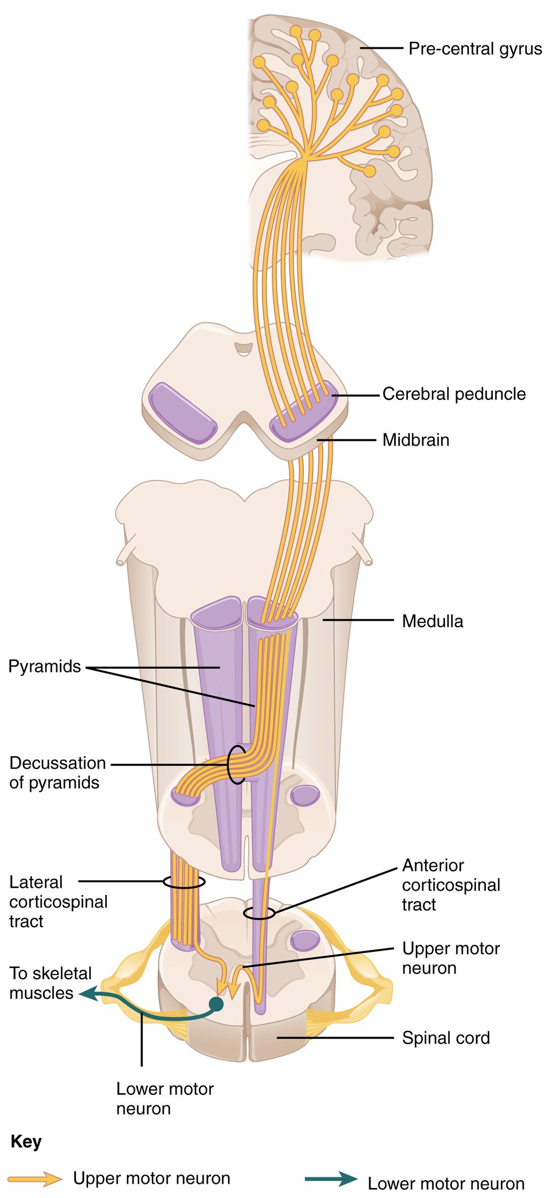
The two spinal parts do different jobs. The lateral tract ends mostly in the cervical and lumbar enlargements, on lower motor neurons for the limbs, especially the hand and fingers. Some of its axons synapse straight onto those motor neurons, which gives you the fine, individual finger movements used to write or play an instrument. The anterior tract ends on both sides of the cord at cervical and upper thoracic levels, on lower motor neurons for the neck and trunk.
What the crossing means
Because the main crossing is in the lower medulla, the rule for motor damage is:
- Above the pyramidal decussation (cortex, internal capsule, midbrain, pons, upper medulla), damage weakens the opposite side of the body.
- Below it (in the spinal cord), damage weakens the same side of the body, from that level down.
| Lateral corticospinal tract | Anterior corticospinal tract | |
|---|---|---|
| Share of the axons | About 85–90% | About 10–15% |
| Where it crosses | Pyramidal decussation, lower medulla | Mostly in the spinal cord, at the level where it ends |
| White column | Lateral | Anterior |
| How far down it runs | The whole length of the cord | Mainly cervical and upper thoracic levels |
| Muscles it controls | Limbs, especially hand and fingers | Neck and trunk |
The corticobulbar tract
Your face, jaw, tongue and throat have no spinal cord segments. Their lower motor neurons sit in the cranial nerve motor nuclei of the brainstem, called the bulb in older anatomy. Upper motor neurons in the face area of the motor cortex reach them through the corticobulbar tract, which travels with the corticospinal tract through the internal capsule and peels off at each brainstem level.
Most cranial nerve motor nuclei receive corticobulbar axons from both hemispheres. The big exception is the part of the facial nerve nucleus that drives the lower face: it gets its input mainly from the opposite hemisphere. The part that drives the forehead gets input from both. This produces a pattern worth knowing:
- Damage to the face area of the left motor cortex weakens the lower right face: the right corner of the mouth droops. The right forehead still wrinkles, because the right hemisphere also drives it.
- Damage to the right facial nerve itself, as in Bell's palsy, weakens the whole right half of the face, forehead included, because it is the lower motor neuron and there is no second route.
Brainstem motor tracts
Stand on a moving bus and your trunk and legs adjust constantly, without a single conscious command. Much of that work runs through upper motor neurons in the brainstem, not the cortex. Their tracts are the brainstem motor tracts, named, like all tracts, from origin to destination:
- Vestibulospinal tract: from the vestibular nuclei of the medulla and pons, which receive balance signals from the balance organs deep in the skull through the vestibulocochlear nerve. It does not cross. It drives the extensor muscles of the trunk and legs that hold you up against gravity, and it corrects your posture when you tip.
- Reticulospinal tracts: from the reticular formation of the pons and medulla. They adjust muscle tone and posture, and help start rhythmic movements such as walking and prepare the trunk before you move a limb.
- Tectospinal tract: from the superior colliculus of the midbrain (tectum = roof, the back of the midbrain). It crosses. It turns the head and neck toward a sudden sight or sound.
- Rubrospinal tract: from the red nucleus of the midbrain (rubr- = red), named for its pinkish color in fresh tissue. It crosses. It helps drive flexor muscles of the arm. In humans it is small, and most of its job has been taken over by the corticospinal tract.
These tracts, together with the basal nuclei circuits you will meet below, were once grouped as the extrapyramidal system (extra- = outside): everything in motor control outside the pyramidal tract. The term is dated, because the two systems work together at every level, but you will still hear it, especially as "extrapyramidal symptoms", the stiffness, tremor and restless movements some drugs that block dopamine cause.
How the basal nuclei shape movement
The basal nuclei do not send axons to the spinal cord. They shape movement by working on the cortex, in the loop you met with the brain regions: cortex → striatum → globus pallidus → thalamus → back to the cortex. The output neurons of the globus pallidus fire all the time and inhibit the thalamus, which is a brake on movement. Two routes through the loop act on that brake in opposite directions (Figure 2):
- Direct pathway (go). The cortex excites striatal neurons that inhibit the output part of the globus pallidus. Inhibiting an inhibitor releases the brake: the thalamus is freed to excite the motor cortex, and the chosen movement goes ahead.
- Indirect pathway (stop). Other striatal neurons act through the outer part of the globus pallidus and the subthalamic nucleus, and the net result is to excite the output of the globus pallidus. The brake presses harder on the thalamus, and competing movements are held back.
Dopamine from the substantia nigra acts on the striatum in two ways at once: it excites the direct-pathway neurons (through one type of dopamine receptor protein) and inhibits the indirect-pathway neurons (through another). Both effects favor movement.
That explains the two diseases you met with the brain regions:
- Parkinson's disease: dopamine neurons of the substantia nigra die. The direct pathway weakens and the indirect pathway strengthens, so the brake stays on. Movements become slow and small and are hard to start, muscles are stiff, and a tremor appears at rest.
- Huntington's disease: the striatal neurons of the indirect pathway die first. The "stop" signal fails, the brake lifts, and unwanted jerky, dance-like movements break through.
How the cerebellum shapes movement
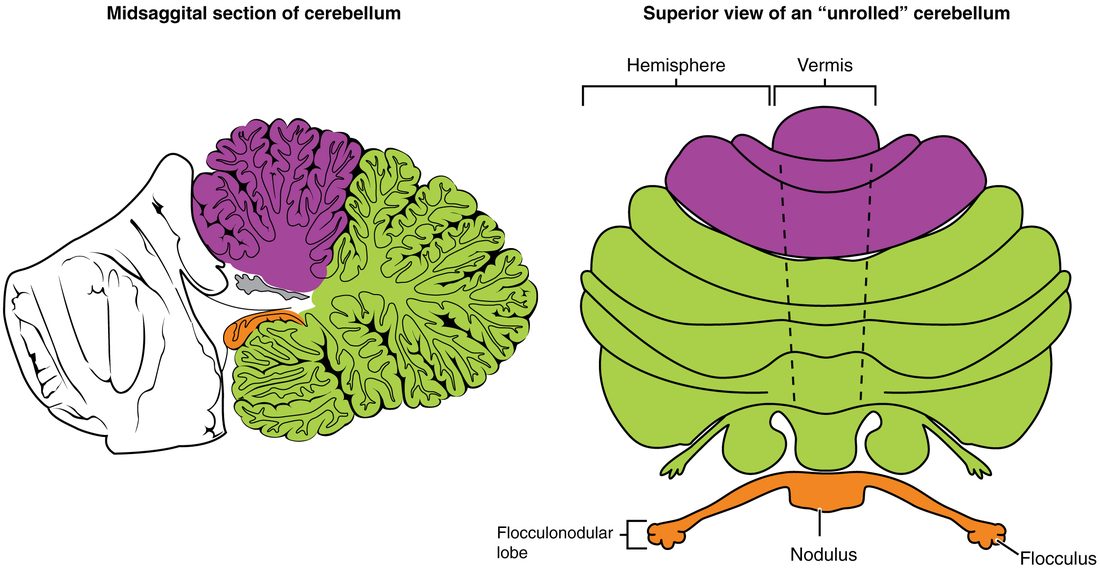
You met the cerebellum as the comparator: it receives a copy of the motor command, compares it with what the muscles and joints report, and sends corrections. Its three functional divisions do this for three different jobs (Figure 3).
The parts you can see
- The vermis (Latin for worm) is the narrow middle strip that joins the two sides.
- Each cerebellar hemisphere is the large lateral part on either side of it.
- The flocculonodular lobe (flocculus = small tuft of wool; nodulus = small knot) is a small lobe on the underside, made of a midline nodule and a flocculus on each side.
Three functional divisions
| Vestibulocerebellum | Spinocerebellum | Cerebrocerebellum | |
|---|---|---|---|
| Region | Flocculonodular lobe | Vermis and the strip of each hemisphere next to it | Lateral parts of the hemispheres |
| Main input | Balance signals from the vestibular nuclei | Proprioceptors, through the spinocerebellar tracts | Cerebral cortex, relayed through the pons |
| Job | Balance and steady eye movements | Posture and correcting movements while they happen | Planning and timing skilled, learned movements |
| Sends output to | Vestibular nuclei | Brainstem motor tracts and, through the thalamus, the motor cortex | Thalamus, then the motor and premotor cortex |
| Damage causes | Unsteady standing, jerky eye movements | A staggering trunk and wide-based walk | Clumsy, poorly timed arm and hand movements |
The cerebellar peduncles
Three pairs of white matter stalks, the cerebellar peduncles (pedunculus = little foot), connect the cerebellum to the brainstem. You can remember them by the level each joins:
- The inferior cerebellar peduncle joins the medulla. It brings input in: most of the spinocerebellar signals from the spinal cord and signals from the vestibular nuclei.
- The middle cerebellar peduncle joins the pons and is the largest. It brings the copy of the cortex's motor plan, relayed by the nuclei of the pons.
- The superior cerebellar peduncle joins the midbrain. It carries most of the cerebellum's output, to the red nucleus and to the thalamus on the way to the motor cortex.
Signs of cerebellar damage
Cerebellar damage leaves strength normal but ruins accuracy, and it shows on the same side of the body. (The output crosses in the superior peduncle and the corticospinal tract crosses back, so the two crossings cancel.) Typical signs:
- Dysmetria (dys- = bad, metr- = measure): overshooting or undershooting a target, such as missing your nose with your fingertip.
- Intention tremor: a shake that grows as the hand nears its target, and is absent at rest, the opposite of the tremor in Parkinson's disease.
- Dysdiadochokinesia (diadocho- = in turn, kine- = movement): clumsy, irregular rapid alternating movements, such as flipping the hand over and back.
- Ataxia of the trunk and legs: a wide-based, staggering walk, typical when the vermis is damaged, as by long heavy alcohol use.
Signs of upper versus lower motor neuron damage
Two patients cannot lift their right arm. In one, the arm is stiff, the tendon jerks are brisk and the muscles look normal. In the other, the arm is floppy, the tendon jerks are gone and the muscles are wasting and twitching. Both have weakness, called paresis (Greek for letting go) when partial and paralysis when complete. But the first has an upper motor neuron problem and the second a lower motor neuron problem, and the signs tell you which.
Lower motor neuron damage
Cut the lower motor neuron and you cut the final common path. Whatever the brain or the spinal cord circuits want, nothing reaches the muscle.
- Weakness in a specific pattern: only the muscles supplied by that nerve, root or segment.
- Flaccidity (flaccid = floppy): muscle tone falls (hypotonia), because the steady low-level firing that produces tone has no route to the muscle. The limb is limp: flaccid paralysis.
- Weak or absent tendon jerks, because their outgoing path is gone.
- Marked atrophy within weeks: muscle fibers that lose their nerve supply waste away.
- Fasciculations (fasciculus = little bundle): brief visible twitches under the skin as a sick lower motor neuron fires by itself and its whole motor unit contracts at once.
Polio, which destroys ventral horn neurons, a cut peripheral nerve, a compressed root and Bell's palsy all cause lower motor neuron signs.
Upper motor neuron damage
Damage upper motor neurons and the lower motor neurons survive, but they lose their descending control. The circuits in the spinal cord still work and are now released from the brain's restraint.
- Weakness of whole movements across a region, such as a whole arm and leg on one side, not single muscles.
- Spasticity (spast- = to pull): a stiffness that increases the faster the limb is moved, then may give way suddenly, like a folding pocket knife. The limb shows spastic paralysis. Tone is raised (hypertonia).
- Hyperreflexia (hyper- = over): tendon jerks that are abnormally brisk and may spread to neighboring muscles. Sometimes a quick stretch sets off clonus, a run of rhythmic jerks, for example at the ankle.
- The Babinski sign, described below.
- Little atrophy: only the slow thinning of disuse, because the muscle keeps its nerve supply.
- No fasciculations, because the lower motor neurons are healthy.
Two points often trip students up. First, right after sudden upper motor neuron damage, the affected limbs are usually floppy with absent tendon jerks for days to weeks, a period of shock in the cord below; spasticity and hyperreflexia appear later. Second, the stiffness comes mainly from losing the brainstem motor tracts, which run alongside the corticospinal axons and are damaged with them in most real injuries. Damage limited to the pyramids themselves causes weakness of fine finger movement and a Babinski sign, but little spasticity.
The Babinski sign
The plantar reflex (plant- = sole of the foot) is tested by stroking the outer edge of the sole firmly with a blunt object, from the heel toward the little toe and then across the ball of the foot toward the big toe. It is a superficial reflex, one set off by stimulating the skin rather than by stretching a muscle.
- Normal (adult): the toes curl downward (plantar flexion).
- Babinski sign: the big toe extends upward (dorsiflexion), and the other toes often fan out. In an adult it means damage somewhere along the corticospinal tract.
In infants the upward big toe is normal, because their corticospinal axons are not yet fully myelinated. It usually changes to the adult downward response by 1 to 2 years of age, as walking develops.
UMN vs LMN signs side by side
| Upper motor neuron damage | Lower motor neuron damage | |
|---|---|---|
| Where the damage is | Motor cortex, internal capsule, brainstem or descending tracts in the cord | Ventral horn, cranial nerve motor nucleus, ventral root or peripheral nerve |
| Weakness pattern | Whole movements over a region (for example one side of the body) | Individual muscles supplied by that segment, root or nerve |
| Muscle tone | Increased: spasticity (after an early floppy phase) | Decreased: flaccidity |
| Tendon jerks | Increased (hyperreflexia), sometimes clonus | Decreased or absent |
| Plantar reflex | Babinski sign (big toe up) | Normal (toes down) or absent |
| Atrophy | Mild, from disuse only | Marked, within weeks |
| Fasciculations | Absent | Often present |
| Examples | Loss of blood flow to the motor cortex or internal capsule; multiple sclerosis | Polio; a cut or compressed nerve; Bell's palsy |
Putting it together: localizing weakness
Worked example: two patients with a weak right leg
Patient 1. A man suddenly cannot use his right arm and leg. Three weeks later both are stiff, his right knee jerk is brisk, his right big toe goes up when the sole is stroked, and the lower right side of his face droops while his forehead wrinkles normally.
- UMN or LMN? Spasticity, hyperreflexia and a Babinski sign are upper motor neuron signs.
- Which side of the brain? The right side of the body is weak, and the face is involved too, so the damage is above the pyramidal decussation and above the facial nucleus in the pons: in the left hemisphere or internal capsule.
- Why is the forehead spared? The forehead part of the facial nucleus receives corticobulbar axons from both hemispheres; the lower face receives them mainly from the opposite one.
Patient 2. A woman has a knife wound that cut the left half of her spinal cord at T10.
- Motor, below the cut. The lateral corticospinal tract has already crossed in the medulla, so the left tract carries commands for the left side. Result: upper motor neuron weakness of the left leg (floppy at first, spastic later, with a Babinski sign).
- Motor, at the cut. The ventral horn neurons at T10 on the left are destroyed: a narrow band of lower motor neuron weakness in the left trunk muscles supplied by that segment.
- Sensory. As you worked out in the sensory pathways: fine touch, vibration and position lost on the left below T10; pain and temperature lost on the right from a segment or two below.
Answer. Patient 1 has upper motor neuron damage in the left cerebral hemisphere. Patient 2 has damage to one half of the cord, with a left weak leg, left loss of position sense and right loss of pain sensation.