Every sensation starts with a sensory receptor turning some form of energy into an electrical signal, and ends when that signal reaches your cerebral cortex. This page covers the sensory receptor types (by stimulus, by location and by structure), how a stimulus becomes a train of action potentials, why some sensations fade while others last, and the two main ascending pathways, the dorsal column–medial lemniscus pathway and the spinothalamic tract: which neurons carry the signal, where each pathway crosses to the other side, and how that lets you locate damage from a patient's symptoms.
Sensation and perception
You put on a watch in the morning and feel it on your wrist. By mid-morning you no longer notice it, although it has not moved. Meanwhile, sensory receptors in your large arteries report your blood pressure to your brainstem every second of the day, and you never feel a thing.
Those examples separate two ideas:
- Sensation is the detection of a stimulus and the delivery of signals about it into the CNS. It happens whether or not you are aware of it.
- Perception is the conscious awareness and interpretation of a sensation, produced by the cerebral cortex. "Something is pressing on my wrist; it is my watch" is a perception.
Much of what your sensory receptors report never becomes a perception. Signals about blood pressure, blood chemistry and muscle length are handled below the level of awareness, by the brainstem, the hypothalamus and the cerebellum.
General and special senses
Sensory systems are sorted into two groups:
- The general senses use sensory receptors scattered widely through the body. They include somatosensation (somat- = body), meaning touch, pressure, vibration, temperature, pain and the sense of body position, from the skin, muscles and joints, and the visceral senses, which report on internal organs: stretch of the stomach or bladder, organ pain.
- The special senses have their sensory receptors gathered in dedicated organs in the head: smell, taste, vision, hearing and balance. Each has its own later topic, and each travels in cranial nerves you met in the last topic: smell in I, taste in VII and IX, vision in II, and hearing and balance in VIII.
A sensory modality is a kind of sensation: touch, vision and pain are different modalities. A submodality is a variety within one modality: warm and cold within temperature, or sweet and salty within taste.
How does the brain know which modality a signal is? Not from the action potentials themselves, which look the same in every sensory neuron. It knows from which neurons are firing. Each sensory neuron responds best to one kind of stimulus and connects through its own chain of neurons to its own patch of cortex. This is called the labeled line principle. It is why a blow to the eye makes you "see stars": the neurons of the vision pathway are firing, so you perceive light.
Classifying sensory receptors
A sensory receptor is a structure that responds to a particular change in its surroundings, the stimulus, by producing an electrical signal. It may be the ending of a sensory neuron or a separate cell that passes the signal to one. Sensory receptors are classified in three ways.
By the stimulus they respond to
| Type | Stimulus | Examples |
|---|---|---|
| Mechanoreceptor | Physical deformation: touch, pressure, vibration, stretch | Tactile (Meissner) corpuscles, lamellated (Pacinian) corpuscles, stretch-sensitive endings in muscles and artery walls |
| Thermoreceptor | Temperature change | Separate warm and cold free nerve endings in the skin |
| Nociceptor | Stimuli that damage or threaten to damage tissue: intense heat or cold, crushing, chemicals released by injured cells | Free nerve endings in skin, muscles, joints and organs |
| Chemoreceptor | Particular chemicals | Sensory receptors for taste and smell; sensory receptors that track oxygen, carbon dioxide and pH in the blood |
| Photoreceptor | Light | The light-sensitive cells at the back of the eye |
| Osmoreceptor | The solute concentration of body fluids | Neurons in the hypothalamus that trigger thirst when your blood becomes too concentrated |
The word nociceptor comes from noci- (= harm): it detects harm, not "pain". Pain is what the brain makes of its signals, as you will see below.
By where the stimulus comes from
- Exteroceptors (extero- = outside) respond to stimuli from outside the body: sensory receptors in the skin, eyes, ears, nose and tongue.
- Interoceptors (intero- = inside) respond to stimuli inside the body, from internal organs and blood vessels. They serve the visceral senses.
- Proprioceptors (proprio- = one's own) sit in skeletal muscles, tendons and joint capsules and report muscle length, tension and joint angle. Together they produce proprioception, your sense of where your body parts are, which lets you touch your nose with your eyes closed. The sense of limb movement in particular is called kinesthesia (kine- = movement, -esthesia = sensation).
By structure
- Free nerve endings: bare, branching dendrites of sensory neurons. They serve pain, temperature and some touch and itch.
- Encapsulated endings: nerve endings wrapped in connective tissue that shapes what they respond to. Tactile corpuscles, in the dermal papillae, respond to light touch; lamellated corpuscles, deep in the dermis and hypodermis, respond to vibration and deep pressure; the endings around hair follicles respond when a hair bends.
- Sensory receptor cells: separate cells that detect the stimulus and release neurotransmitter onto a sensory neuron. Taste, hearing and balance work this way; the light-detecting cells of the eye work similarly but are specialized neurons. Tactile (Merkel) cells in the epidermis also pass touch signals to a nerve ending.
From stimulus to action potentials
Press lightly on your fingertip, then harder. A sensory receptor has to turn that difference in pressure into a difference in electrical signals. Converting one kind of energy into an electrical signal is called transduction, and it runs in steps.
- The stimulus opens ion channels in the membrane of the sensory ending. In a mechanoreceptor, stretching the membrane pulls mechanically gated channels open; in a thermoreceptor, temperature-sensitive channels open; in a chemoreceptor, a chemical binds a receptor protein.
- Positive ions, mainly Na+, flow in, and the ending depolarizes. Local, graded changes like this are called receptor potentials. One that forms in the ending of a sensory neuron itself is also called a generator potential.
- Like any graded potential, it is bigger for a stronger stimulus and fades with distance. It spreads to the trigger zone, the first stretch of axon with voltage-gated sodium channels.
- If the trigger zone reaches threshold, action potentials fire and travel along the axon toward the CNS. While the depolarization stays above threshold, action potentials keep firing.
Action potentials are all-or-none, so a stronger stimulus cannot make a bigger one. Instead, stimulus strength is coded in two ways:
- Frequency: a larger receptor potential holds the trigger zone further above threshold, so the next action potential fires sooner after each refractory period. A firm press fires more action potentials per second than a light one.
- Number of neurons: a stronger or wider stimulus activates more sensory neurons, including ones with higher thresholds.
In most separate sensory receptor cells, receptor potentials do not trigger action potentials in the cell itself (taste cells are an exception, covered in chemical senses; even there, the action potentials never leave the taste bud). Instead it changes how much neurotransmitter the cell releases onto the sensory neuron, whose firing rate rises or falls to match.
Sensory adaptation
Back to the watch. Its steady pressure is still there, but after a while you stop feeling it. Part of the reason lies in the sensory receptors themselves.
Sensory adaptation is a fall in a sensory receptor's response to a stimulus that stays constant. Sensory receptors differ greatly in how fast they adapt (Figure 1):
- Tonic sensory receptors (slowly adapting) keep firing for as long as the stimulus lasts, with only a small drop in frequency. Nociceptors, proprioceptors, tactile (Merkel) cells and the stretch-sensitive endings in artery walls are tonic. That is useful: pain from an injury, and information about body position and blood pressure, stay reported for as long as they matter.
- Phasic sensory receptors (rapidly adapting) fire a burst when the stimulus starts, fall silent while it holds steady, and often fire again when it stops. Lamellated corpuscles, tactile corpuscles and hair follicle endings are phasic. They signal change: the brush of an insect on your arm, or the vibration of a phone in your pocket.
Some adaptation also happens in the CNS, where synapses along the pathway respond less to an unchanging input. That is why you stop noticing a smell in a room within minutes.
Ascending sensory pathways
A sensory signal from your big toe has to travel more than a meter to reach your cortex. It does so through an ascending pathway: a chain of neurons that carries sensory information up the spinal cord and brainstem. The pathways to conscious perception use three neurons in a row:
- The first-order neuron is the sensory neuron. Its ending is (or contacts) the sensory receptor, its cell body sits in a dorsal root ganglion (or a cranial nerve ganglion), and its axon enters the CNS.
- The second-order neuron has its cell body in the spinal cord or the brainstem. Its axon crosses the midline and climbs to the thalamus.
- The third-order neuron runs from the thalamus to the primary somatosensory cortex in the postcentral gyrus.
Crossing from one side of the CNS to the other is called decussation (decuss- = to cross like the letter X). Because the second-order axon always crosses, the left side of your body is felt by the right hemisphere, and the right side by the left. The two main pathways differ in what they carry and, crucially, in where they cross (Figure 2).
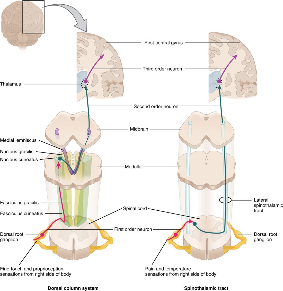
The dorsal column–medial lemniscus pathway
This pathway carries fine, precise touch: two-point discrimination (telling one touch from two close together), vibration, pressure, and conscious proprioception.
- First-order neuron: the sensory axon enters the cord through the dorsal root and turns straight upward in the dorsal column (the posterior column of white matter) on the same side. It does not synapse in the cord. Axons from the lower body (below about T6) form the fasciculus gracilis (gracil- = slender), near the midline; axons from the upper body and arm join lateral to them as the fasciculus cuneatus (cune- = wedge).
- The first-order axons synapse in the lower medulla oblongata, in the nucleus gracilis and nucleus cuneatus.
- Second-order neuron: its axon crosses the midline in the medulla and climbs through the brainstem in a flat ribbon, the medial lemniscus (lemniscus = ribbon), to the thalamus.
- Third-order neuron: from the thalamus, through the internal capsule, to the primary somatosensory cortex.
Because it crosses in the medulla, this pathway runs up the spinal cord on the same side as the body part it serves.
The spinothalamic tract
This pathway carries pain, temperature, itch, and crude touch and pressure (touch you can detect but not locate precisely).
- First-order neuron: the sensory axon enters through the dorsal root and synapses almost at once in the dorsal horn, after rising or falling at most a segment or two.
- Second-order neuron: its cell body lies in the dorsal horn. Its axon crosses the midline in front of the central canal, within a segment or two, and climbs in the spinothalamic tract in the lateral and anterior white columns of the opposite side, through the brainstem to the thalamus. Branches also reach the reticular formation, which is one reason pain is so arousing.
- Third-order neuron: from the thalamus to the primary somatosensory cortex, with other thalamic neurons carrying pain to the insula and the limbic system, where its unpleasantness is processed.
Because it crosses in the spinal cord, this pathway runs up the cord on the side opposite the body part it serves.
| Dorsal column–medial lemniscus | Spinothalamic tract | |
|---|---|---|
| Carries | Fine touch, two-point discrimination, vibration, conscious proprioception | Pain, temperature, itch, crude touch and pressure |
| First-order neuron ends in | Medulla oblongata (nucleus gracilis or cuneatus) | Dorsal horn of the spinal cord |
| Second-order axon crosses in | The medulla oblongata | The spinal cord, within a segment or two of entry |
| Side of the cord it climbs in | Same side as the body part | Opposite side from the body part |
| White matter column in the cord | Dorsal (posterior) column | Lateral and anterior columns |
| Relay in the brain | Thalamus | Thalamus |
| Third-order neuron ends in | Primary somatosensory cortex | Primary somatosensory cortex, plus insula and limbic areas |
| Loss after damage to one side of the cord | On the same side, from the level of damage down | On the opposite side, from a segment or two below the damage down |
Where it ends: the somatosensory cortex
The third-order neurons end in the primary somatosensory cortex, laid out as the sensory homunculus you met in the topic on cortical functions: legs near the top of the hemisphere and in the longitudinal fissure, then trunk, arm and hand, with the face lowest, near the lateral sulcus. The fingertips and lips, with the densest sensory receptors, get the largest areas. Association areas behind it combine touch, position and memory to recognize an object in your hand without looking.
Unconscious proprioception: the spinocerebellar tracts
Not all body position information goes to the cortex. The spinocerebellar tracts carry signals from proprioceptors in the muscles and joints up the lateral columns to the cerebellum, which uses them to coordinate movement. These pathways use two neurons, skip the thalamus, and end on the same side of the cerebellum as the body part. You are never aware of this information directly.
The face: the trigeminal sensory pathway
The face has no dorsal roots. Touch, pain and temperature from the face travel in the trigeminal nerve instead, through the trigeminal sensory pathway, which follows the same three-neuron plan:
- First-order neuron: cell body in the trigeminal ganglion beside the pons. Axons for fine touch end in a trigeminal nucleus in the pons. Axons for pain and temperature turn down and end in a long trigeminal nucleus that runs through the medulla oblongata into the upper cervical cord.
- Second-order neuron: its axon crosses the midline and climbs, next to the medial lemniscus and spinothalamic fibers, to the thalamus.
- Third-order neuron: from the thalamus to the face area of the primary somatosensory cortex, low on the postcentral gyrus.
One detail matters for localizing damage. In the lower brainstem, the uncrossed pain and temperature axons from the face descend on the same side, right beside the already crossed spinothalamic axons from the opposite side of the body. Damage to one side of the medulla can therefore remove pain and temperature from the same side of the face and the opposite side of the body.
Pain and referred pain
Nociception is not pain
Nociception is the neural process of detecting and signaling tissue damage, from nociceptors through the spinothalamic tract. Pain is the unpleasant experience the brain produces. The two usually travel together, but not always: a soldier wounded in battle may feel little pain at first despite intense nociception, and a person can feel real pain in a limb that has been amputated, with no nociceptors to fire at all. The brainstem can also turn down pain signals in the dorsal horn, using endorphins and other transmitters.
Nociceptive signals travel on two kinds of axon:
- Thin, myelinated axons carry fast pain: sharp, pricking and well localized, arriving within a fraction of a second.
- Thinner, unmyelinated axons carry slow pain: burning or aching, poorly localized, arriving a second or more later and lasting longer.
Stub your toe and you feel both: a sharp jolt, then a throbbing ache.
Referred pain
A man having a heart attack often feels pain not in his chest wall alone but down the inner side of his left arm. The heart is not in his arm. This is referred pain: pain from an internal organ felt in an area of skin or muscle.
The best-supported explanation is convergence. Sensory axons from an organ enter the spinal cord through the same dorsal roots as axons from a patch of skin, and in the dorsal horn many of them converge on the same second-order neurons. The brain receives the signal on a pathway that almost always reports skin, so it assigns the pain to the skin of that dermatome.
| Organ | Where the pain is felt | Why there |
|---|---|---|
| Heart | Left chest, left shoulder and the inner left arm; sometimes the jaw | Heart pain axons enter at about T1–T4, the segments that supply the chest and inner arm |
| Diaphragm (irritated from below, as by blood or a diseased gallbladder) | Tip of the shoulder | The diaphragm's sensory axons run in the phrenic nerve to C3–C5, which also supply the skin of the shoulder |
| Appendix, early in appendicitis | Around the navel | Its axons enter at about T10, the navel's dermatome |
| Kidney and ureter | Flank, sweeping toward the groin | Their axons enter at about T10–L1 |
Putting it together: localizing damage
The crossing points let you read the location of damage from the pattern of loss. Work through it step by step.
Case: a knife wound cuts the left half of the spinal cord at the T10 level. Which sensations are lost, and where?
- Dorsal column pathway: it climbs on the same side as the body part and does not cross until the medulla. The cut destroys the left dorsal column, which carries fine touch, vibration and position from the left side of the body below T10. Result: lost on the left, from T10 down.
- Spinothalamic tract: signals from the right side of the body enter on the right, cross within a segment or two and climb on the left. The cut destroys the left spinothalamic tract, carrying pain and temperature from the right. Result: lost on the right, from about T11 or T12 down.
- At the level itself: the left dorsal roots at T10 are also damaged, so there is a narrow band of complete loss on the left at T10.
Answer: the left leg cannot feel vibration or position; the right leg cannot feel pain or temperature. A loss of fine touch on one side with a loss of pain on the other side points to damage to one half of the spinal cord. (The left leg would also be weak, because the motor pathways run on the same side; the next topic covers those.)
Other patterns work the same way:
- Loss of all sensation on one whole side of the body and face: damage above the medulla, where all the pathways have crossed and run together, for example in the opposite thalamus or internal capsule.
- Loss of pain and temperature on one side of the face and the other side of the body: the lower brainstem on the side of the face loss.
- Loss of pain and temperature in a band across both shoulders and arms, with touch kept: damage in the center of the cervical cord, where the crossing spinothalamic axons pass in front of the central canal.
- Loss of vibration and position sense in both legs, with pain and temperature kept: damage to both dorsal columns.