You have 12 pairs of cranial nerves, numbered I to XII from front to back along the underside of the brain. Some carry only sensory signals, some only motor commands, and some both. This page gives the 12 cranial nerves and their functions one by one, where each leaves the skull, a quick bedside test for each, and a full section on the vagus nerve, the one that reaches farthest into the body.
What makes a nerve "cranial"
Try this. Wrinkle your forehead, then clench your teeth, then stick out your tongue. Three different movements, and three different nerves command them. None of those nerves passes through the spinal cord. Each one leaves the brain directly and exits through its own opening in the skull.
A cranial nerve (crani- = skull) is a nerve that attaches to the brain rather than to the spinal cord. There are 12 pairs. Like spinal nerves, they are part of the peripheral nervous system. Unlike spinal nerves, they are not all mixed: a few are purely sensory, a few are purely motor, and the rest carry both.
Each is named in two ways: by a Roman numeral, which gives its order from front (I) to back (XII) along the underside of the brain, and by a name that hints at its job. Look at the underside of the brain in Figure 1 and find them in order.
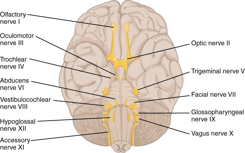
Where they attach
- I and II attach to the forebrain. They are really outgrowths of the brain itself.
- III and IV attach to the midbrain. IV is the only cranial nerve that leaves from the back of the brainstem.
- V attaches to the side of the pons.
- VI, VII and VIII emerge at the border between the pons and the medulla oblongata.
- IX, X and XII leave the medulla oblongata. XI rises mainly from the upper cervical spinal cord, climbs through the foramen magnum and then leaves the skull again with IX and X.
Each cranial nerve has nuclei in the brainstem (or, for I and II, in the forebrain): clusters of cell bodies where its motor axons start and its sensory axons end. Damage to one side of the brainstem therefore usually produces cranial nerve signs on that same side.
Remembering the names and types
Two memory sentences carry most of the load. For the names, in order:
"Old Owls Often Trade Tiny Acorns For Very Good Vanilla And Honey": olfactory, optic, oculomotor, trochlear, trigeminal, abducens, facial, vestibulocochlear, glossopharyngeal, vagus, accessory, hypoglossal.
For the type of each, Sensory, Motor or Both, in the same order:
"Sara Saw Mom Make Bread; Mom Burned Some, But Bob Made More."
A third fact is worth fixing now: four cranial nerves carry parasympathetic axons: III, VII, IX and X. Remember them as "1973" (3, 7, 9, 10).
"Motor" here counts only the nerve's main outgoing axons. Nerves to muscles also carry a few sensory axons from those muscles, which is why some texts call III, IV, VI, XI and XII "mainly motor".
The twelve nerves at a glance
This table is the one to study from. The sections after it add the detail and the reasoning behind each test.
| Nerve | Type | Main functions | Leaves the skull through | Quick test |
|---|---|---|---|---|
| I Olfactory | Sensory | Smell | Cribriform plate of the ethmoid bone | Identify a familiar smell (coffee, soap) through one nostril at a time, eyes closed |
| II Optic | Sensory | Vision | Optic canal | Read an eye chart one eye at a time; count fingers held out to the side |
| III Oculomotor | Motor (with parasympathetic) | Moves the eye up, down and in; raises the upper lid; narrows the opening in the center of the eye; adjusts focus for near objects | Superior orbital fissure | Follow a finger through an H pattern; watch the dark center of each eye narrow when a light shines in |
| IV Trochlear | Motor | One eye muscle that turns the eye down, most strongly when the eye points toward the nose | Superior orbital fissure | Look down and in, toward the nose |
| V Trigeminal | Both | Touch, pain and temperature from the face, scalp front, mouth, teeth and the surface of the eye; chewing muscles | Superior orbital fissure (V1), foramen rotundum (V2), the oval foramen of the sphenoid (V3) | Light touch on forehead, cheek and jaw; clench the teeth and feel the masseter |
| VI Abducens | Motor | The eye muscle that turns the eye outward | Superior orbital fissure | Look straight out to each side |
| VII Facial | Both (with parasympathetic) | Muscles of facial expression; taste from the front two-thirds of the tongue; tears and saliva | Internal acoustic meatus, then the stylomastoid foramen | Raise the eyebrows, shut the eyes tight, smile, puff the cheeks |
| VIII Vestibulocochlear | Sensory | Hearing and balance | Internal acoustic meatus | Hear a finger rub or a whisper at each ear |
| IX Glossopharyngeal | Both (with parasympathetic) | Taste and touch from the back third of the tongue and the throat; one swallowing muscle; saliva from the largest saliva gland; blood pressure and blood gas signals from the neck | Jugular foramen | Touch the back of the throat: the person gags (IX carries the touch in) |
| X Vagus | Both (with parasympathetic) | Throat and voice box muscles for swallowing and speech; parasympathetic control of the heart, lungs and most digestive organs; sensory signals from those organs | Jugular foramen | Say "ah": the back of the roof of the mouth rises evenly; listen for a hoarse voice |
| XI Accessory | Motor | Sternocleidomastoid and trapezius | Jugular foramen | Shrug and turn the head against resistance |
| XII Hypoglossal | Motor | Muscles of the tongue | Hypoglossal canal | Stick out the tongue: it should come out straight |
The sensory nerves: I, II and VIII
I Olfactory nerve
The olfactory nerve (olfact- = smell) is not one cord but about 20 bundles of very short axons. They start in the lining high in the roof of the nose and pass straight up through the holes of the cribriform plate to the forebrain. A blow to the head, most often to the back of the head, can shear them where they cross the plate, and the person loses the sense of smell. Much of what you call flavor is smell, so food then tastes flat too.
Test: with the eyes closed and one nostril blocked, the person names a familiar, non-irritating smell. Sharp irritants such as smelling salts are avoided because they stimulate the trigeminal nerve instead.
II Optic nerve
The optic nerve (opt- = eye, vision) carries visual signals from the back of the eye through the optic canal to the brain. Strictly, it is a tract of the CNS: it is wrapped in meninges and myelinated by oligodendrocytes, not Schwann cells, which is why diseases of CNS myelin such as multiple sclerosis often affect it.
Test: read an eye chart with each eye in turn, and check side vision by counting fingers held out to the sides.
VIII Vestibulocochlear nerve
The vestibulocochlear nerve has two parts, as its name says: a vestibular part (vestibul- = entrance hall) from the balance organs and a cochlear part (cochle- = snail shell) from the hearing organ, both deep in the temporal bone. It passes through the internal acoustic meatus with the facial nerve. Damage causes hearing loss, ringing in the ear, or a spinning dizziness (vertigo) with jerky eye movements.
Test: rub your fingers near each ear in turn, or whisper a number, and ask the person to repeat it. Dizziness on moving the head points to the balance part.
The eye movement nerves: III, IV and VI
Six small muscles move each eye. Three nerves share them unevenly: IV supplies one, VI supplies one, and III supplies the other four. A rule often written as "LR6, SO4, all the rest 3" captures it: the muscle that turns the eye out is supplied by VI, the one whose tendon runs through a pulley by IV, and the rest by III.
- Oculomotor nerve (III) (ocul- = eye): moves the eye up, down and in, and raises the upper lid. Its parasympathetic axons narrow the dark opening at the center of the eye in bright light and thicken the eye's focusing part for near vision. Damage leaves the eye turned down and out, the upper lid drooping, and the dark opening wide and unresponsive to light.
- Trochlear nerve (IV) (trochle- = pulley): supplies the one eye muscle whose tendon passes through a small pulley of cartilage in the orbit. That muscle turns the eye down and slightly out, and twists its top toward the nose. Its downward pull is strongest when the eye already points toward the nose, so you test it by having the person look down and in. Damage causes double vision when looking down, as when reading or walking downstairs, and people often tilt the head to compensate.
- Abducens nerve (VI) (ab- = away, duc- = lead): supplies the eye muscle that turns the eye outward, the movement called abduction. Damage leaves the eye turned in toward the nose and causes double vision when looking toward the damaged side. It has a long path along the base of the skull, so raised pressure inside the skull often affects it first.
Test for all three: the person keeps the head still and follows your finger through a large "H". Each arm of the H stresses different muscles. You also shine a light into each eye and watch the dark center narrow.
The face: V and VII
The face has two nerves that students often confuse. The rule is simple: V feels, VII moves. The trigeminal nerve carries touch, pain and temperature from the face and supplies the chewing muscles. The facial nerve moves the face for expression.
V Trigeminal nerve
The trigeminal nerve (tri- = three, gemin- = twins, so "triplets") is the largest cranial nerve. Near the pons it has a large sensory ganglion, and from it three divisions fan out across the face:
- V1, ophthalmic division: forehead, the skin of the upper lid, the surface of the eye and the nose. Sensory only.
- V2, maxillary division: cheek, upper lip, upper teeth and the palate. Sensory only.
- V3, mandibular division: lower lip, chin, lower teeth, the front of the tongue for touch (not taste), and the motor branch to the muscles of mastication, including the masseter and temporalis.
The face has no spinal dermatomes; these three divisions take their place. In trigeminal neuralgia, a trigeminal root is irritated and a light touch on the cheek or a breeze can set off stabbing, electric pain in one division's area.
Test: touch the forehead, cheek and jaw on each side lightly and compare. Ask the person to clench the teeth and feel both masseters bulge. A wisp of cotton touched to the clear front surface of the eye makes both eyes blink: the touch travels in on V1, and the blink goes out on VII, so this one test checks two nerves.
VII Facial nerve
The facial nerve leaves the brainstem, enters the temporal bone through the internal acoustic meatus, winds through the bone and leaves through the stylomastoid foramen, then fans across the face. It does four jobs:
- Motor to all the muscles of facial expression, including the occipitofrontalis, orbicularis oculi, orbicularis oris, zygomaticus major and buccinator.
- Taste from the front two-thirds of the tongue.
- Parasympathetic to the tear glands and to two of the three pairs of saliva glands (those under the tongue and jaw).
- A little skin sensation near the ear.
In Bell's palsy, the facial nerve swells inside its narrow bony canal, often linked to a viral infection. The whole half of the face goes slack: the brow will not lift, the eye will not close fully, and the mouth droops. Taste from the front of the tongue on that side may be lost.
Test: ask the person to raise the eyebrows, shut the eyes tight while you try to open them, smile, and puff out the cheeks. Compare the two sides.
| V Trigeminal | VII Facial | |
|---|---|---|
| Main job on the face | Feeling: touch, pain, temperature | Moving: facial expression |
| Muscles supplied | Chewing muscles (masseter, temporalis) | Expression muscles (orbicularis oculi and oris, buccinator) |
| Tongue | Touch from the front two-thirds | Taste from the front two-thirds |
| Glands | None of its own | Tear glands and two pairs of saliva glands |
| Leaves the skull | Three openings, one per division | Internal acoustic meatus, then stylomastoid foramen |
| Sign of damage | Numb face, weak chewing, stabbing facial pain | Slack half face, eye will not close |
| Its part in the blink test | Carries the touch in | Carries the blink out |
The throat and tongue: IX, X, XI and XII
IX Glossopharyngeal nerve
The glossopharyngeal nerve (gloss- = tongue, pharyng- = throat) leaves through the jugular foramen. It carries taste and touch from the back third of the tongue and touch from the upper throat, supplies one small muscle that lifts the throat during swallowing, and sends parasympathetic axons to the largest saliva gland, which sits in front of the ear. It also carries signals from pressure sensors and blood gas sensors at the fork of the main artery in the neck. You will meet those sensors again in the chapters on blood pressure and breathing.
X Vagus nerve
The vagus nerve has its own section below. At the throat, its motor axons supply most muscles of the throat and all the muscles of the voice box, so it controls swallowing and speech.
Testing IX and X together: touching the back of the throat triggers gagging. IX carries the touch in and X drives the throat muscles that contract, so a missing gag on one side can mean damage to either. Asking the person to say "ah" tests X alone: the soft back part of the roof of the mouth should rise evenly on both sides. If one vagus nerve is damaged, that side stays low, the hanging tag at the back of the mouth is pulled toward the healthy side, and the voice may be hoarse.
XI Accessory nerve
The accessory nerve (also called the spinal accessory nerve) is odd. Its motor neurons sit in the upper five or so segments of the cervical spinal cord. Its rootlets join into one trunk that climbs up through the foramen magnum into the skull and then leaves again through the jugular foramen. It supplies two neck and shoulder muscles: the sternocleidomastoid, which turns the head, and the trapezius, which shrugs the shoulder. Older texts describe a small "cranial root" as well; its axons actually travel on with the vagus nerve.
Test: the person shrugs both shoulders and turns the head to each side while you push against the movement.
XII Hypoglossal nerve
The hypoglossal nerve (hypo- = below, gloss- = tongue) runs through the hypoglossal canal to the muscles of the tongue. It is purely motor.
Test: the person sticks out the tongue. Each half of the tongue pushes its own side forward. If one hypoglossal nerve is damaged, the healthy side pushes harder, and the tongue points toward the damaged side.
The vagus nerve
Here is a concrete case first. A healthy young man standing in a hot, crowded room sees blood drawn from his arm. His heart suddenly slows, his blood pressure drops, and he faints. The main driver of that sudden slowing is one cranial nerve.
The vagus nerve (cranial nerve X; vagus = wandering) is the only cranial nerve that leaves the head and neck. From the medulla oblongata it passes through the jugular foramen, runs down the neck beside the large blood vessels there, and branches through the thoracic and abdominopelvic cavities. It is both sensory and motor.
- Motor to the throat and voice box: swallowing and speech.
- Parasympathetic to the organs of the chest and abdomen: about three quarters of all parasympathetic axons travel in the vagus nerves. Their acetylcholine slows the heart's pacemaker, so the heart beats more slowly; narrows the airways of the lungs; and increases the muscle activity and gland secretion of the esophagus, stomach and intestines down to about the middle of the colon.
- Sensory: most of the axons in the vagus nerve, often put at about 80%, are afferent. They carry signals from the heart, lungs and digestive organs to the brainstem: how stretched the stomach is, what the blood pressure is doing, how the lungs are inflating. They also carry a little skin sensation from the ear and taste from the very back of the throat.
Because the vagus nerve reaches so much, one strong burst of vagal output affects several organs at once. In the fainting case, a strong vagal burst slows the heart while sympathetic output to the blood vessels falls, so blood pressure drops and blood flow to the brain falls with it.
Putting it together: finding the damaged nerve
Every test above asks one question: does this nerve's function work, on both sides? Put the findings together and they point to one nerve, or to one level of the brainstem where several nerves start close together.
- An eye turned down and out, a drooping upper lid and a wide dark center that ignores light: III.
- Double vision only on looking to one side, with that eye unable to turn outward: VI.
- A whole half of the face slack, forehead included: VII.
- A numb cheek with normal movement: V, maxillary division.
- A tongue that points to one side when stuck out: XII on that side.
- A weak shrug on one side: XI.
- A hoarse voice and a palate that rises only on one side: X.