Your arm is the most mobile limb you have: the shoulder swings through nearly a full circle, the forearm twists, and the fingers make movements fine enough to thread a needle. That range comes from muscles in layers, each group moving the next bone down the chain. This page covers the arm muscles and shoulder muscles you need to name and locate: the muscles that steady and move the scapula, the muscles that move the humerus, the rotator cuff, the muscles that bend, straighten and twist the forearm, and the muscles of the wrist and hand, with the origin, insertion and action of each major one.
A chain of muscle groups
Reach up to a high shelf. Several things happen at once. Muscles on your trunk swing your scapula so the shoulder socket faces upward. Muscles from the scapula and trunk lift your humerus. Muscles on the humerus straighten your elbow. Muscles on the forearm steady your wrist and curl your fingers around the jar.
The upper limb is organized just like that, as a chain. The rule of thumb: a muscle moves the bone beyond the one it sits on.
- Muscles on the trunk position the pectoral girdle (scapula and clavicle).
- Muscles on the trunk and scapula move the humerus at the shoulder.
- Muscles on the arm (and a few on the forearm) move the forearm at the elbow.
- Muscles on the forearm move the wrist and fingers; small muscles in the hand make fine finger movements.
Muscles that position the pectoral girdle
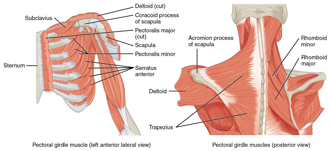
The scapula has only one small joint with the rest of the skeleton, at the clavicle. It is held against the back of the rib cage almost entirely by muscles, which slide it up, down, forward, back and around. These muscles make the girdle a movable but steady base for the arm (Figure 1).
On the back
- Trapezius (trapez- = four-sided): the large, flat diamond covering the upper back and back of the neck. It originates on the occipital bone, the nuchal ligament and the spinous processes of vertebrae C7 to T12, and inserts on the lateral clavicle, the acromion and the spine of the scapula. Its fibers run in three directions, so its parts do different jobs: the upper fibers elevate the scapula (a shrug), the middle fibers retract it (pull it toward the spine) and the lower fibers depress it. Upper and lower fibers together rotate the scapula so the shoulder socket turns upward.
- Rhomboid major and rhomboid minor (rhomb- = diamond): lie under the trapezius. They originate on the spinous processes of the upper thoracic and lowest cervical vertebrae and insert on the medial border of the scapula. They retract the scapula, squeezing your shoulder blades together, and help elevate it.
- A third muscle here, the levator scapulae, runs from the upper cervical vertebrae to the top of the scapula and, as its name says, lifts the scapula.
On the front and side of the chest
- Serratus anterior (serratus = saw-toothed): its finger-like slips originate on the outer surfaces of ribs 1 to 8 or 9 and wrap around the chest wall to insert on the medial border of the scapula, on its rib-side surface. It protracts the scapula (pulls it forward around the chest wall, as in a punch or a push) and rotates it so the socket faces upward, which is needed to raise the arm above the head. It also holds the scapula flat against the rib cage.
- Pectoralis minor (pector- = chest): lies under the large chest muscle. It originates on ribs 3 to 5 and inserts on the coracoid process of the scapula. It depresses the scapula and pulls it forward.
The serratus anterior and the rhomboids are antagonists: one pulls the scapula forward around the chest, the other pulls it back toward the spine. When the serratus anterior is paralyzed, the medial border of the scapula lifts away from the ribs every time the person pushes against a wall, a sign called a winged scapula.
Muscles that move the humerus
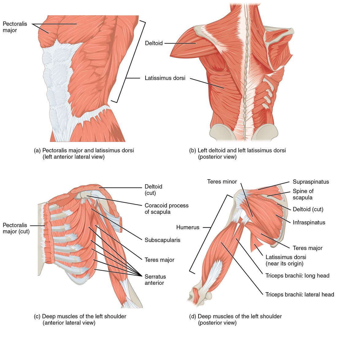
Nine muscles cross the shoulder joint to move the humerus. Two of them start on the trunk (axial muscles); the rest start on the scapula or clavicle. Find them in Figure 2.
The two large trunk muscles
- Pectoralis major: the large, fan-shaped (convergent) muscle of the chest. It originates on the medial clavicle, the sternum and the costal cartilages of the upper ribs, and inserts by a flat tendon on the lateral lip of the intertubercular groove of the humerus. It flexes the arm (raises it forward), adducts it (brings it across the chest) and rotates it medially. It is the prime mover of flexion at the shoulder.
- Latissimus dorsi (latissimus = widest; dorsi = of the back): the broad, flat muscle of the lower back. It originates on the spinous processes of the lower thoracic and lumbar vertebrae, the sacrum, the iliac crest (through a broad sheet of deep fascia) and the lowest ribs, and inserts on the floor of the intertubercular groove. It extends, adducts and medially rotates the arm: the pulling-down stroke of swimming or climbing. It is the prime mover of extension at the shoulder.
The shoulder cap and its helpers
- Deltoid (delt- = triangle): the thick, multipennate muscle that caps the shoulder. It originates on the lateral third of the clavicle, the acromion and the spine of the scapula, the same line the trapezius inserts on, and inserts on the deltoid tuberosity partway down the humerus. It is the prime mover of abduction (raising the arm out to the side). Its front fibers also flex and medially rotate the arm; its back fibers extend and laterally rotate it.
- Teres major (teres = round): runs from the lower corner (inferior angle) of the scapula to the humerus near the latissimus dorsi's insertion. It works with the latissimus dorsi: extension, adduction and medial rotation.
- Coracobrachialis: runs from the coracoid process to the middle of the humerus. It flexes and adducts the arm.
The rotator cuff
The shoulder joint trades stability for range. Its socket, the glenoid cavity, is shallow and far smaller than the head of the humerus, like a golf ball on a tee. Four muscles wrap around the joint from the scapula to the top of the humerus. Their tendons blend with the joint capsule and form a cuff that holds the head of the humerus pressed into the socket while larger muscles swing the arm. These four are the rotator cuff muscles, remembered as SITS:
- Supraspinatus: originates in the supraspinous fossa, above the spine of the scapula, passes over the top of the joint under the acromion and inserts on the greater tubercle. It abducts the arm, working with the deltoid.
- Infraspinatus: originates in the infraspinous fossa, below the spine of the scapula, and inserts on the greater tubercle. It rotates the arm laterally.
- Teres minor: originates on the lateral border of the scapula, just below the infraspinatus, and inserts on the greater tubercle. It also rotates the arm laterally.
- Subscapularis: originates in the subscapular fossa, on the rib-side face of the scapula, and inserts on the lesser tubercle at the front of the humerus. It rotates the arm medially.
Three of the four insert on the greater tubercle; only the subscapularis, the one on the front of the scapula, uses the lesser tubercle. Find all four in the bottom half of Figure 2.
The supraspinatus tendon runs through a tight space between the head of the humerus and the acromion. It is the rotator cuff tendon that tears most often. Most tears come from age-related wear of the tendon itself, and years of repeated overhead work add to the strain. A torn supraspinatus makes raising the arm to the side weak and painful.
Muscles that move the forearm
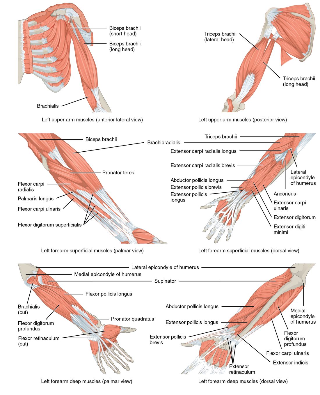
The elbow bends and straightens (the humeroulnar joint), and the radius rolls around the ulna to turn the palm down and up (pronation and supination at the radioulnar joints). Different muscles do each (Figure 3).
Elbow flexors
- Biceps brachii (bi- = two, -ceps = heads; brachi- = arm): has two heads of origin on the scapula. The long head starts on a small bump just above the glenoid cavity, and its tendon runs through the intertubercular groove; the short head starts on the coracoid process. It inserts on the radial tuberosity. It flexes the elbow and is the strongest supinator of the forearm (think of driving a screw in with your right hand). Because it crosses the shoulder too, it weakly flexes the arm.
- Brachialis: lies under the biceps brachii. It originates on the front of the lower half of the humerus and inserts on the coronoid process of the ulna. It flexes the elbow in every forearm position, because the ulna does not rotate.
- Brachioradialis: originates on the lateral edge of the lower humerus and runs down the thumb side of the forearm to insert on the styloid process of the radius. It flexes the elbow, most strongly with the thumb pointing up, as when you lift a mug by its handle.
Elbow extensor
- Triceps brachii (tri- = three): the only large muscle on the back of the arm. Its long head originates on a small bump just below the glenoid cavity; its lateral and medial heads originate on the back of the humerus. All three insert by one tendon on the olecranon of the ulna. It extends the elbow, and its long head, which crosses the shoulder, also extends and adducts the arm.
Pronators and supinator
- Pronator teres: runs diagonally from the medial epicondyle of the humerus (and the ulna) across the front of the forearm to the middle of the radius. It pronates the forearm and helps flex the elbow.
- Pronator quadratus (quadratus = four-sided): a flat square muscle deep across the lower ends of the ulna and radius, near the wrist. It is the main pronator.
- Supinator: wraps around the upper radius from the lateral epicondyle of the humerus and the ulna. It supinates the forearm, turning the palm up, with help from the biceps brachii.
| Front of the arm (flexors) | Back of the arm (extensors) | |
|---|---|---|
| Main muscles | Biceps brachii, brachialis (brachioradialis in the forearm) | Triceps brachii |
| Insert on | Radial tuberosity, coronoid process of the ulna, styloid process of the radius | Olecranon of the ulna |
| Action at the elbow | Flexion | Extension |
| Lever class for that action | Third class: insertion between the elbow and the hand | First class: insertion behind the elbow, load in front |
| Other actions | Biceps brachii supinates and weakly flexes the arm | Long head extends and adducts the arm |
| Relationship | Antagonists of the extensors | Antagonist of the flexors |
Muscles that move the wrist and fingers
The muscles that move the wrist and fingers mostly sit in the forearm and send long tendons down into the hand. This keeps the hand slim while still giving it strong muscles. The forearm has two main groups, easy to remember by their shared origins:
- Flexors on the front (palm side) mostly start from a common tendon on the medial epicondyle of the humerus.
- Extensors on the back mostly start from a common tendon on the lateral epicondyle.
Wrist flexors (front, from the medial epicondyle)
- Flexor carpi radialis (carp- = wrist; radialis = on the radius side): inserts on the bases of the second and third metacarpals. It flexes the wrist and abducts it, bending it toward the thumb.
- Palmaris longus: a thin muscle with a long tendon into the palm. It weakly flexes the wrist. About one person in seven lacks it on one or both sides, with no loss of function, which is why surgeons use its tendon for grafts.
- Flexor carpi ulnaris: inserts on the pisiform, hamate and fifth metacarpal. It flexes the wrist and adducts it, bending it toward the little finger.
Finger flexors
- Flexor digitorum superficialis (digit- = finger): originates on the medial epicondyle and the radius and ulna; its four tendons insert on the middle phalanges of fingers 2 to 5. It flexes the fingers at their middle joints (and the wrist).
- Flexor digitorum profundus (profundus = deep): lies under it, originating on the ulna. Its four tendons pass through splits in the superficialis tendons to insert on the distal phalanges. It is the only muscle that bends the fingertips.
The finger flexor tendons, with a nerve, pass into the palm through the carpal tunnel, under the flexor retinaculum. Swelling of those tendons' sheaths, for example from long hours of repeated gripping, can squeeze the nerve and cause numbness and tingling in the thumb and first fingers: carpal tunnel syndrome.
Wrist and finger extensors (back, from the lateral epicondyle)
- Extensor carpi radialis longus and extensor carpi radialis brevis: run along the thumb side to the second and third metacarpals. They extend the wrist and abduct it.
- Extensor carpi ulnaris: runs along the little-finger side to the fifth metacarpal. It extends the wrist and adducts it.
- Extensor digitorum: its four tendons fan across the back of the hand into fingers 2 to 5. It extends the fingers (and the wrist). You can see the tendons stand out when you spread your fingers.
Each wrist flexor has an extensor partner on the same edge of the forearm: flexor and extensor carpi radialis on the thumb side, flexor and extensor carpi ulnaris on the little-finger side. Contracting both radial muscles bends the wrist toward the thumb; both ulnar muscles bend it toward the little finger. As a whole, the wrist flexors and wrist extensors are antagonists.
| Wrist and finger flexors | Wrist and finger extensors | |
|---|---|---|
| Side of the forearm | Front (palm side) | Back |
| Common origin | Medial epicondyle of the humerus | Lateral epicondyle of the humerus |
| Wrist muscles | Flexor carpi radialis, palmaris longus, flexor carpi ulnaris | Extensor carpi radialis longus and brevis, extensor carpi ulnaris |
| Finger muscles | Flexor digitorum superficialis (middle phalanges) and profundus (distal phalanges) | Extensor digitorum |
| Overuse injury at the origin | Golfer's elbow | Tennis elbow |
Extrinsic and intrinsic muscles of the hand
All the forearm muscles whose tendons reach the hand are the extrinsic muscles of the hand (extrinsic = from outside): they start outside the hand. They give the hand its power, as in a firm grip. The intrinsic muscles of the hand (intrinsic = from within) start and end inside the hand. They make the fine movements: spreading and closing the fingers, and bringing the thumb across the palm to touch the fingertips (opposition). Find them in Figure 4.
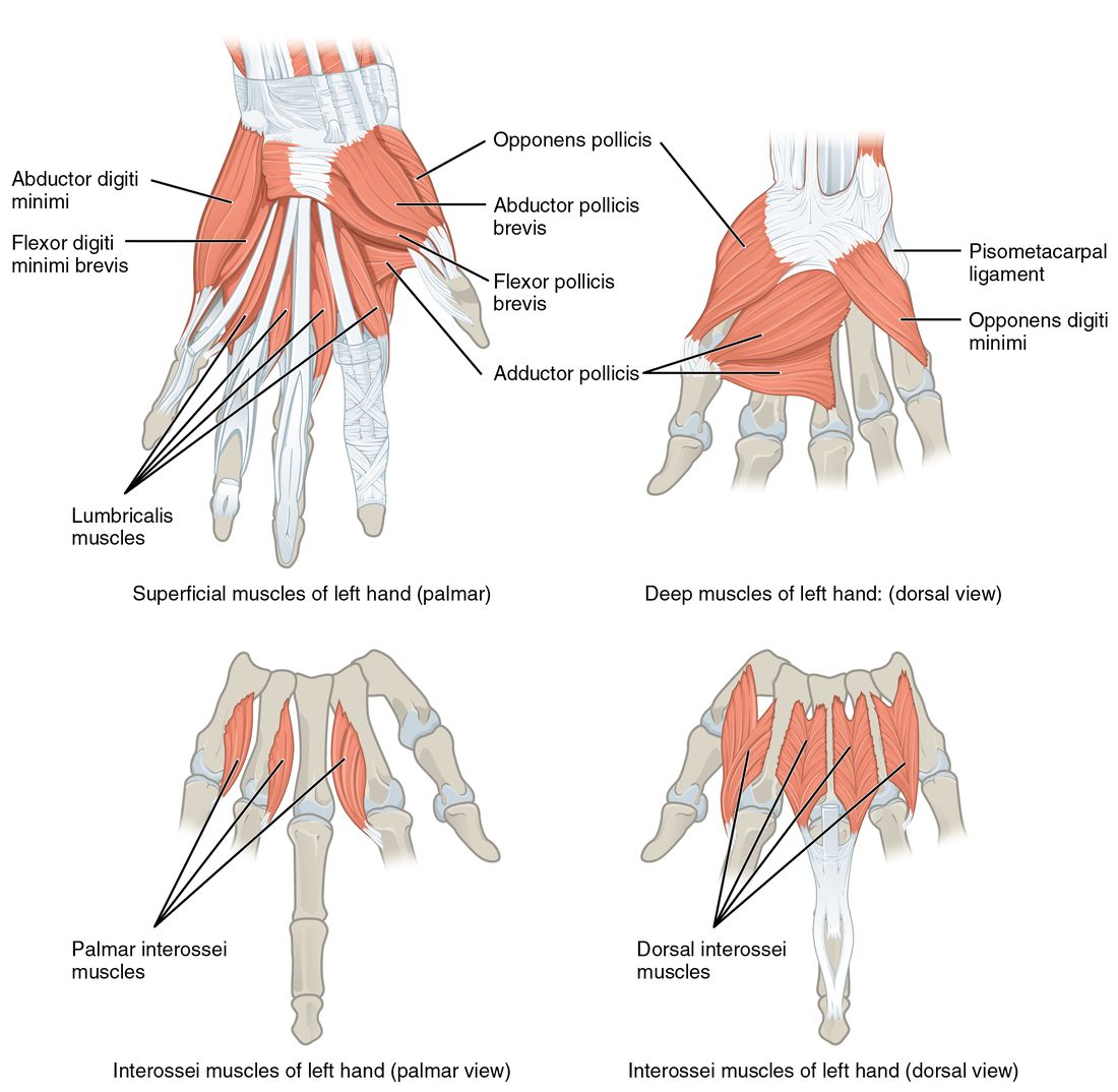
- The thenar eminence (thenar = palm) is the fleshy mound at the base of the thumb. Its muscles abduct, flex and oppose the thumb.
- The hypothenar eminence (hypo- = below) is the smaller mound on the little-finger side of the palm. Its muscles move the little finger, including the abductor digiti minimi you decoded in the muscle-naming lesson.
- Between them, small muscles running alongside the flexor tendons and between the metacarpals bend the knuckles while straightening the fingers, and spread and close the fingers.
Wasting of the thenar eminence is a late sign of severe carpal tunnel syndrome, because the nerve squeezed in the tunnel supplies those thumb muscles.
Major upper limb muscles at a glance
| Muscle | Origin | Insertion | Main action |
|---|---|---|---|
| Trapezius | Occipital bone, nuchal ligament, spinous processes C7–T12 | Lateral clavicle, acromion, spine of the scapula | Elevates, retracts, depresses and upwardly rotates the scapula |
| Serratus anterior | Ribs 1–8 or 9 | Medial border of the scapula (rib side) | Protracts the scapula and rotates it upward |
| Pectoralis major | Clavicle, sternum, upper costal cartilages | Intertubercular groove (lateral lip) | Flexes, adducts and medially rotates the arm |
| Latissimus dorsi | Lower thoracic and lumbar spinous processes, iliac crest, lower ribs | Intertubercular groove (floor) | Extends, adducts and medially rotates the arm |
| Deltoid | Lateral clavicle, acromion, spine of the scapula | Deltoid tuberosity | Abducts the arm (prime mover) |
| Supraspinatus | Supraspinous fossa | Greater tubercle | Abducts the arm; holds the humerus in its socket |
| Infraspinatus and teres minor | Infraspinous fossa; lateral border of the scapula | Greater tubercle | Laterally rotate the arm |
| Subscapularis | Subscapular fossa | Lesser tubercle | Medially rotates the arm |
| Biceps brachii | Above the glenoid cavity (long head); coracoid process (short head) | Radial tuberosity | Flexes the elbow; supinates the forearm |
| Brachialis | Front of the lower humerus | Coronoid process of the ulna | Flexes the elbow |
| Triceps brachii | Below the glenoid cavity (long head); back of the humerus | Olecranon | Extends the elbow |
| Flexor carpi radialis / ulnaris | Medial epicondyle | Metacarpals 2–3 / pisiform, hamate and metacarpal 5 | Flex the wrist (and abduct / adduct it) |
| Extensor digitorum | Lateral epicondyle | Phalanges of fingers 2–5 | Extends the fingers |
Summary
Muscles move the bone beyond the one they sit on. The trapezius, rhomboids, serratus anterior and pectoralis minor position the scapula. The pectoralis major flexes the arm and the latissimus dorsi extends it; both adduct and medially rotate it. The deltoid is the prime mover of abduction. The rotator cuff (supraspinatus, infraspinatus, teres minor, subscapularis) holds the head of the humerus in the shallow glenoid cavity; three insert on the greater tubercle, the subscapularis on the lesser. The biceps brachii, brachialis and brachioradialis flex the elbow; the triceps brachii extends it; the pronator teres and pronator quadratus pronate and the supinator and biceps supinate. Wrist and finger flexors start on the medial epicondyle, extensors on the lateral epicondyle. The extrinsic hand muscles in the forearm give grip strength; the intrinsic muscles, including the thenar and hypothenar eminences, make the fine movements.