Thirty-one pairs of spinal nerves connect your spinal cord to your trunk and limbs. Every one of them carries signals both ways: sensory signals in from the skin, muscles and joints, and motor commands out to the muscles. This page shows how a nerve is built, how each spinal nerve forms from two roots and then splits into branches, how the branches weave into the cervical, brachial, lumbar and sacral plexuses, which major nerves come out of them, and how the skin is mapped into dermatomes, one strip for each spinal nerve.
What a nerve is made of
Start with something you can picture: a thick electrical cable. Inside the outer jacket are several smaller bundles, and each bundle holds many single wires, each with its own thin coating. A nerve is built the same way.
A nerve is a bundle of axons in the peripheral nervous system, wrapped in connective tissue. Three wrappings give a nerve its structure (Figure 1):
- Endoneurium (endo- = within, neur- = nerve): a delicate layer of loose connective tissue around each single axon and its Schwann cells.
- Perineurium (peri- = around): a sleeve of flattened cells and dense connective tissue around each bundle of axons. A bundle is called a fascicle (fascicle = little bundle), the same word you met for bundles of muscle fibers. The cells of the perineurium are joined by tight junctions, so they control what passes from the blood into the fascicle.
- Epineurium (epi- = upon): a tough outer layer of dense irregular connective tissue around the whole nerve. It binds the fascicles together, carries the nerve's blood vessels and fat, and lets the nerve bend and stretch at a joint without tearing.
If the three layers sound familiar, they should. Skeletal muscle is wrapped the same way, and the names follow the same pattern.
| Nerve | Skeletal muscle | |
|---|---|---|
| Around the whole organ | Epineurium | Epimysium |
| Around each bundle (fascicle) | Perineurium | Perimysium |
| Around each single unit | Endoneurium, around one axon | Endomysium, around one muscle fiber |
| What the single unit is | An axon, a process of a nerve cell | A whole muscle cell |
| Where the cell bodies are | Outside the nerve, in a ganglion or the CNS | Inside the fibers themselves |
A nerve holds only axons. The cell bodies of those axons sit elsewhere: in a ganglion (for sensory neurons) or in the gray matter of the CNS (for motor neurons). A cut nerve therefore loses the far part of each axon, but the cell body survives, and in the PNS the axon can slowly regrow along its endoneurial tube.
Nerves are named by the direction of their signals. A sensory nerve carries only afferent signals, a motor nerve only efferent ones, and a mixed nerve carries both. Every spinal nerve is a mixed nerve.
How a spinal nerve forms
You already know the two roots attached to each segment of the spinal cord. The dorsal root carries sensory axons in, and its swelling, the dorsal root ganglion, holds their cell bodies. The ventral root carries motor axons out from the ventral horn.
Just beside the cord, inside the intervertebral foramen, the two roots join. The result is a spinal nerve (Figure 2). Once the roots join, sensory and motor axons run side by side in the same nerve. That is why every spinal nerve is mixed.
A spinal nerve is short, only about a centimeter long. As soon as it leaves the foramen it splits into branches.
Thirty-one pairs, named by level
Spinal nerves are named for the region of the vertebral column where they leave:
- 8 cervical (C1–C8). There are 8 cervical nerves but only 7 cervical vertebrae, because C1 leaves above the first vertebra and C8 leaves below the seventh. So in the neck, each nerve leaves above the vertebra with the same number.
- 12 thoracic (T1–T12). From here down, each nerve leaves below the vertebra with the same number.
- 5 lumbar (L1–L5).
- 5 sacral (S1–S5), through the openings in the sacrum.
- 1 coccygeal (Co1).
The cord itself ends near the L1–L2 vertebrae, so the lower roots run down inside the vertebral canal as the cauda equina before they reach their own foramina.
Rami: where each spinal nerve goes
A ramus (plural rami; ramus = branch) is one of the branches a spinal nerve splits into. Two are large, and both are mixed:
- The dorsal ramus turns backward. It supplies the deep muscles of the back, such as the erector spinae, and the skin of the back near the midline.
- The ventral ramus is larger. It runs around to the front and sides of the trunk and supplies the muscles and skin there, and it supplies both pairs of limbs.
Small extra branches also leave each spinal nerve: one returns through the foramen to supply the meninges, and short branches link every spinal nerve to a chain of autonomic ganglia beside the vertebrae. The thoracic and upper lumbar nerves have an extra link of this kind. You will meet those links in the autonomic nervous system chapter.
Watch the difference between roots and rami. Roots are the two attachments to the cord: one purely sensory, one purely motor. Rami are branches after the roots have joined, so every ramus carries both kinds of axon.
| Roots (dorsal and ventral) | Rami (dorsal and ventral) | |
|---|---|---|
| Position | Between the cord and the spinal nerve | Beyond the spinal nerve, outside the foramen |
| Kind of axons | Dorsal root sensory only; ventral root motor only | Both are mixed |
| Cell bodies | Sensory cell bodies in the dorsal root ganglion | None |
| Damage to one causes | Dorsal root: numbness only. Ventral root: weakness only | Numbness and weakness in the area that ramus supplies |
The thoracic ventral rami stay separate
In the thoracic region, each ventral ramus runs alone around the body wall between two ribs. These are the intercostal nerves (inter- = between, cost- = rib), T1 to T11; T12 runs below the last rib and is called the subcostal nerve. They supply the intercostal muscles, the abdominal wall muscles and the skin over them, in neat horizontal bands.
Nerve plexuses
Everywhere else, the ventral rami do not stay separate. In the neck and the limb regions they branch, join and swap axons with their neighbors, forming a network called a nerve plexus (plexus = braid). Out of each plexus come named nerves that carry axons from several spinal cord segments.
This mixing has a useful result. Most limb muscles receive axons from two or three segments, so damage to one root weakens a muscle but rarely paralyzes it. Only the dorsal rami and the thoracic ventral rami skip plexuses.
There are four main plexuses on each side (Figure 3):
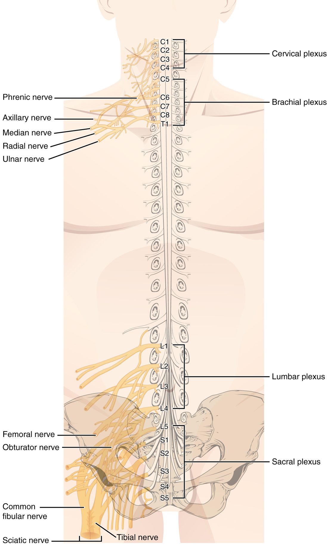
| Cervical plexus | Brachial plexus | Lumbar plexus | Sacral plexus | |
|---|---|---|---|---|
| Ventral rami | C1–C4 | C5–T1 | L1–L4 | L4–S4 |
| Region supplied | Neck, skin of the side of the head and neck, diaphragm | Shoulder, arm, forearm and hand | Lower abdominal wall, front and inner thigh | Buttock, back of thigh, leg and foot |
| Key nerves | Phrenic nerve | Axillary, musculocutaneous, radial, median and ulnar nerves | Femoral and obturator nerves | Sciatic nerve (tibial and common fibular), gluteal nerves |
Some texts extend the cervical plexus to C5, and the lumbar and sacral plexuses are often grouped as one lumbosacral plexus because L4 feeds both.
The brachial plexus in five steps
The brachial plexus (brachi- = arm) is the one most often tested. Its axons are sorted in five stages, from the cord outward: roots (the ventral rami C5–T1), trunks (upper, middle and lower), divisions (each trunk splits into an anterior and a posterior division), cords (lateral, posterior and medial, named for where they lie around the main artery of the armpit), and finally the terminal branches, the named nerves. The posterior cord gives rise to the axillary and radial nerves, the nerves of the back of the limb.
A memory aid: "Really Tired? Drink Cold Beverages" for roots, trunks, divisions, cords, branches.
The major nerves and what each one does
For each nerve, learn three things: where it comes from, what it supplies, and what a patient looks like when it is damaged. The damage pattern is how clinicians recognize it.
From the cervical plexus
- Phrenic nerve (C3–C5; phren- = diaphragm): runs down through the thoracic cavity to the diaphragm, and is its only motor supply. The rhyme "C3, 4 and 5 keep the diaphragm alive" explains why an injury high in the neck, above C3, stops breathing, while an injury below C5 spares the diaphragm.
From the brachial plexus
- Axillary nerve (C5–C6): wraps around the surgical neck of the humerus to supply the deltoid and teres minor and the skin over the shoulder. A fracture of the surgical neck or a dislocated shoulder can stretch it, weakening abduction of the arm.
- Radial nerve (C5–T1): the nerve of the back of the arm and forearm. It supplies the triceps brachii and the wrist and finger extensors. It spirals around the shaft of the humerus, so a mid-shaft fracture can injure it. The result is wrist drop: the hand hangs and the person cannot extend the wrist.
- Median nerve (C6–T1): runs down the middle of the front of the forearm and through the carpal tunnel. It supplies most wrist and finger flexors, most of the thenar muscles of the thumb, and the skin of the thumb, index, middle and half of the ring finger on the palm side. Squeezing it in the carpal tunnel causes numbness and tingling in those fingers and a weak thumb (carpal tunnel syndrome).
- Ulnar nerve (C8–T1): passes behind the medial epicondyle of the humerus, where it is the "funny bone" you tingle when you knock your elbow. It supplies most of the small muscles inside the hand and the skin of the little finger and half the ring finger. Damage weakens finger spreading and grip and can curl the ring and little fingers into a claw.
- The musculocutaneous nerve (C5–C7) supplies the biceps brachii, brachialis and coracobrachialis, the elbow flexors at the front of the arm.
From the lumbar plexus
- Femoral nerve (L2–L4): passes under the inguinal ligament into the femoral triangle and supplies the quadriceps femoris (knee extension), the iliopsoas and sartorius, and the skin of the front of the thigh. Damage makes the knee buckle and climbing stairs hard.
- The obturator nerve (L2–L4) passes through the obturator foramen to the adductors of the medial thigh.
From the sacral plexus
- Sciatic nerve (L4–S3): the largest nerve in your body, about as thick as your thumb. It leaves the pelvis below the piriformis, runs down the back of the thigh, supplies the hamstrings, and usually divides above the knee, near the popliteal fossa, into two branches.
- Tibial nerve: continues down the back of the leg to the gastrocnemius, soleus and the other posterior leg muscles (plantar flexion) and to the sole of the foot.
- Common fibular nerve (also called the common peroneal nerve): wraps around the neck of the fibula just below the knee, right under the skin, and then splits into branches for the front and side of the leg (dorsiflexion and eversion) and the skin of the top of the foot. A blow to the side of the knee, a tight cast or crossing the legs for hours can press on it. The result is foot drop: the toes drag and the person lifts the knee high with each step. This course calls it the fibular nerve from here on.
Sciatica is pain that runs along the path of the sciatic nerve: from the buttock down the back of the thigh, often into the leg and foot. Despite the name, the problem is usually not in the sciatic nerve itself. Most often a herniated disc in the lower lumbar spine presses on an L5 or S1 root before it joins the sacral plexus.
Why injections go where they go
The sciatic nerve runs through the lower half of the buttock, closer to the midline than to the side. That is why intramuscular injections avoid that area and use the ventrogluteal site on the side of the hip instead.
Dermatomes
Here is a concrete case first. A woman develops a painful, blistering rash in a stripe that starts at her spine, wraps around her side and ends at her navel, on one side only. The stripe has that exact shape because it traces the skin supplied by one spinal nerve.
A dermatome (derm- = skin, -tome = cut, section) is the strip of skin whose sensory axons all travel in one spinal nerve, and so enter the cord through one dorsal root. On the trunk, dermatomes run as horizontal bands. On the limbs they run lengthwise, because the limbs grow out sideways from the trunk before birth and draw their bands out with them.
A few landmarks are worth knowing by heart (Figure 4):
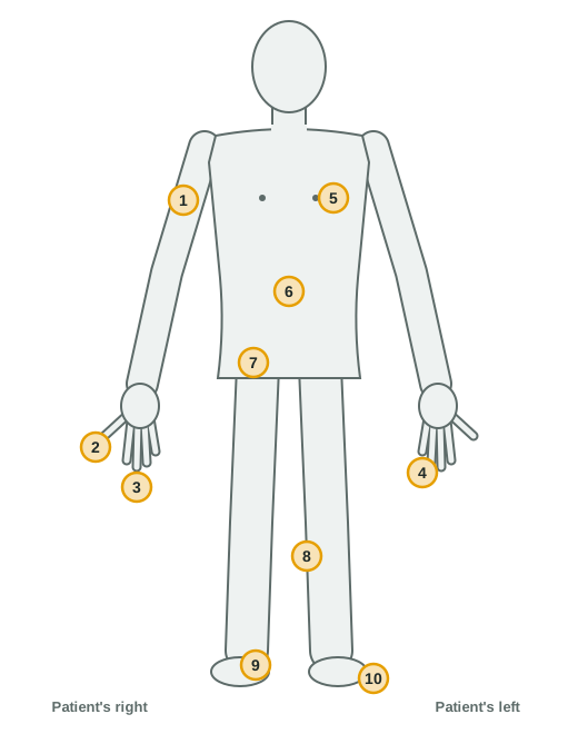
| Landmark | Dermatome |
|---|---|
| Outer side of the upper arm, over the deltoid | C5 |
| Thumb | C6 |
| Middle finger | C7 |
| Little finger | C8 |
| Nipple line | T4 |
| Navel (umbilicus) | T10 |
| Groin (inguinal region) | L1 |
| Inner side of the leg below the knee | L4 |
| Top of the foot and the big toe | L5 |
| Outer edge of the foot, little toe and heel | S1 |
Two landmarks anchor the trunk: T4 at the nipple line and T10 at the navel ("bellybut-TEN"). The thoracic bands between and below them are evenly spaced. The face has no spinal dermatome: its skin is supplied by a nerve that leaves the brain directly, covered in the next topic. C1 usually has no dermatome at all.
Neighboring dermatomes overlap
Dermatome maps draw sharp borders, but real dermatomes overlap: each patch of skin is supplied mainly by one spinal nerve and partly by the nerves above and below. So if a single dorsal root is cut, the matching dermatome does not go completely numb. Sensation there is reduced, and a band of complete numbness appears only when two or three neighboring roots are damaged. Maps from different studies also disagree on the exact borders, so treat the landmarks as the reliable part.
Using dermatomes
- Finding a damaged root. Numbness and pain along a dermatome point to one spinal nerve or root. Burning pain down the back of the thigh into the outer edge of the foot suggests the S1 root; into the big toe, L5.
- Shingles. After chickenpox, the virus stays dormant in the sensory cell bodies of a dorsal root ganglion. If it reactivates, it travels out along that ganglion's axons, so the rash appears in that ganglion's dermatome, on one side.
- Muscles too. Each spinal cord segment also supplies its own group of muscles. Testing a few key movements (for example, elbow flexion for C5 and ankle dorsiflexion for L4) checks the motor side of each segment the way dermatomes check the sensory side.
Putting it together: a dorsal root versus a ventral root
Because the two roots carry different axons, damage to each gives a different picture.
- Dorsal root cut (for example, L5 on the right): signals from the L5 dermatome no longer reach the cord. Sensation over the top of the right foot is reduced. Strength is normal, because the motor axons leave through the ventral root.
- Ventral root cut at the same level: the muscles supplied by L5 are weak, because their motor axons are cut, but the skin feels normal.
- Spinal nerve or ramus damaged, beyond the point where the roots join: both numbness and weakness, in the area that nerve supplies.
This is the logic a clinician uses at the bedside: the pattern of numbness and weakness tells you where along the path the damage lies.