Naming places on the body
"Pain in the right lower quadrant." "Bruising over the left femoral region." "Blood drawn from the antecubital region." Each phrase points to one exact spot on the body in a few words. This page teaches the regional terms for the body surface, then the two ways of dividing the abdomen: the four abdominal quadrants and the nine abdominopelvic regions. Every one of these terms, like the directional terms, assumes the anatomical position and uses the patient's own right and left.
Regional terms
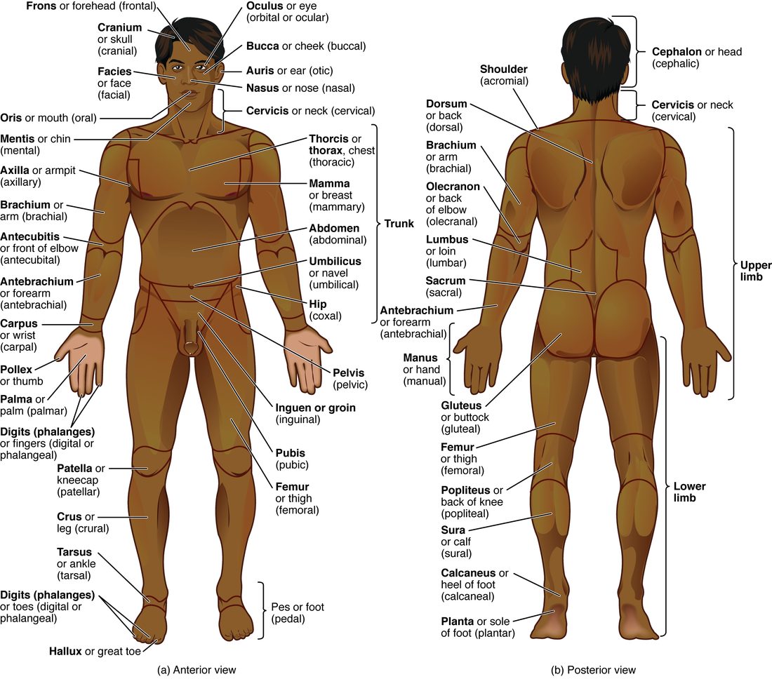
Regional terms are the names for areas of the body surface. Most are adjectives built from Latin or Greek roots, and you can decode them with the word parts you already know. They let you say "the brachial region" instead of "the upper part of the arm, between the shoulder and the elbow". Figure 1 shows them on the front and the back of the body.
Head and neck
- Cephalic (Greek kephale = head): the whole head.
- Frontal: the forehead. Orbital (Latin orbita = circle, track): the eye area. Otic (ot/o = ear): the ear. Nasal (Latin nasus = nose): the nose. Buccal (Latin bucca = cheek): the cheek. Mental (Latin mentum = chin): the chin.
- Cervical (from the Latin word for neck): the neck.
Trunk, front
- Thoracic (thorax = chest): the chest. Mammary (Latin mamma = breast): each breast.
- Abdominal: the belly, between the chest and the hips. Umbilical (Latin umbilicus = navel): the navel.
- Coxal (Latin coxa = hip): the hip. Inguinal (Latin inguen = groin): the groin, the crease where the thigh meets the trunk. Pubic: the area just above the external genitals.
Trunk, back
- Upper back: often called the dorsal region, although dorsal also names a direction.
- Lumbar (Latin lumbus = loin): the lower back, between the lowest ribs and the hips.
- Gluteal (Greek gloutos = buttock): the buttock.
Upper limb
Here anatomy uses everyday words more strictly than you do. In anatomy the arm is only the part between the shoulder and the elbow. The part between the elbow and the wrist is the forearm.
- Axillary (Latin axilla = armpit): the armpit.
- Brachial (Latin brachium = arm): the arm, shoulder to elbow.
- Antecubital (ante- = in front of, Latin cubitus = elbow): the front of the elbow, the crease where the elbow bends.
- Antebrachial (ante- = before; brachium = arm): the forearm, elbow to wrist.
- Carpal (Greek karpos = wrist): the wrist. Palmar: the palm. Digital (Latin digitus = finger or toe): the fingers.
Lower limb
The same strictness applies. In anatomy the leg is only the part between the knee and the ankle. The part between the hip and the knee is the thigh.
- Femoral (from the Latin word for thigh): the thigh, hip to knee.
- Patellar: the front of the knee. Popliteal (Latin poples = back of the knee): the hollow behind the knee.
- Crural (Latin crus = leg): the leg, knee to ankle, on the front. Sural (Latin sura = calf): the calf, on the back.
- Tarsal: the ankle. Pedal (Latin pes, pedis = foot): the foot. Plantar (Latin planta = sole): the sole. Calcaneal: the heel.
| Upper limb | Lower limb | |
|---|---|---|
| Segment nearest the trunk | arm (brachial), shoulder to elbow | thigh (femoral), hip to knee |
| Bend in the middle, front | front of elbow (antecubital) | front of knee (patellar) |
| Bend in the middle, back | back of elbow | back of knee (popliteal) |
| Segment below the bend | forearm (antebrachial), elbow to wrist | leg (crural), knee to ankle |
| Last joint | wrist (carpal) | ankle (tarsal) |
| End segment | hand: palm (palmar), fingers (digital) | foot (pedal): sole (plantar), toes (digital) |
Why clinicians use them
Many blood vessels take the name of the region they cross. You can feel a pulse on the inner side of the brachial region, in the groin at the top of the femoral region, and in the popliteal region behind the knee. Blood is most often drawn from a vein in the antecubital region, where large veins lie just under the skin. A term like "left femoral region" is shorter than a description and means the same spot to every reader.
The four abdominal quadrants
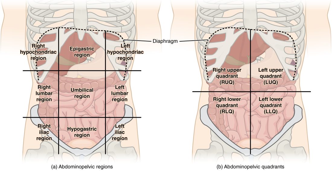
Look at Figure 2. The abdomen has no bones on its front to use as landmarks, so clinicians draw imaginary lines on it. The quick way uses two lines that cross at the navel:
- a vertical line along the midsagittal plane, dividing right from left;
- a horizontal line along a transverse plane through the navel, dividing upper from lower.
The lines make four abdominal quadrants (Latin quadrans = a fourth part), named for the patient's own sides:
- Right upper quadrant: most of the liver and the gallbladder.
- Left upper quadrant: most of the stomach and the spleen.
- Right lower quadrant: the appendix, the start of the wide final part of the intestine, and the right ovary in females.
- Left lower quadrant: the last part of the colon as it heads toward the pelvis, and the left ovary in females.
Coiled loops of intestine lie in all four quadrants, and the pancreas and kidneys lie deep, toward the back wall, so each crosses more than one quadrant. The quadrants are for quick, shared description, such as "tender in the right lower quadrant", in the emergency room and on the ambulance.
The nine abdominopelvic regions
For more exact location, anatomists divide the abdomen into nine abdominopelvic regions, like a tic-tac-toe grid. Two vertical lines run down from the middle of each collarbone. Two horizontal lines cross the abdomen: the upper one just below the lower edge of the ribs, and the lower one across the bony ridge at the top of each hip bone.
The grid makes three rows of three. Right and left are the patient's.
| Patient's right | Middle | Patient's left | |
|---|---|---|---|
| Top row | right hypochondriac | epigastric | left hypochondriac |
| Middle row | right lumbar | umbilical | left lumbar |
| Bottom row | right iliac | hypogastric | left iliac |
Decode each name and you know where it is:
- Hypochondriac (hypo- = below, chondr/o = cartilage): below the cartilages of the lower ribs. The right one holds much of the liver; the left one holds the spleen and part of the stomach.
- Epigastric (epi- = upon, over; gastr/o = stomach): over the stomach, in the upper middle. It holds much of the stomach and part of the liver.
- Lumbar (lumbus = loin): the sides of the middle row, level with the lower back. These regions are also called the lateral regions, or flanks.
- Umbilical (umbilicus = navel): the center square, around the navel. It holds coiled loops of intestine.
- Iliac (Latin ilia = flanks): the lower corners, over the bony ridge at the top of each hip bone. These regions are also called the inguinal regions. The right iliac region holds the appendix.
- Hypogastric (hypo- = below, gastr/o = stomach): below the stomach, in the lower middle, just above the pubic area. It holds the urinary bladder when it is full, and the uterus. It is also called the pubic region.
Quadrants and regions compared
| Four quadrants | Nine regions | |
|---|---|---|
| Lines drawn | one vertical, one horizontal, crossing at the navel | two vertical, two horizontal |
| Planes used | midsagittal and a transverse plane through the navel | two parasagittal and two transverse planes |
| Areas made | 4 | 9 |
| Center of the grid | the navel is where the lines cross | the navel sits in the umbilical region |
| Main users | clinicians, for quick description | anatomists, and clinicians for finer location |
| Example | appendix: right lower quadrant | appendix: right iliac region |
Pain that moves: putting it together
Jordan's pain is a classic pattern. At first, the swollen appendix stretches, and pain signals from an organ's wall are vague. The brain cannot place them exactly, so they are felt near the midline, around the navel, in the umbilical region. Hours later, the inflamed appendix irritates the parietal layer of the peritoneum lining the abdominal wall next to it. Pain signals from that lining are sharp and precisely placed. The pain now sits over the appendix, in the right lower quadrant (the right iliac region), and it worsens when he moves, because each movement shifts the inflamed tissue against the lining.
The rule: the region where pain is felt narrows down which organs to check, but pain from an organ's wall can be felt away from the organ. Location is a clue, not a diagnosis.