Your spinal cord is about as thick as your finger and about 45 cm long, and it carries every signal between your brain and your body below the head. This page covers spinal cord anatomy from the outside in, and the spinal cord cross section: where the cord runs and where it ends, its two enlargements and the cauda equina, the gray matter horns and white matter columns you see when you cut it across, the dorsal and ventral roots that connect it to the body, and an overview of the ascending and descending tracts that run through it.
Where the spinal cord runs
The spinal cord is the part of the CNS inside the vertebral column. It starts at the foramen magnum, where it continues from the medulla oblongata, and runs down the vertebral canal, the tunnel formed by the stacked vertebral foramina. Along the way it is wrapped in the same three meninges as the brain:
- The dura mater forms a loose tube, the dural sac, that is not attached to the vertebrae. Between it and the bone lies the epidural space, a real space filled with fat and veins.
- The subarachnoid space, full of CSF, surrounds the cord along its whole length.
- The pia mater clings to the cord's surface. Small side extensions of the pia anchor the cord to the dura on each side, and a thin strand of pia continues below the cord's end to anchor it to the coccyx.
Where the cord ends
In an adult the spinal cord does not reach the bottom of the vertebral column. It tapers to a cone and ends at about the level of the first or second lumbar vertebra (Figure 1). This cone-shaped end is the conus medullaris (conus = cone, medulla = marrow).
Why does it stop short? Early in fetal life the cord fills the whole canal. After that, the vertebral column grows faster and longer than the cord. At birth the cord ends at about the third lumbar vertebra; by adulthood, at about the first or second.
The cord is divided into 31 segments, each giving off a pair of roots on each side: 8 cervical, 12 thoracic, 5 lumbar, 5 sacral and 1 coccygeal. Because the cord is shorter than the column, a segment does not sit level with the vertebra of the same name. The lower segments are crowded together near the conus, so the roots from the lumbar and sacral segments have to run down inside the canal before they reach their exit holes between the vertebrae.
Below the conus, the canal is filled with these long roots, hanging down in the CSF. The bundle looks like a horse's tail and is called the cauda equina (cauda = tail, equus = horse).
The dural sac and its subarachnoid space do not stop at the conus. They continue down to about the second sacral vertebra. That leaves a pocket of CSF around the cauda equina, which is where a lumbar puncture needle goes. Floating roots slide aside from a needle; the cord itself is out of reach.
The two enlargements
The cord is not the same thickness along its length. It is wider in two regions (Figure 1):
- The cervical enlargement, in the lower neck (roughly cord segments C4 to T1), supplies the upper limbs.
- The lumbar enlargement (roughly cord segments L1 to S2, lying behind the lower thoracic vertebrae) supplies the lower limbs.
The cause is the number of neurons. Your arms and legs have far more muscle and skin than a matching stretch of trunk, so the segments that supply them hold many more motor neurons and receive many more sensory axons. More neurons means more gray matter, and a wider cord.
The spinal cord in cross section
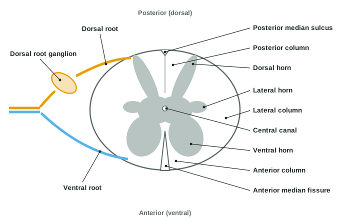
Cut the cord straight across and you see the arrangement in Figure 2. It is the reverse of the cerebrum: gray matter inside, white matter outside.
Finding front and back
Two midline grooves tell you which way the section faces:
- The anterior median fissure is a deep, wide groove at the front.
- The posterior median sulcus is a shallow groove at the back.
The gray matter horns
The gray matter forms a shape like a butterfly or the letter H. Each wing has projections called horns:
- The dorsal horn (also called the posterior horn) reaches toward the back. It is sensory: many incoming sensory axons, such as those for pain and temperature, end here, on interneurons and on neurons that send signals up to the brain. Others, such as those for touch and limb position, send a branch here and turn straight up the posterior column.
- The ventral horn (also called the anterior horn) reaches toward the front. It is motor: it holds the cell bodies of the motor neurons whose axons run out to skeletal muscles. It is broad and blunt, and it is widest in the two enlargements.
- The lateral horn is a small bump at each side, present only in the thoracic and upper lumbar segments (T1 to L2). It holds the cell bodies of autonomic motor neurons of the sympathetic division, which control smooth muscle, cardiac muscle and glands.
The two halves of the gray matter are joined across the middle by a bar of gray matter. In its center runs the central canal, a tiny channel lined by ependymal cells, left from the hollow of the neural tube. It connects to the fourth ventricle above, and in many adults it is partly closed.
The white matter columns
The white matter on each side is divided by the horns and roots into three white columns (also called funiculi; funiculus = little cord):
- The posterior column lies between the dorsal horn and the posterior median sulcus.
- The lateral column lies at the side, between the dorsal and ventral horns.
- The anterior column lies between the ventral horn and the anterior median fissure.
Each column contains several tracts, bundles of axons running up or down the cord, which come later on this page.
| Dorsal (posterior) horn | Ventral (anterior) horn | Lateral horn | |
|---|---|---|---|
| Points toward | The back | The front | The side |
| Present at | Every level | Every level; largest in the enlargements | Only T1 to L2 |
| Main neurons | Interneurons and neurons that relay sensation up | Motor neurons to skeletal muscle | Sympathetic motor neurons |
| Division | Sensory | Motor (somatic) | Motor (autonomic) |
| Connects to | Axons entering through the dorsal root | Axons leaving through the ventral root | Axons leaving through the ventral root |
How the proportions change from top to bottom
The cross section looks different at different levels, for two reasons that follow from what the cord does:
- White matter is thickest near the top. Ascending axons join the cord at every level on their way up, so the higher you go, the more of them there are. Descending axons leave the white matter at every level as they reach their target segments, so the higher you go, the more of them are still present. Both effects make the cervical cord the thickest in white matter and the sacral cord the thinnest.
- Gray matter is largest in the enlargements, for the reason above: the limbs need more neurons.
The dorsal and ventral roots
On each side of every segment, two roots connect the cord to the body (Figure 2). Each root starts as a fan of small rootlets along the cord.
- The dorsal root (also called the posterior root) enters the back of the cord and carries sensory axons in. Along it sits a swelling, the dorsal root ganglion, which holds the cell bodies of those sensory neurons. They are pseudounipolar: one process runs out to the body and the other runs in through the dorsal root into the cord.
- The ventral root (also called the anterior root) leaves the front of the cord and carries motor axons out: axons of ventral horn motor neurons to skeletal muscle, and, at T1 to L2, axons of lateral horn neurons. A few sacral segments, S2 to S4, send out autonomic axons too, as the autonomic chapter shows.
So sensation comes in at the back, and commands go out at the front. The ventral root has no ganglion, because its cell bodies lie inside the cord.
Just beyond the dorsal root ganglion, the dorsal and ventral roots join into one nerve that carries both sensory and motor axons. The next topic follows these nerves out into the body.
| Dorsal (posterior) root | Ventral (anterior) root | |
|---|---|---|
| Direction of signals | Into the cord | Out of the cord |
| Axons carried | Sensory | Motor (somatic, and autonomic at T1–L2 and S2–S4) |
| Where the cell bodies are | Dorsal root ganglion, outside the cord | Ventral and lateral horns, inside the cord |
| Has a ganglion? | Yes | No |
| If cut | Sensation from the area it supplies dulled; strength normal | Muscles it supplies weakened; sensation normal |
Cutting a single root dulls rather than abolishes, because neighboring roots overlap. Each band of skin gets sensory axons mainly from one segment and partly from the segments just above and below, and most muscles get motor axons from two or more segments. A band of skin goes fully numb only when its own dorsal root and both neighbors are cut. The next topic maps these bands.
Diseases show the split clearly:
- Polio virus destroys ventral horn motor neurons. Muscles weaken and waste away, but sensation stays normal.
- After chickenpox, the virus can lie dormant for decades in a dorsal root ganglion. When it reactivates as shingles, it travels out along that ganglion's sensory axons, so the painful rash appears in a band of skin on one side of the body, the area that one dorsal root supplies.
Ascending and descending tracts: an overview
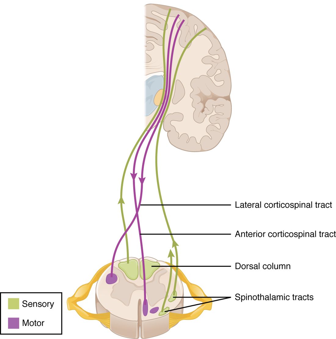
The white columns are made of tracts. A tract is a bundle of axons in the CNS that share an origin, a destination and a job. The spinal cord's tracts fall into two groups (Figure 3):
- Ascending tracts carry sensory information up the cord to the brain: touch, vibration, limb position, pain and temperature.
- Descending tracts carry motor commands down from the brain to the neurons of the ventral and lateral horns.
Three general rules hold for tracts:
- Their names give origin, then destination. A tract named "spino-" plus a brain region starts in the spinal cord and ends in that region, so it is ascending. A tract named for a brain region plus "-spinal" starts in the brain and ends in the cord, so it is descending.
- They sit in predictable columns. The posterior column is almost all ascending. The lateral and anterior columns hold both ascending and descending tracts.
- Most cross the midline somewhere along their path. That is how the left side of the brain ends up feeling and moving the right side of the body. Where each one crosses differs, and that difference matters when you work out what an injury on one side of the cord does.
The individual tracts, where they cross, and how to use them to locate an injury come with the sensory and motor pathways later in this chapter.
Summary
The spinal cord runs from the foramen magnum to the conus medullaris at about L1–L2 in adults, wrapped in the meninges; below the conus the cauda equina of lumbar and sacral roots hangs in the CSF-filled dural sac, which reaches S2, so a lumbar puncture below L2 misses the cord. The cervical and lumbar enlargements hold the extra neurons for the limbs. In cross section, gray matter forms dorsal (sensory), ventral (somatic motor) and, from T1 to L2, lateral (sympathetic) horns around the central canal, and white matter forms posterior, lateral and anterior columns; the anterior median fissure marks the front and the posterior median sulcus the back. Sensory axons enter through the dorsal root, whose ganglion holds their cell bodies, and motor axons leave through the ventral root. The white columns hold ascending tracts carrying sensation up and descending tracts carrying commands down, named from origin to destination, and most cross the midline.