Your brain is soft enough to dent with a finger, yet it survives running, jumping and the odd bump on the head. Three things protect it besides the skull: three membranes wrapped around it, a clear fluid it floats in, and a barrier in the walls of its capillaries. This page covers the meninges and cerebrospinal fluid flow: the three meningeal layers and the spaces between them, the ventricles of the brain, how cerebrospinal fluid (CSF) is made, where it flows and where it leaves, the blood–brain barrier, and the three kinds of bleeding around the brain that the layers explain.
The meninges: three layers
Between the skull and the brain lie three membranes, together called the meninges (singular meninx; meninx = membrane). They continue down the vertebral canal around the spinal cord. From outside in (Figure 1):
- Dura mater (dura = tough, mater = mother). A thick sheet of dense connective tissue. Around the brain it has two layers. The outer one is fused to the inside of the skull. The inner one faces the brain. In places the two layers split apart to enclose large channels that collect venous blood from the brain; you will meet these channels with the veins of the head. The inner layer also folds down between parts of the brain: one fold drops into the longitudinal fissure between the hemispheres (the falx cerebri; falx = sickle), and another forms a tent between the cerebrum and the cerebellum (the tentorium cerebelli; tentorium = tent). These folds limit how far the brain can shift.
- Arachnoid mater (arachn- = spider). A thin, delicate layer pressed against the inside of the dura. From its under surface, fine strands of collagen reach down to the pia like a spider's web.
- Pia mater (pia = tender). A very thin layer stuck to the surface of the brain. It follows every gyrus down into every sulcus, carrying small blood vessels with it.
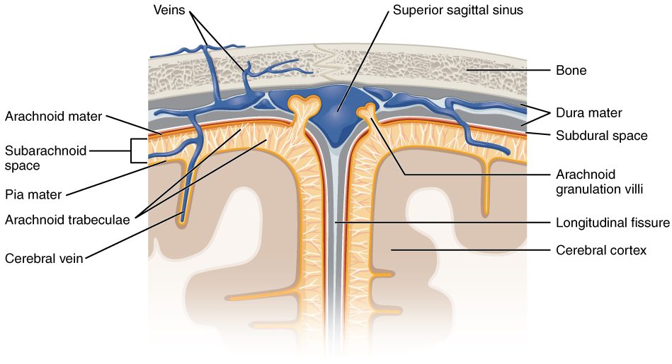
A useful memory aid, from outside in: dura is tough, arachnoid is web-like, pia is tender.
The spaces around the meninges
Three named spaces lie between and around these layers. Only one of them is a real, fluid-filled space in the skull of a healthy person.
- The epidural space (epi- = upon) lies outside the dura. In the skull it is only a potential space, because the dura is fused to the bone; it opens only if something, such as blood, forces the two apart. Around the spinal cord it is a real space, filled with fat and veins, because there the dura is not attached to the vertebrae. This is where anesthetists inject an epidural during labor.
- The subdural space (sub- = under) lies between the dura and the arachnoid. It is normally a potential space too, since the arachnoid lies pressed against the dura.
- The subarachnoid space lies between the arachnoid and the pia. It is a real space, crossed by the arachnoid's web-like strands and filled with cerebrospinal fluid. The large arteries and veins on the surface of the brain run through it.
| Epidural space | Subdural space | Subarachnoid space | |
|---|---|---|---|
| Lies between | Bone and dura mater | Dura mater and arachnoid mater | Arachnoid mater and pia mater |
| In the skull | Potential space only | Potential space only | Real space |
| Around the spinal cord | Real space with fat and veins | Potential space only | Real space |
| Normally contains | Nothing in the skull; fat and veins in the spine | Nothing | Cerebrospinal fluid and the large surface blood vessels |
The ventricles of the brain
Recall that the neural tube was hollow. In the adult brain that hollow center remains as four connected, fluid-filled chambers, the ventricles of the brain (ventriculus = little belly), lined by ependymal cells. Trace them in Figure 2.
- Two lateral ventricles, one deep in each cerebral hemisphere. Each is a long C-shaped space that curves from the frontal lobe back and down into the temporal lobe.
- Each lateral ventricle opens into the third ventricle through a small hole, the interventricular foramen (foramen = opening).
- The third ventricle is a narrow slit in the midline of the diencephalon, with the thalamus and hypothalamus forming its walls.
- The cerebral aqueduct (aqua = water, ductus = channel) is a narrow canal through the midbrain. It connects the third ventricle to the fourth.
- The fourth ventricle is a tent-shaped space between the pons and medulla in front and the cerebellum behind.
- The fourth ventricle opens into the subarachnoid space through three holes: one median aperture (apertura = opening) in the midline of its roof and two lateral apertures, one on each side. Below, it continues as the thin central canal of the spinal cord.
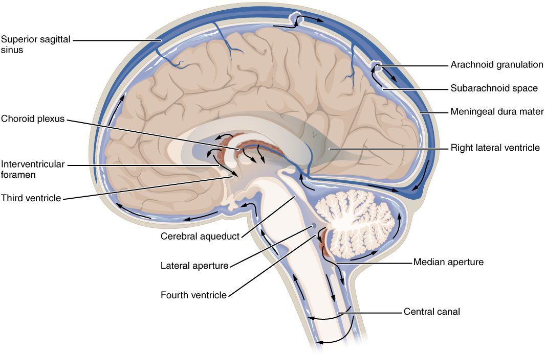
The apertures are the only way out of the ventricles into the subarachnoid space (the central canal below is a dead end, and in adults it is often closed). Remember that, because it explains what happens when CSF flow is blocked.
Cerebrospinal fluid: what it is and how it is made
Cerebrospinal fluid (CSF; cerebr- = brain, spin- = spine) is the clear, colorless fluid that fills the ventricles and the subarachnoid space. You have about 150 mL of it at any moment, but you make about 500 mL a day, so the whole volume is replaced three to four times daily.
Where it is made
Most CSF is made by the choroid plexus (choroid = membrane-like, plexus = braid), a cauliflower-like tuft of capillaries covered by a layer of modified ependymal cells. There is a choroid plexus in each of the four ventricles; the largest are in the lateral ventricles.
It is made by secretion, not simple leakage, in these steps:
- Blood plasma filters out of the choroid plexus capillaries, which are leaky, into the tissue under the covering cells.
- The covering cells are joined by tight junctions, so fluid cannot slip between them. Instead, the cells use active transport to move sodium ions into the ventricle, and chloride and bicarbonate ions follow.
- Water follows the solute through aquaporins in the cells' membranes.
- The result is a fluid made by the cells, with a controlled composition.
Because the covering cells are joined by tight junctions, the choroid plexus acts as a blood–CSF barrier. That is why CSF is not simply plasma:
- It has almost no protein (less than 1% of the amount in plasma) and very few cells.
- Its glucose is about two thirds of the blood level.
- Its ion concentrations differ slightly from plasma's, with less potassium.
What CSF does
- Buoyancy. A brain that weighs about 1,400 g in air weighs only about 25 to 50 g floating in CSF. Without it, the brain's own weight would crush the nerves and vessels on its underside.
- Cushioning. A blow to the head moves the skull first; the fluid layer spreads the force before the brain strikes the bone.
- A stable chemical environment. Neurons fire correctly only when the ions around them are held steady, and CSF exchanges freely with the fluid between brain cells.
- Carrying away wastes from the brain as the fluid drains out.
How CSF flows and where it leaves
CSF flows because it is made continuously at one end and removed at the other. Its path:
- The choroid plexuses of the lateral ventricles secrete CSF.
- It flows through the interventricular foramina into the third ventricle, where its choroid plexus adds more.
- It flows down the cerebral aqueduct into the fourth ventricle, where more is added.
- It leaves the fourth ventricle through the median and lateral apertures into the subarachnoid space. A little also flows down the central canal of the spinal cord.
- In the subarachnoid space it flows around the brainstem, down around the spinal cord and up over the surface of the cerebrum.
- It returns to the blood.
The classic route for the last step is the arachnoid granulations : knobs of arachnoid mater that push up through the dura into the large venous channels (Figure 1). CSF pressure is normally a little higher than the pressure of the venous blood in those channels, so CSF flows across into the blood. The flow is one way: if venous pressure rises above CSF pressure, the granulations collapse rather than letting blood back in.
CSF also leaves by other routes. Some flows out along the sheaths of the nerves that leave the skull, especially the nerves of smell through the roof of the nose, and some drains through fine vessels in the dura, reaching the tissue-fluid drainage vessels of the head and neck. Studies in animals, and newer imaging studies in people, show these routes carry a substantial share. How the load is split in an adult human is not yet settled.
When CSF cannot drain: hydrocephalus
CSF production does not slow down much when pressure rises, so if the fluid cannot leave, it builds up. Excess CSF that enlarges the ventricles is hydrocephalus (hydro- = water, cephal- = head).
Suppose a tumor pinches the cerebral aqueduct shut.
- The lateral and third ventricles keep making CSF, but it cannot pass the aqueduct.
- CSF builds up above the block, so the lateral and third ventricles swell.
- The fourth ventricle, below the block, stays normal size.
- The swelling ventricles press the brain outward against the skull, and pressure inside the skull rises.
This is obstructive hydrocephalus: the block is inside the ventricles. When the ventricles are open but the fluid cannot get back into the blood, for example after bleeding or infection clogs the subarachnoid space and granulations, all four ventricles enlarge. This is communicating hydrocephalus.
In an infant, whose skull bones are not yet fused, hydrocephalus makes the head grow abnormally large. In an adult, the skull cannot expand, so pressure rises instead, causing headache, vomiting and drowsiness. Surgeons treat it by placing a thin tube, a shunt, that drains CSF from a ventricle to the abdomen.
Sampling CSF: lumbar puncture
A lumbar puncture (spinal tap) samples CSF from the subarachnoid space in the lower back. In an adult the spinal cord ends at about the level of the first or second lumbar vertebra, but the subarachnoid space, full of CSF, continues down to about the second sacral vertebra. Below the end of the cord it holds only nerve roots floating in fluid.
So the needle is placed between the third and fourth, or fourth and fifth, lumbar vertebrae. It passes through the skin, the ligaments between the vertebrae, the epidural space, the dura and the arachnoid into the subarachnoid space. Floating roots slide away from the needle, so the cord is not at risk.
Doctors measure the pressure (normally about 10 to 25 cm of water in an adult lying on one side) and test the fluid. Cloudy fluid full of white blood cells, with low glucose, points to a bacterial infection of the meninges. Blood in the fluid points to bleeding into the subarachnoid space.
The blood–brain barrier
Here is a puzzle. Levodopa, a drug for Parkinson's disease, works because the brain turns it into dopamine. Why not just give dopamine? Because dopamine injected into the blood never reaches the brain. Levodopa does. The difference is the blood–brain barrier, a set of features of the brain's capillaries that controls what passes from blood into brain tissue (Figure 3).
What makes the barrier
In most of the body, capillary walls have gaps between their endothelial cells, and some have pores through them. Brain capillaries differ:
- Their endothelial cells are sealed edge to edge by continuous tight junctions, so nothing can pass between the cells.
- The cells have no pores and carry very few transport vesicles, so little passes through them in bulk either.
- The cells carry pumps that push many foreign molecules that do get in straight back out into the blood.
- A basement membrane surrounds them, and the end-feet of astrocytes cover almost the whole outer surface. Signals from the astrocytes keep the endothelial cells making their tight junctions and pumps.
So the real barrier is the endothelium. Everything that enters brain tissue has to go through the endothelial cells themselves.
What gets through
- Small, lipid-soluble molecules dissolve straight through the cell membranes: oxygen, carbon dioxide, alcohol, caffeine and most general anesthetics.
- Water diffuses across the endothelial cells, and water channels (aquaporins) in the astrocyte end-feet move it on into brain tissue.
- Glucose and amino acids cross only through specific carrier proteins in the endothelial membranes. Levodopa is an amino acid, so it rides a carrier protein across. Dopamine is not carried, and it is too water-soluble to dissolve through, so it stays in the blood.
- Proteins, most ions, most water-soluble drugs and many antibiotics are kept out, or pass only slowly.
That is the trade-off. The barrier protects neurons from swings in blood chemistry, toxins and many microbes, but it also blocks many drugs that could treat brain disease. It is also why a brain infection is hard to treat.
Where the barrier is missing
A few small areas around the ventricles have leaky capillaries with no barrier. There, neurons can sample the blood directly: parts of the hypothalamus monitor the blood's water balance, and an area on the floor of the fourth ventricle detects toxins in the blood and triggers vomiting.
Bleeding around the brain
The meningeal layers decide where blood can collect after an injury, and each location behaves differently. Compare the three in Figure 4.
Epidural hematoma
A hematoma (hemat- = blood, -oma = mass) is a collection of clotted blood outside a vessel. An epidural hematoma lies between the skull and the dura.
- Cause: usually a blow to the side of the head that fractures the thin temporal bone and tears the artery running in a groove on its inside, the middle meningeal artery.
- Shape: the dura is fused to the skull, and most firmly at the sutures. Arterial blood has to peel it off the bone, so the clot stays compact and bulges inward as a lens shape that stops at the sutures.
- Course: arterial pressure makes it grow fast, over minutes to hours. The classic story is a brief knock-out, a "lucid interval" of feeling fine, then rapidly worsening drowsiness as the clot compresses the brain. Only a minority of patients show that pattern, so its absence does not rule out an epidural hematoma.
Subdural hematoma
A subdural hematoma lies between the dura and the arachnoid.
- Cause: tearing of the bridging veins, which cross the subdural space from the brain's surface to the venous channels in the dura. A sudden speeding up or slowing down of the head moves the brain relative to the skull and stretches them.
- Shape: nothing holds the arachnoid to the dura, so the blood spreads thinly over the brain's surface as a crescent. It can cross suture lines but stops at the dural folds.
- Course: venous pressure is low, so it may build slowly over hours to weeks. The brain shrinks with age, stretching the bridging veins, so older adults can bleed after a minor fall. Heavy alcohol use and blood-thinning drugs raise the risk too. A chronic subdural hematoma can show up weeks later as confusion, headache or weakness.
Subarachnoid hemorrhage
In a subarachnoid hemorrhage (hemorrhage = bursting forth of blood), blood enters the subarachnoid space and mixes with the CSF, spreading around the brain and into its sulci.
- Cause: head trauma is the most common cause overall. When it happens without injury, it is usually a ballooned weak spot in one of the large arteries at the base of the brain that bursts.
- Signs: a sudden, explosive headache, often called the worst of the person's life, with a stiff neck because blood irritates the meninges.
- Finding: blood in CSF from a lumbar puncture, when a scan does not show it.
| Epidural hematoma | Subdural hematoma | Subarachnoid hemorrhage | |
|---|---|---|---|
| Where the blood is | Between skull and dura | Between dura and arachnoid | In the subarachnoid space, mixed with CSF |
| Vessel usually torn | Middle meningeal artery | Bridging veins | An artery on the surface or at the base of the brain |
| Typical cause | Blow to the side of the head with a skull fracture | Sudden acceleration or deceleration; minor falls in older adults | Trauma; without trauma, a burst weak spot in an artery |
| Speed | Fast (arterial pressure) | Often slow, hours to weeks (venous pressure); an acute one after severe trauma can be rapid | Sudden |
| Shape on a scan | Lens-shaped; stops at sutures | Crescent; crosses sutures, stops at dural folds | Blood outlining the sulci and spaces around the brain |
| Classic signs | Knocked out, lucid interval, then rapid decline | Gradual confusion, headache, drowsiness; an acute one may cause coma at once | Sudden worst-ever headache, stiff neck |
Summary
The meninges wrap the brain and spinal cord: the tough dura mater (two layers in the skull, with folds between brain parts), the web-like arachnoid mater and the tender pia mater on the brain surface. The epidural and subdural spaces are potential spaces in the skull; the subarachnoid space is real and holds CSF and the surface vessels. The four ventricles (two lateral, the third and the fourth) are the neural tube's hollow, joined by the interventricular foramina and the cerebral aqueduct and opening through the median and lateral apertures. The choroid plexuses secrete about 500 mL of CSF a day; it flows through the ventricles into the subarachnoid space and returns to venous blood, classically through the arachnoid granulations. A block causes hydrocephalus. A lumbar puncture samples CSF below the end of the cord. The blood–brain barrier is brain capillary endothelium sealed by tight junctions and kept that way by astrocytes; lipid-soluble molecules cross, glucose and amino acids need carrier proteins, and most other substances are kept out. Epidural hematomas are arterial and lens-shaped, subdural hematomas venous and crescent-shaped, and subarachnoid hemorrhage puts blood into the CSF.