Meninges, ventricles and cerebrospinal fluid
1Why this matters
Jordan, 16, is hit on the side of the head by a baseball. He is knocked out for a minute, then gets up, says he feels fine and wants to keep playing. An hour later he is drowsy and hard to wake, and his left arm is getting weak. A scan shows a lens-shaped clot between his skull and the tough outer membrane around his brain: a torn artery has been bleeding into a space that is normally closed.
2What this builds on
3Quick check before you start
1. What does a tight junction do between two epithelial or endothelial cells?
- Seals the gap so substances cannot pass between the cells
- Lets ions pass directly from one cell to the next
- Anchors the cells' cytoskeletons together against stretching
Show the answer
Tight junctions seal neighboring cells together near their surface, so substances must go through the cells rather than between them. Gap junctions let ions pass cell to cell; desmosomes anchor cells against pulling.
- Correct: Seals the gap so substances cannot pass between the cells:
- Lets ions pass directly from one cell to the next:
- Anchors the cells' cytoskeletons together against stretching:
2. Which glial cells line the fluid-filled spaces inside the brain and spinal cord?
- Astrocytes
- Ependymal cells
- Oligodendrocytes
Show the answer
Ependymal cells line the brain's internal fluid spaces and the central canal of the spinal cord. Astrocytes support neurons and wrap capillaries; oligodendrocytes make myelin in the CNS.
- Astrocytes:
- Correct: Ependymal cells:
- Oligodendrocytes:
3. Which part of the brain lies between the diencephalon and the pons?
- The medulla oblongata
- The cerebellum
- The midbrain
Show the answer
From top to bottom the brainstem is midbrain, pons, medulla oblongata, and the midbrain sits just below the diencephalon. The cerebellum lies behind the brainstem.
- The medulla oblongata:
- The cerebellum:
- Correct: The midbrain:
4Anatomy
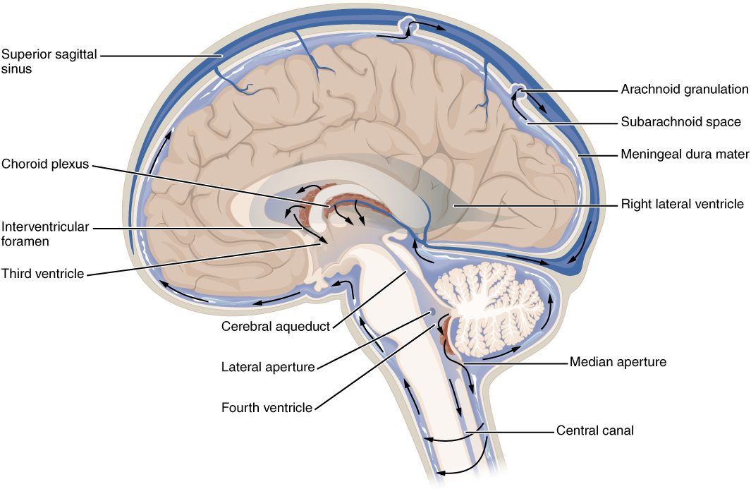
With labels hidden, select a box to reveal its label.
5How it works, step by step
- Cells covering the choroid plexus in the lateral ventricles pump sodium into the ventricle, and chloride and bicarbonate follow.Water follows the solute, and new CSF collects in the lateral ventricles.
- CSF is secreted continuously, so it keeps pushing on the fluid ahead of it.CSF flows through the interventricular foramina into the third ventricle, then down the cerebral aqueduct into the fourth ventricle.
- The only exits from the fourth ventricle into the subarachnoid space are the median and lateral apertures.CSF flows out into the subarachnoid space and spreads around the brain and spinal cord.
- CSF pressure in the subarachnoid space is a little higher than the pressure of the blood in the venous channels of the dura.CSF flows through the arachnoid granulations into the venous blood, balancing what the choroid plexus makes.
- If a tumor pinches the cerebral aqueduct shut, CSF made above the block cannot leave.The lateral and third ventricles swell, pressure inside the skull rises, and the fourth ventricle stays normal: obstructive hydrocephalus.
6Core concepts
7A common mistake
The wrong idea: The blood–brain barrier is a wall of astrocytes wrapped around the brain's capillaries.
What actually happens: The barrier itself is the endothelial cells of the brain's capillaries, sealed edge to edge by continuous tight junctions, with no pores and few transport vesicles. Astrocyte end-feet cover the outside of the capillary and send signals that keep the endothelial cells in this sealed state, but molecules could slip between the end-feet. Everything that enters brain tissue has to pass through the endothelial cells themselves.
8Check yourself
Anything you miss goes into your review queue.
1. Name the pinned layer.
- Dura mater
- Arachnoid mater
- Pia mater
- Web-like arachnoid strands
Show the answer
The dura mater is the thick outer meningeal layer against the inside of the skull. In the skull it has two layers, which split to enclose the venous channel in the midline.
- Correct: Dura mater: Correct. The thick layer against the bone is the dura mater.
- Arachnoid mater: The arachnoid mater is the thin layer just inside the dura, with web-like strands reaching down to the pia.
- Pia mater: The pia mater is the thin layer stuck to the brain surface, following it into the sulci.
- Web-like arachnoid strands: The web-like strands cross the subarachnoid space below the arachnoid; the pinned layer is the thick sheet against the bone.
2. Put the places cerebrospinal fluid passes through in order, from where most of it is made to where it returns to the blood.
- Choroid plexus of a lateral ventricle
- Third ventricle
- Cerebral aqueduct
- Fourth ventricle
- Subarachnoid space
- Arachnoid granulations
Show the answer
CSF made in the lateral ventricles passes through the interventricular foramina to the third ventricle, down the cerebral aqueduct to the fourth ventricle, out through the apertures into the subarachnoid space, and back into venous blood at the arachnoid granulations.
- Correct order: 1. Choroid plexus of a lateral ventricle 2. Third ventricle 3. Cerebral aqueduct 4. Fourth ventricle 5. Subarachnoid space 6. Arachnoid granulations
3. A small tumor pinches the cerebral aqueduct shut. Predict the change in each over the following days.
| Variable | Change |
|---|---|
| Size of the lateral ventricles | — |
| Size of the fourth ventricle | — |
| Pressure inside the skull | — |
| Rate of CSF secretion by the choroid plexuses | — |
Show the answer
A block at the cerebral aqueduct traps CSF made in the lateral and third ventricles. Those ventricles swell and pressure in the skull rises, while the fourth ventricle below the block stays normal and secretion continues: obstructive hydrocephalus.
- Size of the lateral ventricles: up. The choroid plexuses above the block keep secreting CSF, which cannot pass the aqueduct, so it builds up and swells the ventricles above it.
- Size of the fourth ventricle: no change. The fourth ventricle lies below the block. Its own CSF still drains out through the apertures, so it stays normal size.
- Pressure inside the skull: up. Fluid is added to a closed bony box faster than it leaves, so the swelling ventricles press the brain against the skull and pressure rises.
- Rate of CSF secretion by the choroid plexuses: no change. Secretion is driven by the choroid plexus cells' ion pumping and changes little as pressure rises, which is why the fluid keeps building up.
4. Jordan, 16, is hit on the side of the head by a baseball and knocked out briefly. He wakes and seems fine, then an hour later becomes drowsy. A scan shows a lens-shaped clot that stops at the skull's sutures. Which vessel was most likely torn?
- A bridging vein
- An artery at the base of the brain
- A capillary of the choroid plexus
- The middle meningeal artery
Show the answer
A blow to the thin temporal bone can tear the middle meningeal artery, which runs on the inside of the skull. Its arterial blood peels the dura off the bone, making a compact, lens-shaped epidural hematoma that stops at the sutures, where the dura is most firmly attached. Arterial pressure makes it grow fast.
- A bridging vein: Torn bridging veins cause a subdural hematoma, which spreads as a thin crescent and usually builds more slowly.
- An artery at the base of the brain: A burst artery at the base of the brain bleeds into the subarachnoid space, mixing blood with CSF rather than forming a lens-shaped clot against the skull.
- A capillary of the choroid plexus: The choroid plexus lies inside the ventricles, far from the skull, and does not cause a clot between skull and dura.
- Correct: The middle meningeal artery: Correct. A torn middle meningeal artery causes an epidural hematoma.
5. Which of these would cross the blood–brain barrier most easily?
- A large protein molecule circulating in the plasma
- Dopamine injected into a vein
- A small, lipid-soluble anesthetic gas
- Sodium ions dissolved in the plasma
Show the answer
Brain capillary endothelial cells are sealed by tight junctions, so substances must pass through the cells. Small lipid-soluble molecules dissolve straight through the cell membranes, so an anesthetic gas crosses easily.
- A large protein molecule circulating in the plasma: Large proteins cannot pass between the sealed cells, and brain endothelium carries very few transport vesicles, so they barely enter.
- Dopamine injected into a vein: Dopamine is water-soluble and has no carrier protein in the barrier, so it stays in the blood. Its precursor levodopa, an amino acid, is carried across instead.
- Correct: A small, lipid-soluble anesthetic gas: Correct. Small lipid-soluble molecules cross the barrier freely.
- Sodium ions dissolved in the plasma: Charged ions cannot dissolve through the lipid membrane and cannot slip between the sealed cells, so they cross only slowly through specific channels and pumps.
6. Which structure is the barrier itself in the blood–brain barrier?
- The layer of astrocyte end-feet that wraps the outside of each brain capillary
- The pia mater covering the brain surface
- The capillary endothelial cells sealed by continuous tight junctions
- The cells covering the choroid plexus
Show the answer
The barrier is the brain capillary endothelium: cells sealed edge to edge by tight junctions, with no pores and few vesicles. Everything entering brain tissue has to pass through these cells.
- The layer of astrocyte end-feet that wraps the outside of each brain capillary: Astrocyte end-feet cover the capillaries and keep the endothelium in its barrier state, but molecules could slip between them; they are not the barrier itself.
- The pia mater covering the brain surface: The pia mater is a meningeal layer on the brain's surface, not part of every capillary wall.
- Correct: The capillary endothelial cells sealed by continuous tight junctions: Correct. The sealed endothelial cells are the barrier.
- The cells covering the choroid plexus: The choroid plexus covering cells form the blood–CSF barrier, a separate barrier between blood and CSF.
7. A scan shows that both lateral ventricles and the third ventricle are enlarged, but the fourth ventricle is normal. Where is the blockage most likely to be?
- At the arachnoid granulations
- At the median and lateral apertures
- At one interventricular foramen
- In the cerebral aqueduct
Show the answer
Ventricles above a block enlarge and those below stay normal. The third ventricle is enlarged and the fourth is not, so the block lies between them: the cerebral aqueduct.
- At the arachnoid granulations: A failure of return at the arachnoid granulations would back fluid up into all four ventricles, including the fourth.
- At the median and lateral apertures: A block at the apertures would enlarge the fourth ventricle too, since it lies above them.
- At one interventricular foramen: A block at one interventricular foramen would enlarge only that lateral ventricle, not the third ventricle.
- Correct: In the cerebral aqueduct: Correct. A block in the cerebral aqueduct enlarges the lateral and third ventricles and spares the fourth.
9Summary
Three meninges cover the brain and spinal cord: the tough dura mater, the web-like arachnoid mater and the tender pia mater. In the skull the epidural and subdural spaces are only potential spaces, while the subarachnoid space holds CSF and the surface vessels. The four ventricles, left from the neural tube's hollow, are joined by the interventricular foramina and the cerebral aqueduct and open through the median and lateral apertures. The choroid plexuses secrete about 500 mL of CSF a day; it flows through the ventricles into the subarachnoid space, floats and cushions the brain, and returns to venous blood, classically through the arachnoid granulations. A blocked path causes hydrocephalus, and a lumbar puncture samples CSF below the end of the cord. The blood–brain barrier is the sealed endothelium of brain capillaries. Epidural hematomas are fast arterial bleeds, subdural hematomas usually slower venous bleeds, and subarachnoid hemorrhage puts blood into the CSF.