Control of breathing
1Why this matters
Theo, 19, breathes hard and fast for a minute at the edge of a pool so he can swim farther underwater. Halfway across, without ever feeling the urge to breathe, he blacks out. A lifeguard pulls him out and he recovers. His breathing is driven mainly by carbon dioxide, not oxygen, and he had lowered his carbon dioxide before the dive.
2What this builds on
3Quick check before you start
1. Which part of the brainstem lies directly above the spinal cord?
- The midbrain
- The medulla oblongata
- The pons
Show the answer
The medulla oblongata is the lowest part of the brainstem, continuous with the spinal cord. The pons lies above it and the midbrain above that.
- The midbrain:
- Correct: The medulla oblongata:
- The pons:
2. Which of these crosses the blood–brain barrier most easily?
- Hydrogen ions
- Plasma proteins
- Carbon dioxide
Show the answer
Small, lipid-soluble molecules such as carbon dioxide and oxygen cross the blood–brain barrier freely. Charged ions such as hydrogen ions, and large proteins, cross it poorly.
- Hydrogen ions:
- Plasma proteins:
- Correct: Carbon dioxide:
3. When arterial PCO2 rises, what happens to blood pH?
- It falls
- It rises
- It does not change
Show the answer
More carbon dioxide forms more carbonic acid, which releases hydrogen ions, so pH falls.
- Correct: It falls:
- It rises:
- It does not change:
4Anatomy
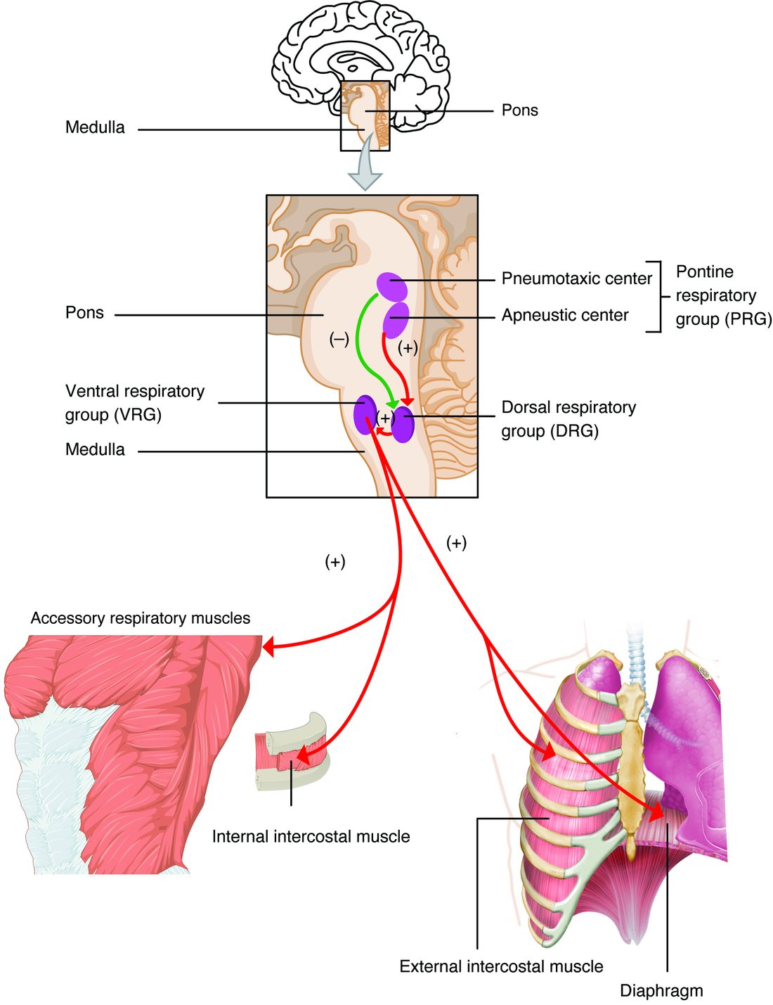
With labels hidden, select a box to reveal its label.
5How it works, step by step
- Your cells make carbon dioxide faster than you breathe it out, and arterial PCO2 rises.Carbon dioxide crosses the blood–brain barrier and forms hydrogen ions in the brain's fluid, and more carbon dioxide and hydrogen ions reach the carotid bodies.
- Central chemoreceptors in the medulla sense the extra hydrogen ions; the carotid bodies fire faster.Signals reach the respiratory centers, from the central chemoreceptors directly and from the carotid bodies through the glossopharyngeal nerves.
- The respiratory centers increase their output.The phrenic and intercostal nerves drive the diaphragm and intercostal muscles harder and more often, so breaths get deeper and faster.
- Alveolar ventilation rises.More carbon dioxide is breathed out, so arterial PCO2 and pH return toward normal and the chemoreceptors' drive fades: negative feedback.
6Core concepts
7A common mistake
The wrong idea: You breathe mainly in response to low oxygen.
What actually happens: Minute to minute, breathing is set mainly by carbon dioxide, through the hydrogen ions it forms. A rise in arterial PCO2 of a few mm Hg sharply increases ventilation, while arterial PO2 can fall from about 95 to 60 mm Hg with little effect, because the carotid bodies fire only weakly until PO2 drops below about 60; meanwhile hemoglobin stays nearly saturated on the flat part of the dissociation curve, so little oxygen is lost. Only the peripheral chemoreceptors sense low oxygen. That is why hyperventilating before a dive is dangerous: it removes the carbon dioxide signal, and oxygen can fall to the point of blacking out before the urge to breathe returns.
8Check yourself
Anything you miss goes into your review queue.
1. Where is the respiratory rhythm most likely generated?
- The pre-Bötzinger complex in the medulla oblongata
- The pneumotaxic center in the upper part of the pons
- The carotid bodies at the fork of each carotid artery
- The primary motor cortex of the frontal lobe
Show the answer
Neurons of the pre-Bötzinger complex, near the top of the ventral respiratory group in the medulla oblongata, fire in rhythmic bursts on their own, even when isolated from the rest of the brain. Many textbooks give the dorsal respiratory group this role; current evidence points to the pre-Bötzinger complex.
- Correct: The pre-Bötzinger complex in the medulla oblongata: Correct. This cluster in the medulla generates the rhythm.
- The pneumotaxic center in the upper part of the pons: The pneumotaxic center fine-tunes the rhythm by switching off inspiration earlier. Breathing continues when the pons is cut away.
- The carotid bodies at the fork of each carotid artery: The carotid bodies are peripheral chemoreceptors. They change breathing but do not generate its rhythm.
- The primary motor cortex of the frontal lobe: The motor cortex can override breathing voluntarily, but you keep breathing in your sleep and in coma, when it is not driving the muscles.
2. Acid produced by metabolism builds up in a patient's blood, and her arterial pH falls quickly. Her arterial PCO2 is normal at first. Which sensors are first to speed her breathing?
- Lung stretch receptors
- Central chemoreceptors in the medulla oblongata
- Irritant receptors in the airways
- The carotid bodies
Show the answer
The carotid bodies sample arterial blood directly and respond within seconds to a fall in pH from any cause. Hydrogen ions cross the blood–brain barrier poorly, so the central chemoreceptors are slow to sense acid that is not carried in as carbon dioxide.
- Lung stretch receptors: Lung stretch receptors respond to inflation of the lungs, not to blood chemistry.
- Central chemoreceptors in the medulla oblongata: Central chemoreceptors respond to hydrogen ions in the brain's fluid, which come mainly from carbon dioxide crossing the barrier. Hydrogen ions from the blood cross only slowly.
- Irritant receptors in the airways: Irritant receptors respond to dust, smoke and fumes in the airways, not to blood pH.
- Correct: The carotid bodies: Correct. The peripheral chemoreceptors sense the low arterial pH first.
3. During a panic attack, a woman breathes fast and deep for several minutes. Predict the change in each variable, compared with before the attack.
| Variable | Change |
|---|---|
| Arterial PCO2 | — |
| Arterial pH | — |
| Blood flow to the brain | — |
| Free calcium in the plasma | — |
| Drive from her central chemoreceptors | — |
Show the answer
Hyperventilation lowers arterial PCO2, which raises pH. Low PCO2 constricts brain arterioles, the alkalosis lowers free calcium, and the central chemoreceptors reduce their drive, which pushes breathing back toward normal once the emotional drive fades.
- Arterial PCO2: down. She is breathing out carbon dioxide faster than her cells make it.
- Arterial pH: up. Less carbon dioxide means less carbonic acid and fewer hydrogen ions.
- Blood flow to the brain: down. Low carbon dioxide constricts the arterioles in the brain, which is why she feels lightheaded.
- Free calcium in the plasma: down. In alkalosis, more calcium binds to albumin and other plasma proteins, leaving less free, which causes tingling and hand spasms.
- Drive from her central chemoreceptors: down. With less carbon dioxide crossing into the brain's fluid, its hydrogen ions fall and the central chemoreceptors fire less.
4. A lowland hiker is driven to a research station at 4,300 m and stays for three weeks. Predict each variable, first in the first hours (compared with sea level) and then after acclimatization (compared with the first hours).
| Variable | Change |
|---|---|
| Ventilation in the first hours | — |
| Arterial PCO2 in the first hours | — |
| Blood pH in the first hours | — |
| Ventilation after one week, compared with the first hours | — |
| Blood pH after one week, compared with the first hours | — |
| Hematocrit after three weeks, compared with the first hours | — |
Show the answer
In the first hours, low oxygen raises ventilation through the carotid bodies, and the fall in PCO2 raises pH, which restrains breathing. Over days, the kidneys correct the pH, the brake comes off and ventilation rises further. Over weeks, more red blood cells raise the blood's oxygen-carrying capacity.
- Ventilation in the first hours: up. Low arterial PO2 stimulates the carotid bodies, which drive the respiratory centers.
- Arterial PCO2 in the first hours: down. The extra ventilation blows off carbon dioxide faster than it is made.
- Blood pH in the first hours: up. Less carbon dioxide means fewer hydrogen ions, and the central chemoreceptors act as a brake on further breathing.
- Ventilation after one week, compared with the first hours: up. The kidneys excrete bicarbonate and the cerebrospinal fluid's bicarbonate falls, so pH returns toward normal, the central brake is released, and the carotid bodies become more sensitive.
- Blood pH after one week, compared with the first hours: down. Losing bicarbonate in the urine brings the raised pH back toward normal.
- Hematocrit after three weeks, compared with the first hours: up. Low oxygen makes the kidneys release more erythropoietin, which speeds erythropoiesis.
5. A patient's alveolar ventilation falls to half its normal value, and his carbon dioxide production does not change. His arterial PCO2 was 40 mm Hg. What will it settle at?
- 20 mm Hg
- 40 mm Hg (unchanged)
- 60 mm Hg
- 80 mm Hg
Show the answer
At constant carbon dioxide production, arterial PCO2 is inversely proportional to alveolar ventilation. Half the ventilation doubles the PCO2: 40 × 2 = 80 mm Hg.
- 20 mm Hg: 20 mm Hg is what happens if alveolar ventilation doubles. Less ventilation raises PCO2.
- 40 mm Hg (unchanged): PCO2 cannot stay the same: the same carbon dioxide production is being cleared by half as much air.
- 60 mm Hg: 60 mm Hg adds half of 40 instead of doubling it. The relationship is inverse proportion, not a 50% rise.
- Correct: 80 mm Hg: Correct. Halving alveolar ventilation doubles arterial PCO2.
6. A patient is breathing 32 times a minute with very shallow breaths. Her arterial PCO2 is 58 mm Hg. How is her breathing best described?
- Hyperventilation, because her breathing rate is high
- Normal ventilation, because the fast rate makes up for the small breaths
- Hypoventilation, because her PCO2 is above 45 mm Hg
- Hyperpnea, because her ventilation matches her metabolism
Show the answer
Hypoventilation and hyperventilation are defined by arterial PCO2, not by rate. Her PCO2 is above 45 mm Hg, so she is hypoventilating. With very shallow breaths, much of each breath only fills the dead space and never reaches the alveoli, so alveolar ventilation is low despite the fast rate.
- Hyperventilation, because her breathing rate is high: A fast rate is not the same as hyperventilation. Hyperventilation lowers PCO2 below 35 mm Hg; hers is high.
- Normal ventilation, because the fast rate makes up for the small breaths: If the fast rate made up for the small breaths, her PCO2 would be normal. At 58 mm Hg it is not.
- Correct: Hypoventilation, because her PCO2 is above 45 mm Hg: Correct. A high PCO2 defines hypoventilation.
- Hyperpnea, because her ventilation matches her metabolism: In hyperpnea, ventilation matches carbon dioxide production and PCO2 stays normal. Hers is high.
7. Mrs. Ruiz has long-term lung disease and a chronically high PCO2. Her SpO2 is 82%. She is given high-flow oxygen, her SpO2 reaches 99%, and her PCO2 rises further. Which explanation is best supported by current evidence?
- Oxygen removed her hypoxic drive, so she stopped breathing
- Oxygen damaged her central chemoreceptors
- Worse V/Q mismatch and the Haldane effect raised her PCO2
- Oxygen blocked carbonic anhydrase in her red blood cells
Show the answer
Studies show ventilation falls only a little when such patients receive oxygen. Most of the rise in PCO2 comes from worsening ventilation–perfusion mismatch, as oxygen reopens constricted arterioles in poorly ventilated lung, and from the Haldane effect, as oxygenated hemoglobin holds less carbon dioxide. The response is to aim for a target saturation, often 88–92%, never to withhold oxygen from a hypoxic patient.
- Oxygen removed her hypoxic drive, so she stopped breathing: This is the older hypoxic-drive explanation that many exams still expect. The evidence shows her ventilation would fall only slightly; it is not the main cause.
- Oxygen damaged her central chemoreceptors: Oxygen does not damage the central chemoreceptors. They are less responsive because they have adapted to her long-term high PCO2.
- Correct: Worse V/Q mismatch and the Haldane effect raised her PCO2: Correct. V/Q worsening and the Haldane effect explain most of the rise.
- Oxygen blocked carbonic anhydrase in her red blood cells: Oxygen does not block carbonic anhydrase. The enzyme works regardless of oxygen level.
9Summary
Breathing is generated in the brainstem. In the medulla oblongata, the ventral respiratory group holds the pre-Bötzinger complex, the likely rhythm generator, and the dorsal respiratory group integrates sensory input and drives inspiration; in the pons, the pontine respiratory group (pneumotaxic center) shortens inspiration, and the apneustic center is described as promoting it. Signals reach the diaphragm through the phrenic nerves and the intercostal muscles through the intercostal nerves. Central chemoreceptors in the medulla respond to hydrogen ions made from carbon dioxide in the brain's fluid; peripheral chemoreceptors in the carotid and aortic bodies respond to low PO2, high PCO2 and low pH. Carbon dioxide is the main drive: breathing holds arterial PCO2 near 40 mm Hg, and low oxygen matters only below about 60 mm Hg. Lung stretch receptors (the Hering–Breuer reflex), irritant receptors, proprioceptors, the cortex and emotions also change breathing. Hyperventilation and hypoventilation are defined by PCO2 below 35 or above 45 mm Hg. Exercise raises ventilation with little change in blood gases (hyperpnea); altitude raises it through the carotid bodies, and acclimatization over days and weeks raises it further and adds red blood cells.