How much air do your lungs hold, and how much of each breath actually does anything useful? This page explains lung volumes and capacities the way a spirometry chart shows them: the four volumes that stack up to fill your lungs, the four capacities built from them, what forced spirometry adds and how it tells obstructive from restrictive disease, and the difference between the air you move each minute and the air that reaches your alveoli.
Measuring breaths: the spirometer
Breathe normally into a mouthpiece connected to a machine that records the volume of air going in and out. Each breath draws a small wave on the screen. Now take the deepest breath you can, and then blow out as far as you can. The wave suddenly swings to a tall peak and a deep trough.
The machine is a spirometer (spir- = breathe, -meter = measuring device), and the test is spirometry (-metry = the process of measuring): measuring the volumes of air you breathe in and out, and how fast you can move them. The trace it draws, volume against time, is a spirogram.
A spirometer measures only changes in volume: the air that crosses your mouth. Anything that never leaves your lungs is invisible to it. That limit matters below.
The four respiratory volumes
Take a healthy young man at rest. Figure 1 shows a spirogram like his. Four non-overlapping respiratory volumes stack up to fill his lungs completely:
- Tidal volume (TV): the air moved in or out in one quiet, resting breath, about 500 mL. The name comes from the tide: the air flows in and out as regularly as the sea.
- Inspiratory reserve volume (IRV): the extra air you can still breathe in after a normal breath in, by inhaling as hard as you can: about 3,000 mL (reserve = held back for later).
- Expiratory reserve volume (ERV): the extra air you can still breathe out after a normal breath out, by exhaling as hard as you can: about 1,200 mL.
- Residual volume (RV): the air left in the lungs after the hardest possible breath out, about 1,200 mL (residual = left behind). You cannot exhale it.
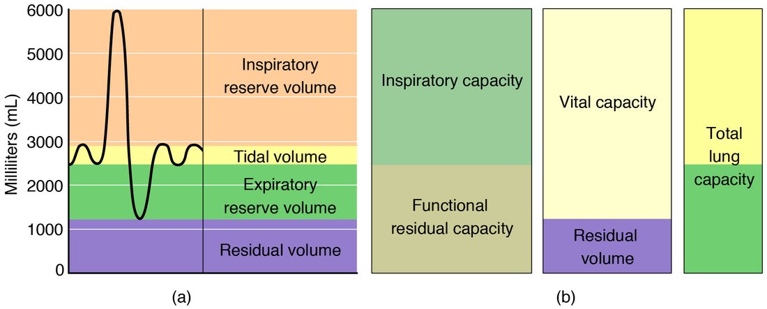
Why can't you blow out the residual volume? Two mechanisms stop you. As the lungs get small, the smallest airways, which have no cartilage in their walls, are squeezed shut by the pressure around them and trap the air beyond them. And the lungs cannot collapse away from the chest wall, because the pleural fluid holds them to it, and the chest wall itself stops getting smaller at its limit. Residual volume keeps the alveoli partly open between breaths, so blood flowing past them meets air all the time, not only during a breath in.
These numbers are typical, not fixed. Volumes are larger in taller people, and a woman of the same age has volumes roughly 20 to 25% smaller than a man. With age, the lungs lose elastic recoil and small airways close earlier, so residual volume rises and the other volumes shrink. Test results are therefore compared with the values predicted for the person's age, sex and height.
The four respiratory capacities
A respiratory capacity is the sum of two or more respiratory volumes. The capacities describe things you can do or measure as one maneuver, such as "breathe in as deeply as you can from a normal breath out."
| Capacity | Volumes it adds | Typical value | What it means |
|---|---|---|---|
| Inspiratory capacity (IC) | TV + IRV | 3,500 mL | The most you can breathe in, starting from the end of a quiet breath out |
| Functional residual capacity (FRC) | ERV + RV | 2,400 mL | The air still in your lungs at the end of a quiet breath out |
| Vital capacity (VC) | TV + IRV + ERV | 4,700 mL | The most you can breathe out after the deepest breath in |
| Total lung capacity (TLC) | TV + IRV + ERV + RV | 5,900 mL | All the air in your lungs after the deepest breath in |
Two shortcuts help: TLC = VC + RV, and TLC = IC + FRC. Vital capacity (vita = life) got its name in the 1800s, when it was used as a measure of how healthy a person's lungs were.
Worked example 1: building the capacities
Problem. A student's spirometry gives TV = 500 mL, IRV = 2,800 mL and ERV = 1,100 mL. A separate test finds RV = 1,300 mL. Find her inspiratory capacity, functional residual capacity, vital capacity and total lung capacity.
- Inspiratory capacity. IC = TV + IRV = 500 + 2,800 = 3,300 mL.
- Functional residual capacity. FRC = ERV + RV = 1,100 + 1,300 = 2,400 mL.
- Vital capacity. VC = TV + IRV + ERV = 500 + 2,800 + 1,100 = 4,400 mL.
- Total lung capacity. TLC = VC + RV = 4,400 + 1,300 = 5,700 mL.
- Check. TLC should also equal IC + FRC: 3,300 + 2,400 = 5,700 mL. It does.
Answer. IC 3,300 mL, FRC 2,400 mL, VC 4,400 mL, TLC 5,700 mL.
Notice which of these a spirometer can find by itself. Any capacity that includes residual volume, that is FRC and TLC, cannot be measured by spirometry alone, because residual volume never passes the mouthpiece. Those need a different test: breathing a gas such as helium and seeing how much it is diluted by the air already in the lungs, or sitting in a sealed box (body plethysmography) that measures volume from pressure changes, using Boyle's law.
Functional residual capacity: the resting balance point
At the end of a quiet breath out, your breathing muscles are relaxed, yet your lungs are far from empty: they still hold the FRC, about 2,400 mL. The FRC is where two opposite pulls balance. Your lungs' elastic recoil pulls them inward, and your chest wall springs outward. With the muscles relaxed, the lungs settle at the volume where these two are equal and opposite.
Anything that changes either pull moves the FRC. Lungs that have lost elastic recoil settle at a larger FRC; stiff, scarred lungs (fibrosis) settle at a smaller one. Lying down lets the abdominal organs push the diaphragm up, which lowers FRC by several hundred milliliters.
Forced spirometry: FEV1 and FVC
How much air you can move matters, but so does how fast. In the most common lung test, you breathe in as deeply as you can and then blow out as hard and as fast as you can, until nothing more comes out. Two numbers come from this one breath:
- Forced vital capacity (FVC): the total volume blown out in the forced breath. In healthy lungs it is close to the vital capacity.
- FEV1 (forced expiratory volume in 1 second): the volume blown out in the first second.
Healthy lungs empty fast. A young adult blows out about 80% of the FVC in the first second, so the FEV1/FVC ratio is about 0.8. The ratio falls a little with age. A ratio below about 0.70 means air is leaving the lungs too slowly.
Worked example 2: reading the FEV1/FVC ratio
Problem. Use the three curves in Figure 2. The normal person blows out 4.0 L in the first second and 5.0 L in total. The first patient blows out 1.6 L in the first second and 3.9 L in total. The second blows out 2.2 L in the first second and 2.5 L in total. Find each FEV1/FVC ratio.
- Write the ratio. FEV1/FVC = volume in the first second ÷ total volume.
- Normal. 4.0 ÷ 5.0 = 0.80.
- First patient. 1.6 ÷ 3.9 = 0.41. Well below 0.70: air leaves too slowly.
- Second patient. 2.2 ÷ 2.5 = 0.88. Normal or high, even though the FVC is only half the normal value.
Answer. 0.80, 0.41 and 0.88. The first patient's pattern is obstructive; the second patient's points to restriction, which a measured total lung capacity would confirm.
Obstructive and restrictive disease
Lung diseases that change these numbers fall into two broad patterns.
Obstructive disease (ob- = against, struct- = build, as in building a barrier) narrows the airways, so air has trouble getting out. You met the reason in airway resistance: resistance rises steeply as an airway's radius falls. Asthma is the example you know; the long-term, smoking-related lung diseases that come later in this chapter are others. During a forced breath out, the narrowed airways close early, so air is trapped behind them, and residual volume and FRC rise.
Restrictive disease (restrict- = hold back) stops the lungs from expanding fully, so air has trouble getting in. The lungs themselves may be stiff (low compliance), as in fibrosis; the chest wall may be deformed, as in severe scoliosis or kyphosis, or loaded, as in severe obesity; or the breathing muscles may be weak, as in muscular dystrophy or myasthenia gravis. The airways are open, so what air there is leaves quickly.
| Obstructive disease | Restrictive disease | |
|---|---|---|
| Basic problem | Narrowed airways: air has trouble getting out | Lungs or chest cannot expand fully: air has trouble getting in |
| Examples | Asthma; smoking-related airway and alveolar damage | Fibrosis of the lungs; severe scoliosis or obesity; weak breathing muscles |
| Airway resistance | Raised | Normal |
| FEV1 | Much lower than predicted | Lower, in proportion to the smaller FVC |
| FVC | Normal or somewhat lower | Lower |
| FEV1/FVC ratio | Low (below about 0.70) | Normal or high |
| Total lung capacity | Normal or raised | Low: this defines restriction |
| Residual volume and FRC | Raised (air trapping) | Usually normal or low |
Spirometry alone can suggest restriction, through a low FVC with a normal ratio, but it cannot prove it. Only a measured total lung capacity, which includes the residual volume, confirms that the lungs really are small.
Minute ventilation
Suppose you take 12 breaths a minute, each of 500 mL. In one minute you move 12 × 500 = 6,000 mL of air in and out.
That is your minute ventilation (also called the pulmonary ventilation rate): the total volume of air moved into or out of the lungs each minute. It is the tidal volume times the respiratory rate, the number of breaths per minute (12 to 20 in a resting adult):
Minute ventilation = tidal volume × respiratory rate
Worked example 3: minute ventilation
Problem. A resting adult has a tidal volume of 450 mL and a respiratory rate of 14 breaths/min. During brisk walking, her tidal volume rises to 1,200 mL and her rate to 20. Find her minute ventilation in both states.
- Write the equation. Minute ventilation = TV × respiratory rate.
- At rest. 450 mL/breath × 14 breaths/min = 6,300 mL/min = 6.3 L/min.
- Walking. 1,200 × 20 = 24,000 mL/min = 24 L/min.
- Compare. 24 ÷ 6.3 = 3.8. Tidal volume rose 2.7-fold and rate 1.4-fold.
Answer. 6.3 L/min at rest and 24 L/min walking, almost four times as much.
Dead space
Not all of that air reaches the alveoli. Follow one 500 mL breath in (Figure 3). At the end of the previous breath out, the conducting zone, from the nose to the terminal bronchioles, is full of stale air that has just come out of the alveoli. The breath in pushes that stale air back into the alveoli first. Then fresh air follows, and the last 150 mL of fresh air stops in the conducting zone, where no gas exchange happens, and simply leaves again with the next breath out.
Air that is moved but does not take part in gas exchange is dead space. There are two kinds:
- Anatomical dead space is the volume of the conducting zone, about 150 mL in an adult. A useful rule of thumb is about 1 mL per pound (2.2 mL per kg) of ideal body weight. It is set by the size of the airways, so it barely changes from breath to breath.
- Alveolar dead space is the volume of alveoli that receive air but little or no blood flow, so no gas exchange can happen in them. In a healthy person standing upright it is close to zero. It rises when blood flow to part of a lung is blocked, for example by a pulmonary embolism, a blood clot lodged in a pulmonary artery.
Total dead space (also called physiological dead space) is the sum: anatomical plus alveolar. In health, it is essentially the same as the anatomical dead space.
Equipment adds dead space too. A snorkel, a long breathing tube or a face mask adds its own volume to the conducting zone.
Alveolar ventilation
Alveolar ventilation (also called the alveolar ventilation rate) is the volume of fresh air that reaches the alveoli each minute. It is what counts for gas exchange. Subtract the dead space from each breath, then multiply by the rate:
Alveolar ventilation = (tidal volume − dead space) × respiratory rate
Worked example 4: alveolar ventilation at rest
Problem. A resting adult breathes 12 times a minute with a tidal volume of 500 mL. His dead space is 150 mL. Find his minute ventilation and his alveolar ventilation.
- Minute ventilation. 500 × 12 = 6,000 mL/min = 6.0 L/min.
- Fresh air reaching the alveoli per breath. 500 − 150 = 350 mL.
- Alveolar ventilation. 350 × 12 = 4,200 mL/min = 4.2 L/min.
- Share wasted. 150 ÷ 500 = 0.30, so 30% of each breath is dead space.
Answer. Minute ventilation 6.0 L/min; alveolar ventilation 4.2 L/min.
Why depth matters more than rate
Dead space is a fixed cost paid on every breath. Shallow breaths pay it more often for less fresh air.
Worked example 5: same minute ventilation, different alveolar ventilation
Problem. Three people each have a minute ventilation of 6,000 mL/min and a dead space of 150 mL. Person A takes 12 breaths of 500 mL. Person B takes 24 breaths of 250 mL. Person C takes 6 breaths of 1,000 mL. Find each alveolar ventilation.
- Check minute ventilation. 12 × 500 = 24 × 250 = 6 × 1,000 = 6,000 mL/min for all three.
- Person A. (500 − 150) × 12 = 350 × 12 = 4,200 mL/min.
- Person B (fast and shallow). (250 − 150) × 24 = 100 × 24 = 2,400 mL/min.
- Person C (slow and deep). (1,000 − 150) × 6 = 850 × 6 = 5,100 mL/min.
Answer. 4.2, 2.4 and 5.1 L/min. The same total air movement delivers more than twice as much fresh air to the alveoli when breaths are deep and slow as when they are fast and shallow.
This is why rapid, shallow breathing is dangerous. A patient with broken ribs who breathes 32 times a minute with 200 mL breaths moves 6.4 L/min, a normal-looking minute ventilation, but only (200 − 150) × 32 = 1,600 mL of fresh air reaches the alveoli each minute. Oxygen in the alveoli falls and carbon dioxide builds up.
Deeper breaths are not free either: they stretch the lungs and chest wall further, which takes more muscle work against elastic recoil. At rest, a rate of about 12 to 15 breaths a minute falls where the total work of breathing is lowest.