Lung volumes and ventilation rates
1Why this matters
Mr. Adebayo, 58, broke four ribs in a fall. Every breath hurts, so he takes tiny breaths, 32 of them a minute. The air he moves each minute looks normal, about 6.4 liters, yet he is drowsy and his blood carbon dioxide is climbing. The problem is not how much air he moves. It is how little of each breath gets past his airways to his alveoli.
2What this builds on
3Quick check before you start
1. Where in the airways does gas exchange with the blood happen?
- Along the whole airway, from the nose onward
- Only in the respiratory zone: respiratory bronchioles, alveolar ducts and alveoli
- Only in the trachea and the main bronchi
Show the answer
The conducting zone, from the nose to the terminal bronchioles, only carries, warms and cleans air. Gas exchange happens only in the respiratory zone, where alveoli sit next to capillaries.
- Along the whole airway, from the nose onward:
- Correct: Only in the respiratory zone: respiratory bronchioles, alveolar ducts and alveoli:
- Only in the trachea and the main bronchi:
2. During a quiet breath in, what makes air flow into the lungs?
- The diaphragm contracts, the chest enlarges, and alveolar pressure falls below atmospheric pressure
- Air is pushed in because atmospheric pressure rises during each breath
- The lungs actively expand themselves by contracting muscle in the alveolar walls
Show the answer
Contraction of the diaphragm and external intercostals enlarges the chest. By Boyle's law, the larger volume lowers alveolar pressure below atmospheric pressure, and air flows in down that pressure gradient.
- Correct: The diaphragm contracts, the chest enlarges, and alveolar pressure falls below atmospheric pressure:
- Air is pushed in because atmospheric pressure rises during each breath:
- The lungs actively expand themselves by contracting muscle in the alveolar walls:
3. An airway narrows to half its radius. What happens to its resistance to airflow?
- It doubles
- It rises about 16-fold
- It halves
Show the answer
Resistance varies with the fourth power of the radius, so halving the radius raises resistance 2 × 2 × 2 × 2 = 16 times.
- It doubles:
- Correct: It rises about 16-fold:
- It halves:
4Anatomy
With labels hidden, select a box to reveal its label.
5How it works, step by step
- Pain makes a patient breathe faster with much smaller breaths: tidal volume falls from 500 to 200 mL while the rate rises.Minute ventilation, tidal volume times rate, stays about normal.
- Each breath must first fill the conducting zone, about 150 mL of anatomical dead space, whatever the breath's size.Dead space takes up a much larger share of each small breath: 150 of 200 mL instead of 150 of 500 mL.
- Only the part of each breath beyond the dead space reaches the alveoli.Alveolar ventilation, (tidal volume − dead space) × rate, falls sharply, here from 4.2 to 1.6 L/min.
- Less fresh air reaches the alveoli each minute, while the tissues keep using oxygen and making carbon dioxide at the same rate.Oxygen in the alveolar air falls and carbon dioxide builds up, so the blood leaving the lungs carries less oxygen and more carbon dioxide.
6Core concepts
7A common mistake
The wrong idea: Minute ventilation tells you how much fresh air reaches the alveoli, so breathing faster always means better ventilation.
What actually happens: Minute ventilation counts all the air moved, including the roughly 150 mL of each breath that only fills the conducting airways and leaves again. What reaches the alveoli is alveolar ventilation, (tidal volume − dead space) × rate. Fast, shallow breathing can keep minute ventilation normal while alveolar ventilation collapses: 24 breaths of 250 mL deliver only 2.4 L/min of fresh air, against 4.2 L/min from 12 breaths of 500 mL.
8Check yourself
Anything you miss goes into your review queue.
1. A patient's tidal volume is 500 mL, inspiratory reserve volume 3,000 mL, expiratory reserve volume 1,100 mL and residual volume 1,200 mL. What is her vital capacity?
- 3,500 mL
- 5,800 mL
- 4,600 mL
- 2,300 mL
Show the answer
Vital capacity = TV + IRV + ERV = 500 + 3,000 + 1,100 = 4,600 mL. Residual volume is not part of it, because it can never be breathed out.
- 3,500 mL: 3,500 mL is TV + IRV, the inspiratory capacity.
- 5,800 mL: 5,800 mL adds the residual volume too. That sum is total lung capacity.
- Correct: 4,600 mL: Correct. 500 + 3,000 + 1,100 = 4,600 mL.
- 2,300 mL: 2,300 mL is ERV + RV, the functional residual capacity.
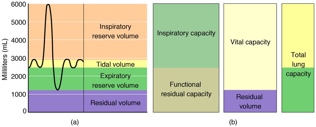
2. Using the spirogram, estimate this person's inspiratory capacity: the most air he could breathe in starting from the end of a quiet breath out.
- About 500 mL
- About 6,000 mL
- About 4,800 mL
- About 3,500 mL
Show the answer
A quiet breath out ends at about 2,500 mL, the bottom of the tidal band. The deepest breath in reaches 6,000 mL. The difference, 6,000 − 2,500 = 3,500 mL, is the inspiratory capacity (TV + IRV).
- About 500 mL: About 500 mL is the tidal volume alone, a quiet breath, not the deepest possible one.
- About 6,000 mL: 6,000 mL is the total lung capacity, which counts air already in the lungs at the start.
- About 4,800 mL: About 4,800 mL is the vital capacity, measured from the fullest breath out, not from a quiet one.
- Correct: About 3,500 mL: Correct. From about 2,500 up to 6,000 mL is about 3,500 mL.
3. A resting man breathes 12 times a minute with a tidal volume of 500 mL. His dead space is 150 mL. What is his alveolar ventilation?
- 6.0 L/min
- 4.2 L/min
- 1.8 L/min
- 7.8 L/min
Show the answer
Alveolar ventilation = (tidal volume − dead space) × rate = (500 − 150) × 12 = 350 × 12 = 4,200 mL/min = 4.2 L/min.
- 6.0 L/min: 6.0 L/min is the minute ventilation, 500 × 12, which still includes the dead space.
- Correct: 4.2 L/min: Correct. 350 mL of fresh air reaches the alveoli on each of 12 breaths.
- 1.8 L/min: 1.8 L/min is the dead space ventilation, 150 × 12: the air wasted each minute.
- 7.8 L/min: 7.8 L/min adds the dead space instead of subtracting it: (500 + 150) × 12.
4. A resting person switches from 12 breaths of 500 mL per minute to 24 breaths of 250 mL per minute. Her anatomical dead space is 150 mL. Predict the change in each variable.
| Variable | Change |
|---|---|
| Minute ventilation | — |
| Anatomical dead space | — |
| Share of each breath that is dead space | — |
| Alveolar ventilation | — |
Show the answer
Doubling the rate and halving the depth keeps minute ventilation the same, but the dead space is a fixed cost on every breath. With smaller breaths, a larger share of each one is wasted, and alveolar ventilation falls by almost half.
- Minute ventilation: no change. 12 × 500 and 24 × 250 both equal 6,000 mL/min, so the total air moved each minute is unchanged.
- Anatomical dead space: no change. Anatomical dead space is the volume of the conducting airways, set by their size, not by how she breathes.
- Share of each breath that is dead space: up. 150 mL is now 60% of a 250 mL breath, instead of 30% of a 500 mL breath.
- Alveolar ventilation: down. (250 − 150) × 24 = 2,400 mL/min, down from (500 − 150) × 12 = 4,200 mL/min, because the fixed dead space is paid on twice as many smaller breaths.
5. Ms. Park, 34, has episodes of wheezing. On spirometry her FEV1 is 1.6 L and her FVC is 3.9 L. Which pattern does this show?
- Obstructive: narrowed airways slow the breath out
- Restrictive: her lungs and chest wall cannot expand fully
- Normal: her FVC is close to the typical value
- Neither pattern can be judged without her residual volume
Show the answer
FEV1/FVC = 1.6 ÷ 3.9 = 0.41, far below about 0.70. She can blow out a nearly normal total volume, but slowly, because narrowed airways raise resistance. That is obstruction, typical of asthma.
- Correct: Obstructive: narrowed airways slow the breath out: Correct. A low FEV1/FVC ratio is the mark of obstruction.
- Restrictive: her lungs and chest wall cannot expand fully: Restriction shrinks the FVC but leaves the FEV1/FVC ratio normal or high. Her ratio is very low.
- Normal: her FVC is close to the typical value: A near-normal FVC does not make the test normal; the speed of emptying, FEV1/FVC, is badly reduced.
- Neither pattern can be judged without her residual volume: Residual volume is needed to confirm restriction, but a low FEV1/FVC ratio shows obstruction from spirometry alone.
6. A swimmer breathes through a snorkel that holds 150 mL. His dead space without it is 150 mL. If he keeps breathing 12 times a minute with 500 mL breaths, what happens to his alveolar ventilation, and what is the best way for him to restore it?
- It falls from 4.2 to 2.4 L/min; breathe more deeply
- It is unchanged, because the snorkel is outside his body
- It falls from 4.2 to 2.4 L/min; breathe faster with the same small breaths
- It rises, because the snorkel adds extra air to each breath
Show the answer
The snorkel adds its volume to the dead space: 150 + 150 = 300 mL. Alveolar ventilation falls from (500 − 150) × 12 = 4.2 L/min to (500 − 300) × 12 = 2.4 L/min. Deeper breaths help most, because each extra milliliter of tidal volume goes straight to the alveoli once the dead space is filled.
- Correct: It falls from 4.2 to 2.4 L/min; breathe more deeply: Correct. Deeper breathing restores alveolar ventilation most efficiently.
- It is unchanged, because the snorkel is outside his body: Any tube the breath must pass through adds dead space, wherever it is. The stale air left in the snorkel is breathed back in first.
- It falls from 4.2 to 2.4 L/min; breathe faster with the same small breaths: The fall is right, but faster breathing with the same small breaths pays the larger dead space on every extra breath, so it restores alveolar ventilation poorly.
- It rises, because the snorkel adds extra air to each breath: The snorkel holds air that is breathed back and forth unused; it adds dead space, not fresh air.
7. A blood clot lodges in a branch of Ms. Reyes's pulmonary artery. The alveoli supplied by that branch still fill with air on every breath. What happens to her dead space?
- Anatomical dead space rises, because the airways widen
- Alveolar dead space rises, so total dead space rises
- Dead space is unchanged, because her airways are normal
- Total dead space falls, because less blood reaches the lungs
Show the answer
Air reaching alveoli with no blood flow cannot exchange gas, so those alveoli become alveolar dead space. Total dead space, anatomical plus alveolar, rises, and she must breathe more to deliver the same useful ventilation.
- Anatomical dead space rises, because the airways widen: Anatomical dead space is the volume of the conducting airways, which a clot in an artery does not change.
- Correct: Alveolar dead space rises, so total dead space rises: Correct. Ventilated alveoli with no blood flow are alveolar dead space.
- Dead space is unchanged, because her airways are normal: Normal airways keep anatomical dead space normal, but dead space also includes alveoli that get air and no blood.
- Total dead space falls, because less blood reaches the lungs: Losing blood flow to ventilated alveoli wastes their air, which raises dead space rather than lowering it.
9Summary
Four respiratory volumes stack up to fill the lungs: tidal volume (about 500 mL, one quiet breath), inspiratory reserve volume (extra air you can breathe in), expiratory reserve volume (extra air you can breathe out) and residual volume (air you can never breathe out). Capacities are sums of volumes: inspiratory capacity (TV + IRV), functional residual capacity (ERV + RV, the resting balance point), vital capacity (TV + IRV + ERV) and total lung capacity (all four). Spirometry cannot measure residual volume or any capacity that includes it. In forced spirometry, obstructive disease lowers the FEV1/FVC ratio below about 0.70 and traps air; restrictive disease lowers total lung capacity and FVC with a normal or high ratio. Minute ventilation is tidal volume × respiratory rate, but only alveolar ventilation, (tidal volume − dead space) × rate, reaches the alveoli, so shallow breathing wastes a larger share of each breath.