The control of breathing rests on a few groups of neurons in your brainstem and on chemoreceptors that sense the chemistry of your blood. You breathe about 12 to 20 times a minute without thinking about it, awake or asleep, and the depth and rate change within seconds when you exercise, climb a mountain or hold your breath. This page explains the respiratory centers that set the rhythm, the central and peripheral chemoreceptors that adjust it, why carbon dioxide rather than oxygen is the main drive to breathe, what hyperventilation and hypoventilation mean, and how breathing changes during exercise and at high altitude.
Automatic, but you can override it
Take a deep breath now, then hold it. You can: the motor cortex can drive your breathing muscles directly, through the corticospinal tract, bypassing the brainstem. That is how you speak, sing, sniff and hold your breath. But you cannot hold it forever. After a minute or so, the urge to breathe becomes overwhelming and you take a breath whether you mean to or not. And tonight, asleep, you will breathe all night with no conscious effort at all.
So breathing has two controllers: a voluntary one in the cerebral cortex, and an automatic one in the brainstem that always wins in the end. The rest of this page is about the automatic one.
The respiratory centers
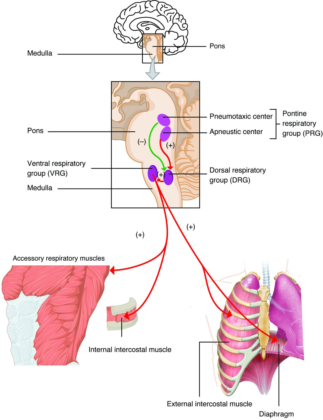
The respiratory centers are groups of neurons in the medulla oblongata and the pons that generate the rhythm of breathing and send it to the breathing muscles. Figure 1 shows where they are.
In the medulla oblongata
- The ventral respiratory group (VRG) (ventr- = belly side) is a column of neurons toward the front of the medulla. Near its upper end lies a small cluster called the pre-Bötzinger complex, whose neurons fire in rhythmic bursts on their own, even when cut off from the rest of the brain in a dish. It is the leading candidate for the generator of the respiratory rhythm, the repeating cycle of inspiration and expiration. The VRG also holds neurons that drive the muscles of forced expiration, and neurons that drive the accessory muscles when breathing is hard.
- The dorsal respiratory group (DRG) (dors- = back) sits toward the back of the medulla. It receives sensory information from the chemoreceptors and the lungs through the glossopharyngeal and vagus nerves, and it sends inspiratory signals to the diaphragm. It integrates what the body reports and adjusts the depth of each breath.
In the pons
- The pontine respiratory group (PRG), also called the pneumotaxic center (pneum- = air, breath; tax- = arrangement, order), sends signals to the medullary groups that switch off inspiration. Stronger signals from it make each breath shorter and shallower, so breathing gets faster. It smooths the change from inspiration to expiration.
- The apneustic center (a- = without, pneus- = breathing: named after "apneusis", long held breaths) lies lower in the pons. In animal experiments, when the pneumotaxic center and the vagus nerves were cut, this region produced long, gasping inspirations. It is described as promoting inspiration. In humans, no single anatomical center matching it has been clearly identified.
From the centers to the muscles
- During quiet breathing, neurons in the medulla fire a burst of signals for about 2 seconds.
- The signals travel down the spinal cord to motor neurons that leave through the phrenic nerves (from spinal segments C3–C5) to the diaphragm and through the intercostal nerves to the external intercostal muscles.
- The diaphragm and external intercostals contract, the thoracic cavity enlarges, and air flows in.
- The burst stops for about 3 seconds. The muscles relax, the lungs recoil, and air flows out. Quiet expiration is passive.
- In forced breathing, VRG neurons also drive the internal intercostals and the abdominal wall muscles during expiration, and the accessory muscles such as the scalenes and sternocleidomastoid during inspiration.
The rhythm is not fixed. Sensory information changes both how deep each breath is and how many you take. The most important information comes from chemoreceptors.
Central chemoreceptors
Central chemoreceptors (chem- = chemical; central = inside the central nervous system) are neurons near the front surface of the medulla oblongata, bathed in the brain's interstitial fluid close to the cerebrospinal fluid. They respond to the hydrogen ion concentration of that fluid, not to oxygen.
Here is how carbon dioxide in your blood reaches them (Figure 2):
- Arterial PCO2 rises.
- Carbon dioxide crosses the blood–brain barrier easily, because it is small and dissolves in lipids. Hydrogen ions and bicarbonate cross it poorly.
- In the brain's fluid, carbon dioxide forms carbonic acid, which releases hydrogen ions. The fluid's pH falls.
- Cerebrospinal fluid holds very little protein, so it buffers poorly, and its pH falls more than blood pH would for the same change.
- The central chemoreceptors sense the rise in hydrogen ions and stimulate the respiratory centers, and breathing gets deeper and faster.
Two consequences follow:
- Central chemoreceptors respond mainly to carbon dioxide. Acid added to the blood by some other cause, such as acids from metabolism, crosses into the brain's fluid only slowly, so the central chemoreceptors are slow to notice it.
- They adapt over a day or two. If PCO2 stays high or low, the choroid plexus and the brain slowly adjust the bicarbonate in the cerebrospinal fluid, which brings its pH back toward normal. The central chemoreceptors then respond less to the lasting change in PCO2.
At steady state, the central chemoreceptors account for most of your breathing response to carbon dioxide, roughly two thirds to four fifths of it.
Peripheral chemoreceptors
Peripheral chemoreceptors are clusters of sensory cells outside the central nervous system, in contact with arterial blood:
- The carotid bodies are two tiny nodules, each smaller than a grain of rice, at the fork where each common carotid artery divides into the internal and external carotid arteries, next to the carotid sinus. They send signals to the medulla through the glossopharyngeal nerves. They are the main peripheral chemoreceptors in humans.
- The aortic bodies are smaller clusters on the aortic arch. They send signals through the vagus nerves. Their role in breathing is small in adults.
The carotid bodies have one of the highest blood flows per gram of any tissue in the body, so the blood inside them stays close to arterial composition. They respond to three things:
- A fall in arterial PO2. This is their unique job: they are the only sensors that make you breathe more when oxygen is low. Their firing rises steeply once arterial PO2 falls below about 60 mm Hg.
- A rise in arterial PCO2. They respond within seconds, faster than the central chemoreceptors.
- A fall in arterial pH from any cause, including acid that is not from carbon dioxide. This is how acids added to the blood by metabolism quickly speed breathing.
They sense the partial pressure of oxygen, not how much oxygen the blood carries. In anemia, or in carbon monoxide poisoning, the blood carries much less oxygen, but the oxygen dissolved in the plasma, and so the arterial PO2, is normal. The carotid bodies are not stimulated much, and breathing does not rise much.
| Central chemoreceptors | Peripheral chemoreceptors | |
|---|---|---|
| Where | Near the front surface of the medulla oblongata | Carotid bodies at the carotid fork; aortic bodies on the aortic arch |
| Fluid they sample | Brain interstitial fluid, close to the cerebrospinal fluid | Arterial blood |
| Main stimulus | Hydrogen ions made from carbon dioxide in the brain's fluid | Low PO2; also high PCO2 and low pH |
| Respond to low oxygen? | No | Yes, strongly below about 60 mm Hg |
| Respond to acid from other causes? | Slowly, because hydrogen ions cross the blood–brain barrier poorly | Yes, quickly |
| Speed | Slower: tens of seconds to minutes | Fast: within seconds |
| Share of the response to carbon dioxide | Most, roughly two thirds to four fifths | The rest, roughly one fifth to one third |
| Pathway to the medulla | Short connections within the brainstem | Glossopharyngeal nerves (carotid bodies) and vagus nerves (aortic bodies) |
Carbon dioxide is the main drive to breathe
Try it in numbers. Raise the carbon dioxide in the air you breathe so that your arterial PCO2 climbs from 40 to about 45 mm Hg, and your ventilation roughly doubles. Now lower the oxygen instead: your arterial PO2 can fall from 95 to about 60 mm Hg before your ventilation rises much at all (Figure 3).
Why does oxygen have so little effect at first? The carotid bodies' own response is shaped that way: their firing rises only a little as arterial PO2 falls from 100 to about 60 mm Hg, then steeply below that. The oxygen–hemoglobin dissociation curve you met earlier shows why little is lost meanwhile. Between a PO2 of 100 and 60 mm Hg the curve is flat: hemoglobin stays about 90% saturated or more, so the blood still carries almost as much oxygen. Below about 60 mm Hg, where oxygen content starts to fall fast, the carotid bodies respond strongly.
Carbon dioxide, by contrast, changes pH directly, and pH affects almost every protein in the body. Minute to minute, then, your breathing is set mainly to hold arterial PCO2 near 40 mm Hg. Oxygen is a backup signal that takes over when PO2 falls low, as at high altitude or in lung disease.
The carbon dioxide loop
Breathing control is a negative feedback loop:
- Stimulus: arterial PCO2 rises (and hydrogen ions rise with it).
- Sensory receptors: central chemoreceptors in the medulla oblongata, and peripheral chemoreceptors in the carotid and aortic bodies.
- Afferent pathway: short connections within the brainstem from the central chemoreceptors; the glossopharyngeal and vagus nerves from the peripheral ones.
- Control center: the respiratory centers in the medulla oblongata and pons.
- Efferent pathway: motor neurons in the phrenic and intercostal nerves.
- Effectors: the diaphragm and the intercostal muscles.
- Response: deeper, faster breathing raises alveolar ventilation, more carbon dioxide is breathed out, and arterial PCO2 and pH return toward normal. The stimulus is reduced: negative feedback.
The low-oxygen loop
- Stimulus: arterial PO2 falls, strongly below about 60 mm Hg.
- Sensory receptors: peripheral chemoreceptors, mainly the carotid bodies. The central chemoreceptors do not sense oxygen.
- Afferent pathway: the glossopharyngeal nerves (and the vagus nerves from the aortic bodies).
- Control center: the respiratory centers.
- Efferent pathway and effectors: the phrenic and intercostal nerves to the diaphragm and intercostal muscles.
- Response: more ventilation raises alveolar PO2 and arterial PO2. The stimulus is reduced.
How much the feedback changes arterial PCO2 depends on alveolar ventilation, which you met with lung volumes. At a steady rate of carbon dioxide production, arterial PCO2 is inversely proportional to alveolar ventilation.
Worked example 1: alveolar ventilation and PCO2
Problem. A resting adult has an alveolar ventilation of 4.2 L/min and an arterial PCO2 of 40 mm Hg. His carbon dioxide production does not change. What will his arterial PCO2 settle at if his alveolar ventilation (a) doubles to 8.4 L/min, or (b) halves to 2.1 L/min?
- Write the relationship. With carbon dioxide production constant, PCO2 × alveolar ventilation stays constant: 40 mm Hg × 4.2 L/min.
- Case (a). Alveolar ventilation is 2 times higher, so PCO2 falls to 40 ÷ 2 = 20 mm Hg.
- Case (b). Alveolar ventilation is half as much, so PCO2 rises to 40 × 2 = 80 mm Hg.
- Link to pH. From the last topic: PCO2 down raises pH; PCO2 up lowers it.
Answer. (a) 20 mm Hg, with a rise in pH. (b) 80 mm Hg, with a fall in pH. Doubling or halving the air reaching your alveoli halves or doubles your carbon dioxide.
Other inputs to breathing
Chemoreceptors are not the only source of input. Other inputs to breathing come from the lungs, the airways, the body and the rest of the brain:
- Lung stretch receptors are sensory receptors in the smooth muscle of the airways that fire as the lungs inflate. Their signals travel up the vagus nerves and inhibit inspiration. This is the Hering–Breuer reflex (the inflation reflex), named after the two physiologists who described it. In adults at rest it is weak; it becomes active when tidal volume is large, about three times normal or more, as in hard exercise, and it limits how far the lungs are inflated. In newborns it is stronger.
- Irritant receptors are sensory receptors among the cells lining the airways. Dust, smoke, cold air or chemical fumes stimulate them. Their signals, carried in the vagus nerves, cause coughing, narrowing of the airways (bronchoconstriction) and rapid, shallow breathing.
- Sensory receptors in the walls of the alveoli, near the capillaries, respond when fluid collects in the lung tissue, causing rapid, shallow breathing and a feeling of breathlessness. You will meet them again with lung disorders.
- Proprioceptors in moving joints and muscles speed breathing as soon as you start to move, before the blood's chemistry has changed.
- The cerebral cortex gives voluntary control for speaking, singing and breath-holding. What finally ends a breath-hold, the breaking point, is mainly the rising PCO2 acting on the chemoreceptors.
- The hypothalamus and limbic system change breathing with emotion, pain and body temperature. Fear and anxiety speed it; a sudden shock of cold water makes you gasp; fever speeds it.
Hyperventilation and hypoventilation
Suppose two patients are each breathing 30 times a minute. One has an arterial PCO2 of 25 mm Hg; the other, whose breaths are very shallow, has 60 mm Hg. Only the first is hyperventilating.
These terms are defined by carbon dioxide, not by breathing rate:
- Hyperventilation (hyper- = over, above) is breathing more than your carbon dioxide production requires, so arterial PCO2 falls below 35 mm Hg. Low PCO2 is hypocapnia (hypo- = under, kapn- = smoke, carbon dioxide).
- Hypoventilation (hypo- = under) is breathing less than your carbon dioxide production requires, so arterial PCO2 rises above 45 mm Hg. High PCO2 is hypercapnia (hyper- = over).
Rapid, shallow breathing can be hypoventilation, because much of each small breath only fills the dead space and never reaches the alveoli. Deep, slow breathing can be hyperventilation. The PCO2 decides.
| Hyperventilation | Hypoventilation | |
|---|---|---|
| Alveolar ventilation compared with carbon dioxide production | More than needed | Less than needed |
| Arterial PCO2 | Below 35 mm Hg (hypocapnia) | Above 45 mm Hg (hypercapnia) |
| Blood pH | Rises: alkalosis if above 7.45 | Falls: acidosis if below 7.35 |
| Arterial PO2 | Normal or slightly high | Falls |
| Common causes | Anxiety and panic, pain, fever, high altitude, early lung problems that stimulate irritant or alveolar-wall sensory receptors | Opioid or sedative overdose, brainstem injury, weakness of the breathing muscles, a stiff or injured chest wall |
| What the person feels | Lightheadedness, tingling around the mouth and in the fingers, muscle spasms in the hands | Headache, drowsiness, confusion; unconsciousness if severe |
Why hyperventilation makes you dizzy and tingly
- PCO2 falls. Low carbon dioxide constricts the arterioles in the brain, so blood flow to the brain falls and you feel lightheaded.
- pH rises. In alkalosis, more calcium binds to albumin and other plasma proteins, so the free calcium in the plasma falls.
- Low free calcium makes nerves and muscles more excitable, causing tingling and cramping spasms of the hands and feet.
Hyperventilating before a dive
Some swimmers breathe hard before diving to stay under longer. It works, dangerously. Hyperventilation lowers PCO2 a lot but barely raises the oxygen in the blood, because hemoglobin is already almost fully saturated. Underwater, PCO2 now takes longer to reach the level that forces a breath, while oxygen keeps falling. The diver can lose consciousness from low oxygen before feeling any urge to surface.
Carbon dioxide retention and oxygen therapy
In some people with severe long-term lung disease, arterial PCO2 stays high for months or years. Their central chemoreceptors adapt, as described above, so they respond less to the high PCO2. When such a person is given a lot of oxygen, their PCO2 can rise further and they can become drowsy.
The older explanation was a "hypoxic drive": their breathing depends on low oxygen stimulating the carotid bodies, and oxygen takes that stimulus away so they stop breathing. The evidence does not support this as the main cause. In studies, their ventilation falls only a little, and briefly, when oxygen is given. The rise in PCO2 comes mainly from two other effects:
- Ventilation–perfusion mismatch gets worse. In poorly ventilated parts of the lung, low alveolar oxygen constricts the arterioles (hypoxic pulmonary vasoconstriction), which sends blood away from them. Extra oxygen reverses that constriction, so more blood flows past alveoli that are barely ventilated, and less carbon dioxide is removed.
- The Haldane effect. With more oxygen on hemoglobin, the blood holds less carbon dioxide at the same PCO2, so for the carbon dioxide it carries, PCO2 rises.
The practical rule follows from this: oxygen is given to such patients carefully, aiming for a target saturation, often 88–92%, and watching their breathing. It is never withheld from a person who is hypoxic.
Breathing during exercise
When you run, your ventilation can rise from about 6 L/min at rest to over 100 L/min. Yet in moderate exercise, your arterial PCO2, PO2 and pH hardly change. This increased breathing that matches increased metabolism, with normal blood gases, is called hyperpnea (hyper- = over, -pnea = breathing). It differs from hyperventilation, because PCO2 stays normal.
If the blood's chemistry does not change, the chemoreceptors cannot be what drives the rise. The drive comes from several sources together:
- At the start of exercise, ventilation jumps within a breath or two. Signals from the motor cortex to the working muscles also reach the respiratory centers (sometimes called central command), and proprioceptors in moving limbs add their input.
- Over the next few minutes, ventilation climbs more slowly to a steady level that matches carbon dioxide production. The chemoreceptors fine-tune it, holding PCO2 steady.
- In very hard exercise, the muscles release extra acid, blood pH falls, and the carotid bodies drive ventilation up faster than carbon dioxide production. Arterial PCO2 then falls below normal.
- When exercise stops, ventilation drops sharply at first, then slowly returns to resting levels.
Breathing at high altitude
At 4,300 m, the height of some mountain peaks in North America, air pressure is only about 60% of that at sea level. The air is still 21% oxygen, but the PO2 of the air you breathe in falls from about 150 mm Hg to about 85 mm Hg, and arterial PO2 falls to about 50 mm Hg.
The first hours
- Low arterial PO2 stimulates the carotid bodies, and ventilation rises.
- The extra ventilation blows off carbon dioxide, so arterial PCO2 falls and blood pH rises.
- Carbon dioxide leaves the brain's fluid too, its pH rises, and the central chemoreceptors now reduce their drive. They act as a brake, so ventilation rises less than the low oxygen alone would cause.
Over days: acclimatization
Acclimatization (ad- = to, clima- = climate) is the set of changes over days and weeks that let you work better in low oxygen:
- Days 1 to 7. The kidneys excrete more bicarbonate in the urine, and the bicarbonate in the cerebrospinal fluid is also lowered. Blood and brain pH move back toward normal, the central brake is released, and the carotid bodies become more sensitive to low oxygen. Ventilation keeps rising, and arterial PO2 improves.
- Within a day or two. Red blood cells make more 2,3-BPG, which shifts the oxygen–hemoglobin dissociation curve to the right and helps unload oxygen in the tissues.
- Weeks. Low oxygen makes the kidneys release more erythropoietin, which speeds erythropoiesis. Red blood cell count and hematocrit rise, so each liter of blood carries more oxygen.
Acute mountain sickness
Acute mountain sickness is a cluster of symptoms that can start 6 to 12 hours after going above about 2,500 m too quickly: headache, nausea, tiredness, dizziness and poor sleep. How low oxygen causes it is not fully understood; changes in blood flow and fluid in the brain are thought to play a part. It usually settles over one to three days as acclimatization proceeds. Going up slowly prevents it. Going down treats it, and severe symptoms such as confusion or poor coordination mean going down at once. Acetazolamide, the carbonic anhydrase blocker from the last topic, makes the kidneys excrete more bicarbonate, which acidifies the blood a little and speeds the rise in ventilation.