Besides the pituitary, thyroid, parathyroid, pancreatic islets and adrenal glands, your body has many other endocrine glands and hormone-making tissues. This page covers the pineal gland and the melatonin it releases at night, the daily clock that times it, the thymus, and the gonads with their sex hormones. It then turns to organs whose main job is something else but which also release hormones: the heart, the kidneys and fat. It ends with two applied topics: how several hormones together control bone growth, and how performance-enhancing substances hijack them.
Hormones from many organs
A heart cell that stretches, a kidney cell short of oxygen and a fat cell swollen with stored triglyceride all release hormones. Endocrine tissue is not confined to the classic glands. Figure 1 shows the glands whose main job is making hormones; this page adds the organs that do it on the side.
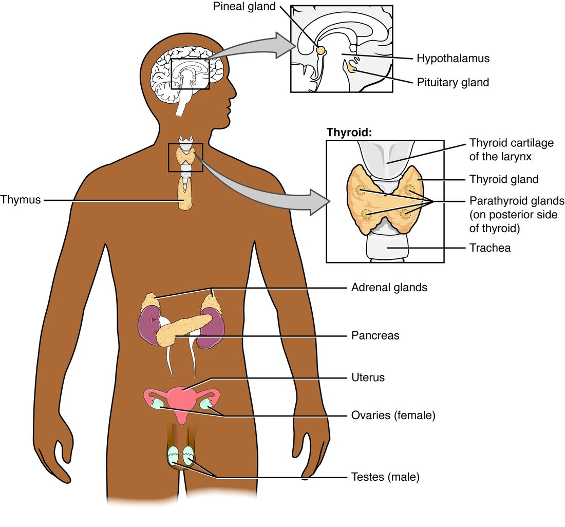
| Organ | Hormone | Released when | Main effect |
|---|---|---|---|
| Pineal gland | Melatonin | It is dark | Signals "night" to the body's clocks |
| Thymus | Thymosins | Mostly in childhood | Help white blood cells mature in the thymus |
| Testes | Testosterone | LH arrives | Male sex characteristics; muscle and bone growth |
| Ovaries | Estrogens, progesterone | FSH and LH arrive | Female sex characteristics; the monthly cycle; bone |
| Heart | Atrial natriuretic peptide (ANP) | Its upper chambers are stretched | Kidneys excrete more sodium and water |
| Kidneys | Erythropoietin (EPO) | Oxygen delivery to the kidney falls | Bone marrow makes more red blood cells |
| Adipose tissue | Leptin | Fat stores are large | Tells the hypothalamus that energy stores are full |
You have already met other part-time endocrine organs: the kidney's renin and calcitriol, the liver's IGF-1 and angiotensinogen, and the skin's first step in making vitamin D. The gut releases a family of hormones of its own, which you will meet with digestion.
The pineal gland and melatonin
Why do you feel sleepy at 10 p.m. but wide awake at noon, even on the same amount of sleep? Part of the answer is a gland the size of a grain of rice. The pineal gland (pine- = pine cone, for its shape) hangs from the back of the roof of the third ventricle, in the epithalamus of the diencephalon. Its hormone-making cells are pinealocytes, and their product is melatonin (melat- from melanin, because it was discovered as a substance that lightens the pigment cells of frog skin; -tonin from serotonin). Pinealocytes make it from serotonin, which in turn comes from the amino acid tryptophan.
Light controls melatonin release through a chain of neurons that has nothing to do with seeing images:
- A small set of retinal ganglion cells contains its own light-sensitive pigment, melanopsin, and responds best to bright blue light.
- Their axons branch off from the visual pathway at the optic chiasm and reach the suprachiasmatic nucleus of the hypothalamus.
- From the hypothalamus, the signal runs down into the spinal cord, out to the superior cervical ganglion of the sympathetic chain, and back up to the pineal gland on sympathetic fibers.
- At night, those sympathetic fibers release norepinephrine onto pinealocytes, which make and release melatonin. Light shuts the pathway down, and melatonin release stops within minutes.
So melatonin in your blood is low all day, starts to climb two or three hours before your usual bedtime, and peaks in the middle of the night. Melatonin binds receptor proteins in the suprachiasmatic nucleus and elsewhere. Its effects are modest: it lowers body temperature slightly, makes sleep a little more likely, and, most importantly, tells the body's clocks what time of night it is. It is a timing signal more than a sleeping pill.
Circadian rhythm: the body's daily clock
A circadian rhythm (circa = about, dies = day) is any body process that rises and falls on a cycle of about 24 hours and keeps cycling even without outside time cues. Your body temperature is lowest around 4 to 5 a.m. Cortisol peaks within an hour of waking. Melatonin peaks around 2 to 4 a.m. Alertness, blood pressure and kidney output all follow daily curves.
The suprachiasmatic nucleus (SCN; supra- = above, chiasm = the optic chiasm) is a pair of tiny clusters of neurons just above the optic chiasm. It is the master clock, or biological clock. Each of its neurons keeps time on its own through a loop of clock genes: their protein products build up over the day, switch off their own genes, then break down and let the cycle start again, roughly every 24 hours. Almost every other cell in the body has the same kind of clock. The SCN keeps them in step through nerve signals, body temperature and hormones such as melatonin and cortisol (Figure 2).
Left in a cave with no clocks or daylight, most people drift to a cycle slightly longer than 24 hours, about 24.2. Morning light resets the clock every day, pulling it back into step with the sun. Light in the evening, including the blue-rich light of screens, pushes the clock later.
When your schedule and your clock disagree, you feel it. After a flight across six time zones, your SCN is still on home time, and it shifts only about an hour a day, so for days you are sleepy and hungry at the wrong times: jet lag. Night-shift workers live in a permanent version of it, and long-term shift work is linked with poorer sleep, weight gain and higher rates of diabetes and heart disease. A small dose of melatonin taken at the new bedtime, and bright light at the right time of day, can speed the shift.
The thymus and thymosin
The thymus (from the Greek name for the gland, thymos, whose origin is uncertain; it may come from the herb thyme) sits in the mediastinum, behind the upper sternum and in front of the heart and the large vessels leaving it. It is large in infants and children, keeps growing until puberty, and then slowly shrinks, its working tissue replaced by fat. By old age little working tissue is left.
The thymus is mainly an organ of the immune system, where these white blood cells mature. Its epithelial cells also release a family of peptides called thymosins, along with related thymic hormones, which help the immature cells develop. How much each of these acts as a true circulating hormone is still unclear; much of their effect seems to be local, inside the thymus. That the thymus matters most early in life is clear: a baby born without one lacks these white blood cells and is prone to severe infections.
The gonads and their hormones
The gonads (gon- = seed, offspring) are the testes and ovaries. Besides making sperm and eggs, they are endocrine glands, and their products are called the sex hormones. All are steroids made from cholesterol, so, like the adrenal cortex hormones, they act through intracellular receptor proteins and change gene expression.
- Testes make testosterone (testo- = testis, -sterone = a steroid), the main androgen. LH from the anterior pituitary drives its release, and testosterone feeds back on the hypothalamus and pituitary. Testosterone drives the male changes of puberty, maintains sperm production and sex drive, and builds muscle and bone.
- Ovaries make estrogens (estr- = frenzy, desire, -gen = producing; mainly estradiol) and progesterone (pro- = for, gest- = carrying, as in pregnancy). FSH and LH drive them, in a pattern that changes across the monthly cycle. Estrogens drive the female changes of puberty and build up the lining of the uterus; progesterone prepares that lining for a pregnancy. Estrogen also protects bone.
Men make some estrogen and women make some androgen, and the difference between the sexes is in the amounts. How the gonadal axes run the reproductive cycles is taught in the reproductive chapter; here, their role in bone growth matters most.
The heart: atrial natriuretic peptide
Give a patient two liters of saline through a drip in an hour and his blood volume rises. More blood returns to his heart and stretches its walls.
Stretched muscle cells in the heart's upper chambers release atrial natriuretic peptide (ANP; natri- = sodium, -uretic = relating to urine). ANP is one of a family of natriuretic peptides, hormones that make the kidneys excrete sodium. Its effects all lower blood volume and pressure:
- The kidneys filter more fluid and reabsorb less sodium, so more sodium and water leave in the urine.
- Renin, aldosterone and ADH release all fall, removing the signals that keep salt and water.
- Vascular smooth muscle relaxes: vasodilation.
As volume falls, the stretch eases and ANP release falls: negative feedback. ANP is the counterweight to the renin–angiotensin–aldosterone system. You will see it at work in long-term blood pressure control.
The kidneys: erythropoietin and hypoxia
Hypoxia (basic)
A climber arrives in a mountain town at 3,500 meters. Each breath carries about a third less oxygen than at sea level, and within hours her tissues are receiving less oxygen than they need. That is hypoxia (hypo- = below, ox- = oxygen, -ia = condition): too little oxygen reaching the tissues. Its close relative is hypoxemia (-emia = blood condition): too little oxygen in the arterial blood.
The two often go together, but not always. At altitude or in lung disease, the arterial blood is low in oxygen, so there is hypoxemia and hypoxia. After a large bleed, each milliliter of blood carries a normal amount of oxygen, but there are too few red blood cells and too little flow, so the tissues become hypoxic without hypoxemia. You will study oxygen delivery in full with the respiratory system.
Erythropoietin
The kidneys detect hypoxia. Cells in the tissue between the kidney's tubes sense oxygen directly: when oxygen falls, a protein that is normally broken down within minutes survives, enters the nucleus and switches on the gene for erythropoietin (EPO; erythr/o = red, -poiesis = making). EPO travels to the red bone marrow, where it keeps more of the cells that make red blood cells alive and dividing. Within days, more new red blood cells enter the blood; over weeks, the red cell count rises and carries more oxygen, and EPO release falls back: negative feedback.
The kidney is a good place for the sensor. It receives about a fifth of the blood the heart pumps but extracts little of its oxygen, so the oxygen level in its tissue tracks how much oxygen the blood can deliver.
When the kidneys fail, EPO release falls, and people whose kidneys have failed slowly over years develop too few red blood cells, feel tired and breathless, and may need injections of synthetic EPO. At altitude, EPO rises within hours, which is one reason athletes train high up.
Adipose tissue: leptin
Fat is not just stored fuel. Adipocytes release leptin (lept- = thin) roughly in proportion to how much fat they hold. Leptin crosses into the hypothalamus and binds receptor proteins on neurons that control eating and energy use. When leptin is high, those neurons reduce food intake and allow energy spending; when leptin falls, as during weight loss or a long fast, they drive hunger and save energy.
Leptin also acts as a "fuel gauge" for reproduction. Very low body fat, as in some endurance athletes and in anorexia nervosa, means low leptin, which turns down GnRH release. Periods can stop, and puberty can be delayed.
A few children are born unable to make leptin. They are constantly hungry and become severely obese, and leptin injections correct it. But most people with obesity have high leptin, not low: their brains respond to it weakly, a state called leptin resistance, so giving more leptin does little.
Hormonal control of bone growth
You learned that long bones grow in length at the epiphyseal plates, where chondrocytes divide and are replaced by bone, and in width by adding bone at the surface. How fast that happens, and when it stops, is set by hormones working together:
| Hormone | Effect on bone growth | Too little | Too much |
|---|---|---|---|
| Growth hormone (through IGF-1) | Drives chondrocyte division at the epiphyseal plates | Short stature (pituitary dwarfism) | Gigantism before the plates close; acromegaly after |
| Thyroid hormones | Needed for growth hormone to work and for bone to mature | Short stature and delayed bone age | Faster growth, early bone maturation |
| Sex hormones (testosterone, estrogens) | Cause the growth spurt of puberty, then close the plates | Long limbs; plates stay open longer | Early growth spurt, then early closure and short adult height |
| PTH and calcitriol | Supply calcium and phosphate for mineralizing bone | Poorly mineralized, soft bone | Bone broken down to raise blood calcium |
| Cortisol (in excess) | Slows cartilage and bone formation | Not a cause of short stature by itself | Slowed growth in children on long-term steroid drugs |
The sex hormones do both jobs in sequence. At puberty, rising testosterone and estrogens speed chondrocyte division, and height climbs fast. At the same time, estrogen makes the plate's cartilage mature and be replaced by bone faster than it forms. Within a few years the plate is gone, leaving the epiphyseal line, and growth in length stops.
Estrogen closes the plates in males too. Much of a boy's testosterone is converted to estrogen in his tissues by an enzyme called aromatase. Men born without working aromatase, or without a working estrogen receptor protein, have normal testosterone but keep growing into their twenties and thirties, with open plates and thin bones. That finding showed that estrogen, not testosterone, is the hormone that ends bone growth in both sexes.
Performance-enhancing substances
Doping is the use of banned substances or methods to improve performance in sport. Most doping agents are hormones or copies of them, and each works, and harms, through the physiology on this page.
Anabolic steroids
Anabolic steroids (ana- = up, -bolic = throwing; building up) are testosterone and synthetic chemicals made to act like it. At high doses they bind androgen receptor proteins in skeletal muscle, increase protein synthesis and cause muscle hypertrophy. The side effects follow from the same hormone:
- Negative feedback shuts down the user's own axis. High androgen suppresses GnRH, LH and FSH. The testes shrink and sperm production falls, and after the drug stops the user can be left with low testosterone for months.
- Some of the excess becomes estrogen, through aromatase, so men can grow breast tissue.
- In women, the voice deepens, facial and body hair grows, and periods stop. Some of these changes are permanent.
- In teenagers, the plates close early, so adult height is lower.
- Heart, blood and liver: the heart muscle thickens, the cholesterol-carrying particles in the blood shift toward a pattern that promotes artery disease, blood clots form more easily, and oral forms can damage the liver. Acne and mood changes, including aggression, are common.
Erythropoietin and growth hormone
Endurance athletes have injected synthetic EPO to raise their red blood cell count and so the oxygen their blood carries. The extra cells make the blood thicker. It flows less easily, and the risk of blood clots, stroke and heart attack rises, especially with the dehydration of a long race. Growth hormone is misused to build muscle and lose fat; excess brings the features of acromegaly and high blood glucose. Because EPO and growth hormone copy the body's own proteins, catching their misuse depends partly on tracking an athlete's blood values over time.
Summary
Many organs release hormones. The pineal gland's pinealocytes release melatonin at night, under control of a pathway from light-sensitive retinal ganglion cells through the suprachiasmatic nucleus and the sympathetic system; light switches it off. The suprachiasmatic nucleus is the master clock of your circadian rhythms, reset each day by light. The thymus, large in childhood, releases thymosins that help white blood cells mature there. The gonads make the sex hormones: testosterone from the testes, estrogens and progesterone from the ovaries. Stretch of the heart's upper chambers releases ANP, which makes the kidneys excrete sodium and water. Hypoxia, too little oxygen reaching tissues, makes the kidneys release erythropoietin, which raises red blood cell production. Fat releases leptin in proportion to its stores. Bone growth needs growth hormone, thyroid hormones and the sex hormones, and estrogen closes the epiphyseal plates in both sexes. Anabolic steroids, EPO and growth hormone are misused in sport, and their harms follow from what each hormone normally does.