How do hormones work? Every hormone is a chemical messenger carried in your blood, but hormones do not all act the same way. Water-soluble vs lipid-soluble is the split that matters most: it decides how a hormone travels in the blood, how long it lasts, where its receptor protein sits, and whether the target cell answers in seconds or in hours. This page opens the endocrine chapter. It shows how the endocrine system is organized, sorts hormones into four chemical classes, follows each kind from the blood to its effect inside the target cell, and ends with what makes a gland release a hormone and how hormones combine their effects.
Two patients, two speeds
A man stung by a bee collapses with a severe allergic reaction. A paramedic injects epinephrine into his thigh. Within a minute or two his blood pressure climbs and his breathing eases. A woman with a flare of a long-term skin disease starts a steroid tablet. Nothing visible happens that day; her rash begins to settle over the next two or three days.
Both drugs copy hormones your own body makes. Epinephrine is the hormone of your adrenal medulla; the steroid copies a hormone from the outer part of your adrenal gland. Both reach every tissue in the blood. The difference in speed comes from their chemistry, and the rest of this page explains why.
The endocrine system
You met the hormone in Chemical signaling: a chemical messenger that cells release into the blood, which carries it to target cells elsewhere in the body. Only cells that carry a receptor protein for that hormone respond. You also met the endocrine gland (endo- = within, -crine = to secrete): a ductless gland that releases its product into the fluid around its cells, from which it diffuses into the blood.
The endocrine system is every hormone-secreting gland and cell in your body, taken together (Figure 1). Its parts are not joined to one another. They are scattered from your head to your pelvis and linked only by the blood. They fall into three groups:
- Glands whose main job is making hormones. The pituitary gland under the brain, the thyroid gland in the neck, and the adrenal glands on top of the kidneys. Four rice-sized glands on the back of the thyroid gland, and a small gland deep in the middle of the brain, also belong here; each gets its own topic in this chapter.
- Organs with a hormone-making part. The pancreas mostly makes digestive juice but contains clusters of hormone-secreting cells. The ovaries and testes make eggs and sperm and also make hormones. The thymus, behind the breastbone, releases hormones that act on developing white blood cells.
- Scattered hormone-secreting cells in organs you would not call glands. Cells in the wall of the stomach and intestine, in the heart's upper chambers, in the kidneys, in the liver, and in fat tissue all release hormones.
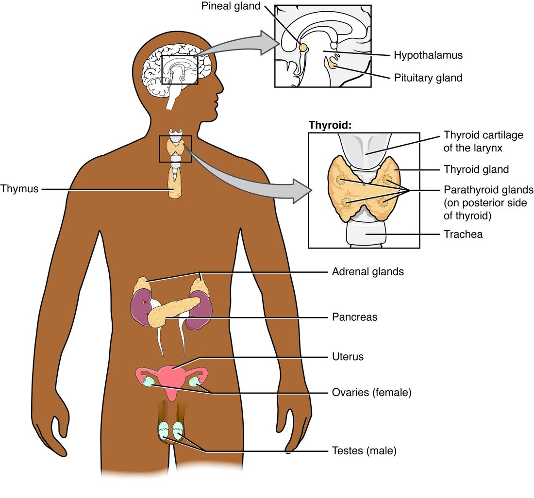
The hypothalamus, which you met with the brain, links the two control systems. Its neurons receive nerve signals like any other neurons, and some of them release hormones into the blood. The next topic follows those neurons into the pituitary gland.
Hormones regulate slow, widespread, lasting processes: growth and development, how your cells use fuel, the balance of water and salts, calcium in the blood, reproduction, and your response to stress. You compared nervous and endocrine signaling in Chemical signaling; this chapter adds the details.
Four chemical classes of hormones
Hormones are grouped by what they are built from (Figure 2).
- Amine hormones (amine = a molecule with a nitrogen-containing amino group) are single amino acids, chemically changed. Epinephrine and norepinephrine from the adrenal medulla are made from the amino acid tyrosine. The thyroid gland's hormones are also made from tyrosine, and a sleep-related hormone from the brain is made from another amino acid, tryptophan.
- Peptide hormones are short chains of amino acids joined by peptide bonds, from a few to a few dozen.
- Protein hormones are longer chains, from about 50 to about 200 amino acids, folded into a shape. Some carry sugar groups (glycoproteins). The line between a long peptide and a short protein is arbitrary, and the two behave alike.
- Steroid hormones are lipids built from cholesterol, with the four fused carbon rings of every steroid. The outer part of the adrenal gland, the ovaries and the testes make them. One group, the androgens (andr- = male, -gen = producing), are steroids that drive male characteristics such as a deeper voice and facial hair; the testes and the adrenal glands make them in both sexes.
For how a hormone behaves, one property matters more than its class: whether it dissolves in water or in lipid.
- Water-soluble hormones: all peptide and protein hormones, and epinephrine and norepinephrine.
- Lipid-soluble hormones: all steroid hormones, and one group of amines, the hormones of the thyroid gland, which are unusually hydrophobic for amines because they carry several large iodine-containing groups.
| Peptide and protein hormones | Steroid hormones | |
|---|---|---|
| Built from | Amino acids | Cholesterol |
| Solubility | Water-soluble | Lipid-soluble |
| How the gland stores it | Made ahead and stored in vesicles; released by exocytosis | Not stored; made on demand and diffuses out as soon as it is made |
| How it travels in blood | Mostly dissolved, free in the plasma | Mostly bound to carrier proteins |
| Half-life in blood | Short: minutes | Long: hours |
| Where its receptor protein is | In the plasma membrane | Inside the cell, in the cytosol or nucleus |
| What it changes | The activity of proteins the cell already has | Which genes are transcribed, so which proteins the cell makes |
| Speed of response | Seconds to minutes | Hours to days |
| Can it be taken as a tablet? | Usually not: digested in the gut like any protein | Yes: absorbed intact |
How hormones travel in blood
Plasma is mostly water. A water-soluble hormone simply dissolves in it. A lipid-soluble hormone does not dissolve well, so most of it rides on a carrier protein: a protein in the plasma that binds the hormone loosely and reversibly. (This is a different job from the membrane carrier proteins you met in facilitated diffusion.) Some carrier proteins bind one hormone tightly; others carry many lipid-soluble molecules loosely.
At any moment, a lipid-soluble hormone exists in two forms that swap back and forth:
- Bound hormone, attached to a carrier protein. For most lipid-soluble hormones this is more than 90% of the total.
- Free hormone, dissolved in the plasma. Only free hormone can leave the capillaries, reach the target cells and bind their receptor proteins. Only free hormone is active.
The bound form acts as a reservoir. As free hormone is used up or cleared, more comes off the carrier proteins, so the free level stays steady between bursts of secretion. Binding also shields the hormone from the enzymes of the liver and from filtering by the kidneys, which clear hormones from the blood.
Half-life
The half-life of a hormone is the time it takes for half of it to be removed from the blood. A short half-life means the level falls fast once the gland stops secreting; a long one means the effect lingers.
- Most peptide and protein hormones, and epinephrine, have half-lives of a few minutes. Epinephrine's is about two minutes.
- Lipid-soluble hormones, protected by carrier proteins, last hours; the thyroid gland's main hormone lasts about a week.
Worked example 1: how fast a hormone is cleared
Problem. A hormone has a half-life of 10 minutes. Its gland suddenly stops secreting when the blood level is 80 units. What is the level 30 minutes later?
- Count the half-lives. 30 minutes ÷ 10 minutes per half-life = 3 half-lives.
- Halve once per half-life. After the first: 80 ÷ 2 = 40 units.
- Second half-life. 40 ÷ 2 = 20 units.
- Third half-life. 20 ÷ 2 = 10 units.
- Check with the shortcut. After n half-lives, the fraction left is (1/2)n. (1/2)3 = 1/8, and 80 × 1/8 = 10 units.
Answer. 10 units, one eighth of the starting level.
Worked example 2: free and bound hormone
Problem. A steroid hormone's total blood concentration is 400 units, and 97% of it is bound to carrier proteins. How much is free? Then a drug displaces some hormone from the carrier proteins, so only 94% is bound. If the total has not yet changed, what is the free level now?
- Find the free fraction. 100% − 97% = 3% free.
- Free hormone before. 3% of 400 = 0.03 × 400 = 12 units.
- Free fraction after. 100% − 94% = 6% free.
- Free hormone after. 0.06 × 400 = 24 units.
Answer. Free hormone doubles, from 12 to 24 units, even though the bound percentage fell only from 97% to 94%. Because free hormone is the active part, a small change in binding can have a big effect. Over the following hours, the extra free hormone is cleared faster and, through negative feedback, the gland slows its secretion, so the free level drifts back toward normal.
How water-soluble hormones act
A water-soluble hormone cannot cross the plasma membrane. It binds a receptor protein on the outer surface of the target cell, and the signal crosses the membrane without the hormone. You saw the basic plan in Chemical signaling: hormone, G protein, second messenger. Here are the parts in full (Figure 3).
The cAMP pathway
- The hormone, the first messenger, binds its receptor protein in the plasma membrane.
- The receptor protein changes shape and switches on a G protein on the inner face of the membrane.
- The G protein switches on adenylyl cyclase (adenylyl = the adenine-containing part of ATP, cyclase = an enzyme that makes a ring), a membrane enzyme.
- Adenylyl cyclase turns ATP into cyclic AMP (cAMP), the second messenger.
- cAMP binds and switches on protein kinase A. A protein kinase (kin- = to move) is an enzyme that moves a phosphate group from ATP onto another protein, which is phosphorylation.
- Protein kinase A phosphorylates target proteins. Some of these are other kinases, which phosphorylate still more proteins: a phosphorylation cascade. Each phosphorylated enzyme or channel becomes more or less active, and the cell's behavior changes.
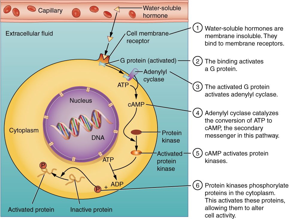
Every step multiplies the signal, which is the signal amplification you met in Chemical signaling. That is why a hormone at a billionth of a mole per liter can change a whole organ's behavior.
Switching the signal off
A signal that could not be stopped would be useless. Three things end it:
- Phosphodiesterase (PDE; phospho- = phosphate, di- = two, -esterase = an enzyme that splits a particular bond) breaks cAMP down to plain AMP, which cannot switch on protein kinase A. Theophylline, an older drug for narrowed airways, related to caffeine, blocks phosphodiesterases in airway smooth muscle, so cAMP lasts longer and the muscle stays relaxed.
- Phosphatases remove the phosphate groups the kinases added, returning the target proteins to their resting state.
- The G protein switches itself off within seconds, and the hormone lets go of the receptor protein as its level in the blood falls.
Other G proteins, other second messengers
The same receptor-plus-G-protein design is wired to different machinery in different cells:
- Inhibitory G proteins switch adenylyl cyclase off, so cAMP falls. You met one example: alpha-2 receptor proteins lower cAMP.
- The calcium pathway. Some G proteins switch on a different membrane enzyme, which splits a phospholipid in the plasma membrane into two second messengers. Inositol trisphosphate (IP3) diffuses into the cytosol and opens calcium channels in the endoplasmic reticulum, so calcium floods out; calcium then binds proteins that change the cell's activity. Diacylglycerol (DAG, di- = two, acyl = fatty acid chain, glycerol) stays in the membrane and, with the calcium, switches on another protein kinase. This is how alpha-1 receptor proteins make smooth muscle contract.
- Receptor proteins that are enzymes. Some protein hormones bind a receptor protein whose inner end is itself a kinase. Binding switches the kinase on directly, with no G protein. Several hormones that control growth and fuel storage work this way.
So the same second messenger can mean different things in different cells. cAMP speeds your heart and relaxes your airways; which one happens depends on which proteins that cell's kinases phosphorylate.
How lipid-soluble hormones act
A lipid-soluble hormone leaves its carrier protein, diffuses out of the capillary, and slips through the phospholipid bilayer of every cell it meets. (The thyroid gland's hormones are the exception: although lipid-soluble, they cross mainly on transporter proteins in the membrane, as the thyroid topic explains.) Only target cells hold an intracellular receptor protein for it (Figure 4).
- Free hormone diffuses through the plasma membrane.
- It binds its receptor protein in the cytosol or in the nucleus. A receptor protein in the cytosol then moves into the nucleus with the hormone attached.
- The hormone–receptor complex binds a hormone response element: a short, specific DNA sequence near the genes that hormone controls.
- The complex acts as a transcription factor, switching transcription of those genes on or off.
- The new mRNA is translated into new proteins, often enzymes, channels or transporters.
- The new proteins change the cell's activity.
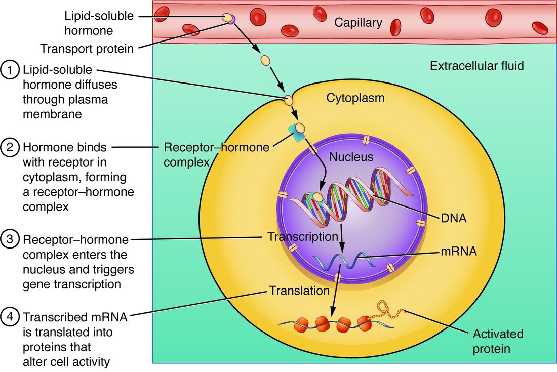
This explains the steroid tablet from the start of the page. Nothing happens for hours, because transcription and translation take time; and the effect lasts for days, because the new proteins last. It also explains why one steroid affects many tissues differently: each cell type has a different set of genes open to the hormone–receptor complex.
| Water-soluble hormone | Lipid-soluble hormone | |
|---|---|---|
| Classes | Peptides, proteins, epinephrine and norepinephrine | Steroids and the thyroid gland's amines |
| Receptor protein | In the plasma membrane | Inside the cell |
| Messenger inside the cell | A second messenger such as cAMP, IP3, DAG or calcium | None: the hormone–receptor complex itself acts on DNA |
| What changes | Existing proteins are switched on or off, often by phosphorylation | New proteins are made |
| Onset | Seconds to minutes | Hours |
| Duration | Minutes | Hours to days |
How target cells set their sensitivity
How strongly a target cell responds depends on three things:
- The level of free hormone in the blood around it.
- How many receptor protein molecules it carries. You met up-regulation and down-regulation in Chemical signaling. A hormone that stays high for a long time makes its target cells remove receptor protein molecules (down-regulation), so each unit of hormone does less. A hormone that stays low makes them add more (up-regulation).
- How tightly the receptor protein binds the hormone, and what it is linked to inside the cell.
Hormones can also change the receptor proteins for other hormones. That is one way hormones interact, covered at the end of this page.
What makes a gland release a hormone
Most hormones are released in pulses rather than at a steady rate, and the amount is set by negative feedback, which you met in Foundations. What starts the release falls into three types of stimulus (Figure 5):
- Humoral stimulus (humor = body fluid): a change in the level of an ion or nutrient in the blood acts on the gland directly. After a meal, the rise in glucose in the blood makes cells in the pancreas release a glucose-lowering hormone. A fall in blood calcium makes the four small glands on the back of the thyroid gland release a calcium-raising hormone.
- Hormonal stimulus: one hormone makes another gland release its hormone. The hypothalamus releases a hormone that makes the pituitary gland release a hormone, which in turn makes the thyroid gland release its own hormones. Chains like this are the subject of the next topic.
- Neural stimulus: nerve fibers trigger release. Sympathetic preganglionic fibers release acetylcholine onto the chromaffin cells of the adrenal medulla, which release epinephrine within seconds. This is the fastest route.
In each case, negative feedback usually stops the release: the hormone's own effect removes the stimulus. When glucose in the blood falls back, the pancreatic cells slow their secretion; when blood calcium rises back, the small glands on the thyroid gland slow theirs. A gland can also respond to more than one kind of stimulus: the hormone-secreting cells of the pancreas answer both the glucose level and autonomic nerves.
How hormones interact
A target cell is usually exposed to several hormones at once. Their effects combine in three standard ways:
- Permissive: one hormone must be present for another to have its full effect. The thyroid gland's hormones make cells build more beta adrenergic receptor proteins. With them present, epinephrine has its full effect; without them, it has much less. The first hormone "permits" the second.
- Synergistic (syn- = together, erg- = work): two hormones with the same effect produce more together than the sum of each alone. Epinephrine and a glucose-raising hormone from the pancreas both make the liver release glucose; together they raise glucose far more than either one's effect added up.
- Antagonistic (anti- = against, agon- = contest): two hormones have opposite effects on the same variable. One pancreatic hormone lowers the glucose in your blood and another raises it; a hormone from the four small glands on the thyroid gland raises blood calcium and one from the thyroid gland itself lowers it.
| Permissive | Synergistic | Antagonistic | |
|---|---|---|---|
| Rule | Hormone A lets hormone B work fully | A and B together give more than A + B separately | A and B push the same variable in opposite directions |
| Usual mechanism | A raises the number of B's receptor proteins or the machinery behind them | A and B act through different pathways on the same end point | A and B switch on opposing processes |
| Example | The thyroid gland's hormones and epinephrine | Epinephrine and a glucose-raising pancreatic hormone | The two pancreatic hormones that lower and raise glucose in the blood |
With these tools, every later topic in the chapter follows the same pattern: which gland, which stimulus, which class of hormone and so which mechanism, which targets, and which feedback stops it.