Sympathetic vs parasympathetic anatomy comes down to three questions: where the neurons leave the central nervous system, where they stop to relay, and how far they reach. This page answers each one. You will follow the two-neuron chain that every autonomic signal travels, trace the sympathetic division from the thoracic and lumbar spinal cord through the sympathetic chain and the collateral ganglia, see why the adrenal medulla counts as part of that division, and then follow the parasympathetic division out of the brainstem and sacral cord to ganglia sitting on the organs themselves.
The two-neuron chain
Touch a hot pan and pull your hand back. One motor neuron runs from your spinal cord all the way to the muscle in your arm. Now think about the sweat that breaks out on your palm a moment later. That signal also starts in your spinal cord, but it changes neurons partway out.
That is the defining feature of the autonomic motor pathway: two neurons in series between the central nervous system (CNS) and the effector. They meet at an autonomic ganglion (ganglion = knot or swelling), a cluster of neuron cell bodies outside the CNS.
- The preganglionic neuron (pre- = before) has its cell body in the brainstem or spinal cord. Its axon, the preganglionic fiber, leaves the CNS and ends in an autonomic ganglion.
- The postganglionic neuron (post- = after) has its cell body in that ganglion. Its axon, the postganglionic fiber, runs on to the effector: cardiac muscle, smooth muscle or a gland.
Preganglionic fibers are thinly myelinated. Postganglionic fibers are unmyelinated, so they conduct more slowly. Figure 1 shows the chain for both autonomic divisions.
Somatic versus autonomic motor pathways
You met the somatic and autonomic nervous systems as the two branches of the motor division. Their wiring differs at every step, as Figure 2 shows side by side.
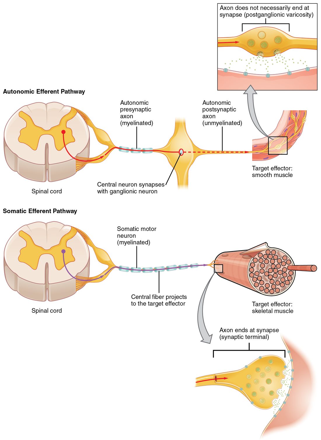
Look at the top inset. The postganglionic axon does not end in one terminal. It runs across the smooth muscle and bulges every few micrometers into swellings packed with vesicles. Each swelling is a varicosity (varix = a swollen vein), and transmitter released from it spreads to many nearby muscle cells. A somatic motor neuron, in contrast, ends at a single neuromuscular junction on each muscle fiber.
| Somatic motor pathway | Autonomic motor pathway | |
|---|---|---|
| Neurons from CNS to effector | One | Two: preganglionic and postganglionic, meeting in a ganglion |
| Where the CNS cell body sits | Ventral horn of the spinal cord, or a brainstem motor nucleus | Lateral horn of the spinal cord (T1–L2), sacral gray matter (S2–S4), or a brainstem nucleus |
| Effectors | Skeletal muscle | Cardiac muscle, smooth muscle, glands |
| Myelin | Heavily myelinated, fast | Thinly myelinated preganglionic fiber, unmyelinated postganglionic fiber, slower |
| Ending on the effector | One neuromuscular junction per muscle fiber | Varicosities strung along the axon |
| Transmitter at the effector | Acetylcholine | Acetylcholine or norepinephrine (next topic) |
| Effect on the effector | Always excites | Excites some effectors and inhibits others |
| If the nerve is cut | The muscle is paralyzed and wastes away | The effector keeps working on its own; the heart keeps beating and the gut keeps moving |
| Conscious control | Mostly voluntary | Mostly involuntary |
Sympathetic division: the thoracolumbar outflow
Every sympathetic preganglionic neuron has its cell body in the lateral horn of the spinal cord, in segments T1 to L2. No sympathetic fibers leave the brain, and none leave the cervical or sacral cord. That is why the sympathetic division is also called the thoracolumbar division (thorac/o = chest, lumb/o = loin).
The preganglionic axon leaves the cord in the ventral root, enters the spinal nerve, and within a centimeter or so turns off into a short connecting branch, the white ramus communicans (ramus = branch, communicans = connecting; plural white rami communicantes). It is white because its preganglionic fibers are myelinated. White rami exist only at T1–L2, because only those segments send preganglionic fibers out.
The sympathetic chain
The white ramus leads to the sympathetic chain (also called the sympathetic trunk): a string of ganglia joined by nerve fibers, one chain on each side of the vertebral column, running from the base of the skull to the coccyx. Its ganglia are the chain ganglia (also called paravertebral ganglia; para- = beside). There are about 22 to 23 on each side. Although fibers enter the chain only from T1–L2, the chain extends above and below, so every spinal nerve still gets sympathetic fibers. In the neck the cervical ganglia fuse into three, the largest being the superior cervical ganglion, which supplies the head.
A preganglionic fiber that enters the chain has three routes (Figure 3):
- Synapse at the same level. It synapses on a postganglionic neuron in the chain ganglion it enters.
- Travel up or down, then synapse. It runs up or down the chain and synapses in a ganglion at a higher or lower level. This is how the head and neck, and the sacral region, get sympathetic supply.
- Pass straight through. It leaves the chain without synapsing, in a splanchnic nerve (splanchn- = the internal organs), and synapses in a collateral ganglion in the abdomen.
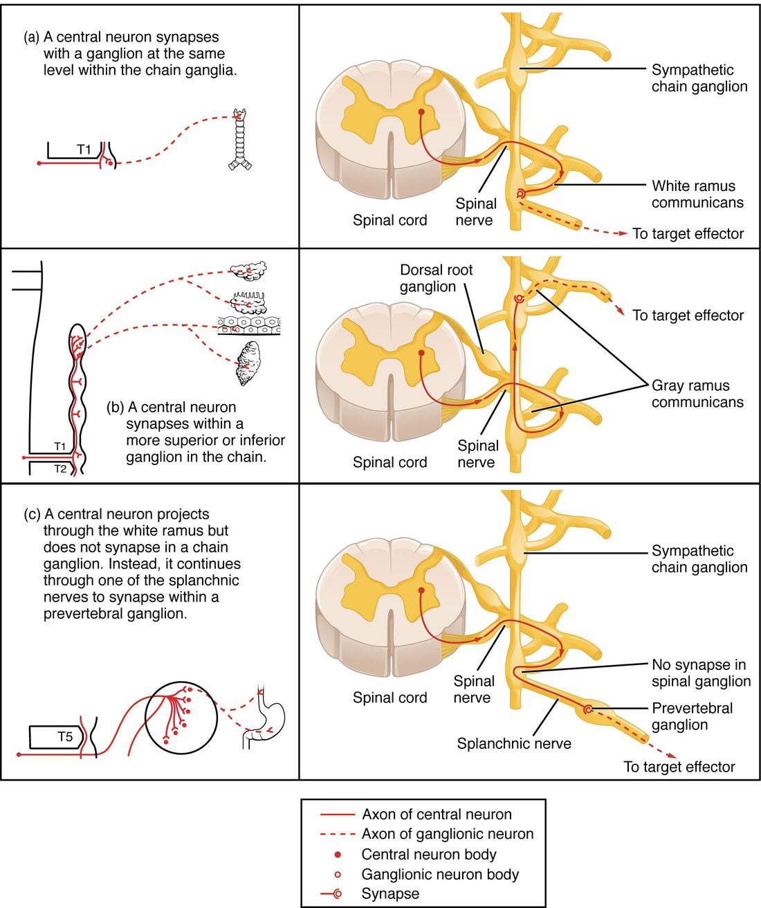
After a synapse in the chain, the postganglionic fiber has two ways out:
- Back into a spinal nerve through a gray ramus communicans (plural gray rami communicantes). It is gray because its postganglionic fibers are unmyelinated. Every spinal nerve, from C1 to the coccyx, has a gray ramus. These fibers ride the spinal nerve out to the skin and limbs, where they supply sweat glands, the arrector pili muscles and the smooth muscle of blood vessel walls.
- In separate sympathetic nerves that run straight from the chain to organs in the head and chest, such as the eye, the heart, the trachea and the lungs.
Here is the full route for the sweat on your palm: lateral horn cell body → ventral root → spinal nerve → white ramus → chain ganglion (synapse) → gray ramus → spinal nerve → sweat gland.
Collateral ganglia and splanchnic nerves
The abdominal and pelvic organs get sympathetic supply through a second set of ganglia. Collateral ganglia (also called prevertebral ganglia; pre- = in front of) are ganglia outside the chain that sit in front of the vertebral column, around the roots of the large arteries that branch from the body's main artery in the abdomen. They are named for those arteries:
- The celiac ganglion (celi- = belly; often a pair, one on each side of its artery) supplies the stomach, liver, pancreas and spleen.
- The superior mesenteric ganglion (mes- = middle, enter- = intestine) supplies most of the intestines.
- The inferior mesenteric ganglion supplies the last part of the colon, the rectum, the urinary bladder and the reproductive organs.
Preganglionic fibers reach them through the splanchnic nerves. The greater splanchnic nerve (from about T5–T9) runs mainly to the celiac ganglion, and the lesser splanchnic nerve (about T10–T11) to the superior mesenteric ganglion. Lumbar splanchnic nerves reach the inferior mesenteric ganglion. Figure 4 puts the whole division together.
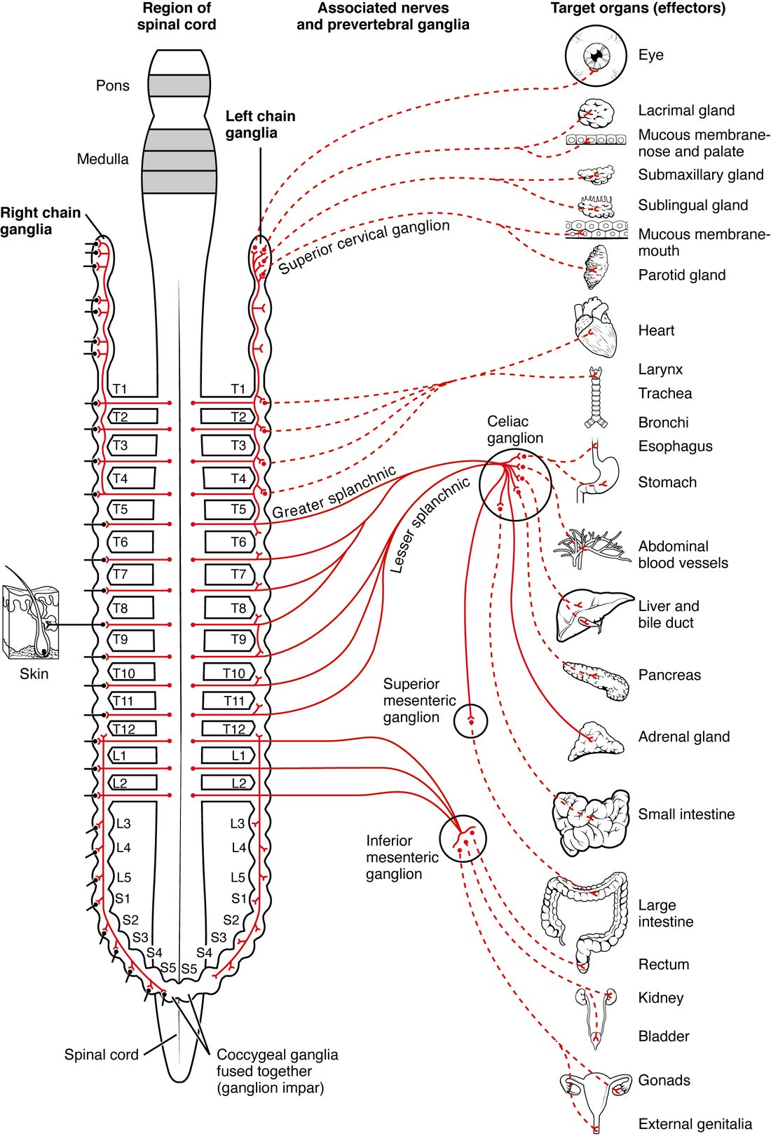
Two features make sympathetic output widespread. First, the ganglia sit near the spinal cord, so preganglionic fibers are short and postganglionic fibers are long. Second, one preganglionic neuron branches to synapse on many postganglionic neurons, often ten or more, spread over several chain levels. This branching is called divergence. A single burst from the lateral horn therefore reaches many organs at once.
The adrenal medulla
A small group of preganglionic fibers takes a route with no second neuron at all. These fibers run through the chain and a splanchnic nerve, pass through the celiac ganglion without synapsing, and end directly on cells in the center of the adrenal gland.
The adrenal medulla (ad- = near, ren- = kidney; medulla = inner part) is the inner core of each adrenal gland, which sits on top of the kidney. Its secreting cells are chromaffin cells (chrom- = color, affin- = attracted to; they stain brown with chromium salts). Chromaffin cells develop from the same embryonic tissue as postganglionic neurons, but they have no axons. When preganglionic fibers stimulate them, they release their product into the blood instead of onto a target. The adrenal medulla is an endocrine gland that behaves like a sympathetic ganglion.
About 80 percent of what chromaffin cells release is epinephrine (epi- = upon, nephr- = kidney; also called adrenaline, from the Latin for the same idea), and about 20 percent is norepinephrine. Epinephrine is a hormone: carried in the blood, it reaches cells all over the body, including cells no sympathetic fiber touches, and it stays in the blood for a few minutes after the nerve signal stops. The path runs: lateral horn → ventral root → white ramus → through the chain → splanchnic nerve → through the celiac ganglion → chromaffin cells → epinephrine into the blood.
Parasympathetic division: the craniosacral outflow
Parasympathetic preganglionic neurons sit at the two ends of the CNS, which is why this is the craniosacral division (crani/o = skull, sacr/o = sacrum):
- Cranial outflow: nuclei in the brainstem send preganglionic fibers out in four cranial nerves, III, VII, IX and X.
- Sacral outflow: neurons in the gray matter of cord segments S2–S4 send preganglionic fibers out in the ventral roots and then in the pelvic splanchnic nerves.
Parasympathetic preganglionic fibers run all the way to terminal ganglia: ganglia that sit close to the target organ or inside its wall. Those inside the wall are called intramural ganglia (intra- = within, mur- = wall). So parasympathetic preganglionic fibers are long and postganglionic fibers are very short, the reverse of the sympathetic pattern. Each preganglionic neuron contacts only a few postganglionic neurons, so parasympathetic effects stay more local. There is no parasympathetic chain, and parasympathetic fibers do not travel in the gray rami, so they do not reach the limbs, the skin or the walls of most blood vessels.
Figure 5 maps the four cranial nerves and the sacral outflow.
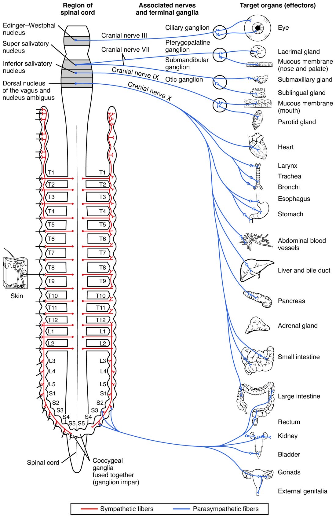
| Nerve | Terminal ganglion | What it supplies |
|---|---|---|
| III, oculomotor | Ciliary ganglion, behind the eye | The ring of iris muscle that narrows the pupil; the ciliary body muscle that focuses for near vision |
| VII, facial | Pterygopalatine and submandibular ganglia | The lacrimal gland (tears), glands of the nose and palate, and two of the glands that make saliva |
| IX, glossopharyngeal | Otic ganglion, below the skull near the ear | The largest gland that makes saliva, in front of the ear |
| X, vagus | Terminal and intramural ganglia in the organ walls | Heart, lungs and airways, esophagus, stomach, liver, pancreas, and the intestines as far as about two thirds of the way along the part of the colon that crosses the abdomen |
| S2–S4, pelvic splanchnic nerves | Intramural ganglia in the pelvic organs | The rest of the colon (the last third of the part that crosses the abdomen, and all of the colon that runs down the left side), the rectum, the urinary bladder and the reproductive organs |
The vagus nerve alone carries about three quarters of all parasympathetic preganglionic fibers.
Sympathetic vs parasympathetic: the anatomy side by side
| Sympathetic division | Parasympathetic division | |
|---|---|---|
| Other name | Thoracolumbar division | Craniosacral division |
| Where preganglionic cell bodies sit | Lateral horn, T1–L2 | Brainstem nuclei (with nerves III, VII, IX, X) and sacral gray matter, S2–S4 |
| Where the ganglia sit | Near the spinal cord: chain ganglia and collateral ganglia | Near or inside the target organ: terminal and intramural ganglia |
| Preganglionic fiber | Short | Long |
| Postganglionic fiber | Long | Short |
| Branching (divergence) | High: one preganglionic neuron reaches many postganglionic neurons | Low: one preganglionic neuron reaches a few |
| Reach | Whole body, including skin, limbs and blood vessel walls | Head, and the organs of the chest, abdomen and pelvis only |
| Link to a hormone gland | Adrenal medulla, stimulated directly by preganglionic fibers | None |
| Resulting pattern of output | Widespread, many organs at once | Local, organ by organ |
Both divisions reach most organs of the chest and abdomen. The next topic explains the transmitters and receptor proteins that let the same organ respond to each division differently.
Common mix-ups
- "Sympathetic fibers come from the whole spinal cord." The fibers are distributed at every level, through the chain and the gray rami, but they leave the CNS only from T1–L2.
- "The white ramus carries postganglionic fibers." White rami carry myelinated preganglionic fibers into the chain; gray rami carry unmyelinated postganglionic fibers back out.
- "Parasympathetic fibers only travel in the vagus nerve." The vagus carries most of them, but nerves III, VII and IX and the sacral outflow carry the rest.