Your eye works like a camera only in outline: a clear front bends light into a sharp image, and a sheet of light-sensitive cells at the back turns that image into nerve signals. This page covers eye anatomy and how vision works, in the order light meets each part: the structures that protect and move the eye, the three layers of its wall, the lens and the fluids inside it, how the eye focuses and why it sometimes cannot, how rods and cones turn light into a signal, and the pathway that carries that signal to your visual cortex. Along the way you will see why a tumor behind the eyes steals the outer half of both visual fields, and why a pupil that ignores light is an emergency.
Accessory structures of the eye
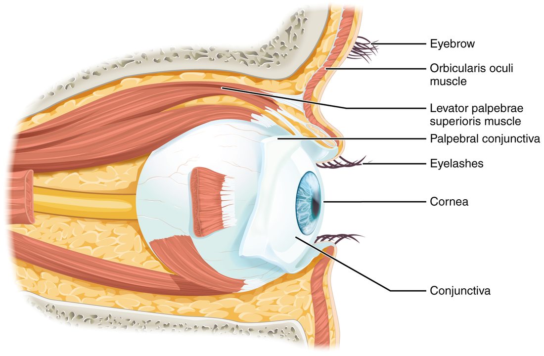
Before light reaches the eyeball, it passes a set of protective and supporting parts. Together these are the accessory structures of the eye: the eyelids, the conjunctiva, the tear-making lacrimal apparatus and the muscles that aim the eye. The eyeball itself sits in the bony orbit, cushioned by fat (Figure 1).
Eyelids
Each eyelid (palpebra; palpebr- = eyelid) is a fold of skin stiffened by a plate of dense connective tissue. Blinking spreads tears across the eye every few seconds and shuts out dust and bright light.
- The levator palpebrae superioris (levator = lifter, palpebrae = of the eyelid, superioris = upper) raises the upper eyelid. The oculomotor nerve (III) supplies it, which is why oculomotor damage makes the lid droop. A thin sheet of smooth muscle under sympathetic control helps hold the lid up too.
- The orbicularis oculi, the ring of muscle around the eye, closes the lids. The facial nerve (VII) supplies it.
Conjunctiva
The conjunctiva (con- = together, junct- = joined) is a thin, clear mucous membrane. It lines the inside of the eyelids and folds back to cover the white of the eye. It stops at the edge of the cornea, the clear window at the front, so it never covers the part you see through. Its goblet cells add mucus to the tear film. When its tiny blood vessels widen, as in conjunctivitis ("pink eye"), the white of the eye turns red.
The lacrimal apparatus and tear flow
The lacrimal gland (lacrim- = tear) is an exocrine gland about the size of an almond, tucked into the upper outer corner of each orbit. Several small lacrimal ducts carry its watery secretion, the tears, onto the surface of the eye under the upper lid. Parasympathetic fibers carried by the facial nerve drive the gland, which is why irritating the eye or crying makes tears pour.
Tears then take a fixed route (Figure 2):
- The lacrimal gland releases tears onto the upper outer part of the eye.
- Each blink sweeps them across the eye toward the inner corner, near your nose.
- They drain into two tiny openings, the lacrimal puncta (punctum = point), one on the edge of each lid at the inner corner.
- Short channels, the lacrimal canaliculi (canaliculus = little channel), carry them to the lacrimal sac.
- The nasolacrimal duct carries them down through the nasolacrimal canal of the skull into the nose, below the lowest nasal concha.
That last step is why crying gives you a runny nose, and why an eye drop can leave a taste in your throat. In everyday speech, "tear duct" usually means this drainage route.
Extrinsic eye muscles
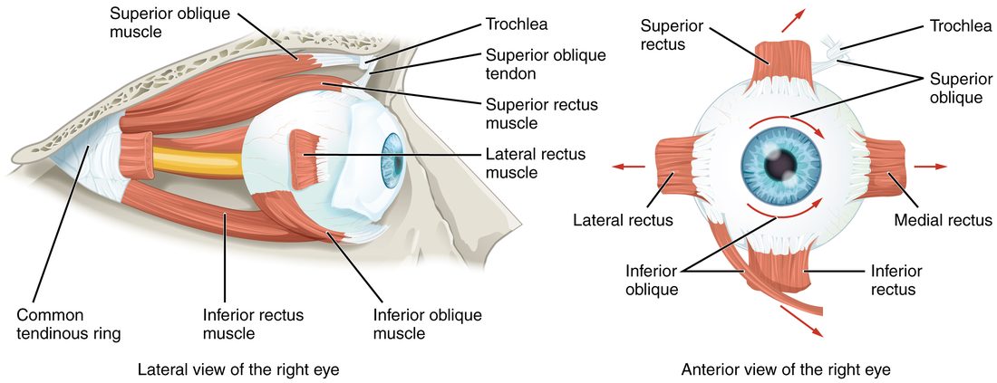
Six skeletal muscles attach to the outside of each eyeball and turn it. They are the extrinsic eye muscles, also called the extraocular muscles (extra- = outside, ocul- = eye). Four are straight muscles, the recti (rectus = straight), which run forward from a common tendon ring at the back of the orbit to the four sides of the eye. Two are oblique muscles, which approach the eye at a slant (Figure 3).
| Muscle | Main movement of the eye | Nerve |
|---|---|---|
| Superior rectus | Up | Oculomotor (III) |
| Inferior rectus | Down | Oculomotor (III) |
| Medial rectus | In, toward the nose | Oculomotor (III) |
| Lateral rectus | Out, away from the nose | Abducens (VI) |
| Superior oblique | Down, most strongly when the eye is turned in; also rotates the top of the eye inward | Trochlear (IV) |
| Inferior oblique | Up, most strongly when the eye is turned in; also rotates the top of the eye outward | Oculomotor (III) |
The superior oblique is the odd one. Its tendon runs forward to the trochlea, a small loop of cartilage at the upper inner edge of the orbit, bends back through it like a rope through a pulley, and attaches to the top of the eye behind its equator. Pulling on it therefore tips the front of the eye downward. You use it to look down while your eyes are turned in: reading a book in your lap, or looking at your feet on the stairs. A person with a trochlear nerve injury has double vision in exactly those moments.
A clinician tests the muscles by asking you to follow a finger in an "H" pattern. Looking straight out to the side tests the lateral and medial recti. Looking up or down with the eye turned out isolates the superior and inferior recti; looking up or down with the eye turned in isolates the two obliques.
The three layers of the eyeball
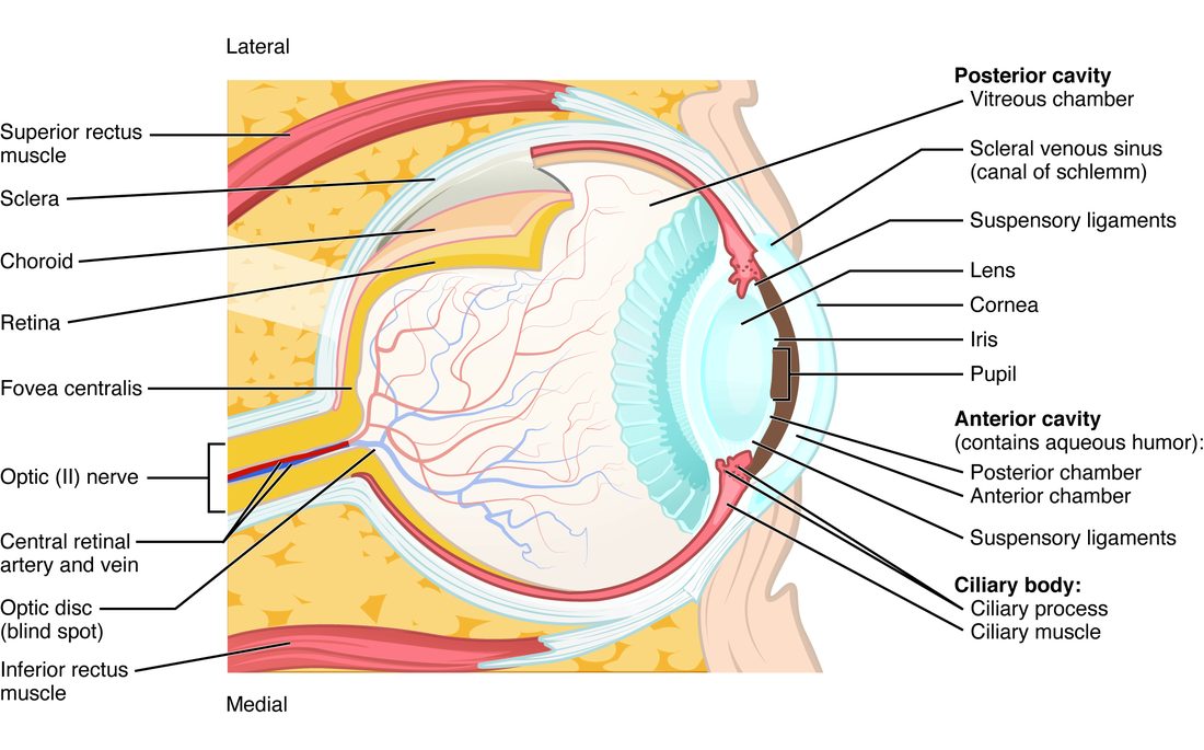
The wall of the eyeball has three layers, or tunics (tunica = coat), from outside in (Figure 4).
The fibrous tunic: sclera and cornea
The fibrous tunic is the tough outer coat of dense connective tissue.
- The sclera (scler- = hard) is the white of the eye and covers most of the eyeball. Its collagen fibers run in a tangle, which makes it strong and opaque. The extraocular muscles attach to it.
- The cornea (corne- = horn) is the clear front sixth. Its collagen fibers are laid in regular sheets, and it has no blood vessels, both of which keep it transparent. Oxygen reaches it by diffusion from the tear film and the fluid behind it. The cornea is packed with pain-sensing nerve endings from the ophthalmic division of the trigeminal nerve, so a speck of dust on it feels huge and makes you blink.
The vascular tunic: choroid, ciliary body and iris
The vascular tunic is the middle layer, rich in blood vessels and dark pigment.
- The choroid (chori- = membrane) lines the back of the eye. Its blood vessels feed the outer part of the retina, and its melanin absorbs light that has passed through the retina, so the light does not scatter around inside the eye and blur the image.
- The ciliary body (cili- = eyelash, for its fringe of folds) is a thickened ring where the choroid meets the iris. It holds the ciliary muscle, a ring of smooth muscle that changes the shape of the lens, and folds called ciliary processes, which secrete the fluid that fills the front of the eye.
- The iris (iris = rainbow) is the colored ring you see through the cornea. Its hole is the pupil. The iris holds two smooth muscles: a circular sphincter muscle that makes the pupil smaller, supplied by parasympathetic fibers of the oculomotor nerve, and a set of radial dilator fibers that make it larger, supplied by sympathetic fibers. Eye color depends on how much melanin the iris holds and how its tissue scatters light.
The neural tunic: retina
The neural tunic is the retina (rete = net), the inner layer. It has two parts. A single layer of pigmented epithelium lies against the choroid. Inside it lies the neural retina, a thin sheet of neurons including the light-sensitive cells. The two parts are only loosely attached; in a retinal detachment they separate, and the detached area stops seeing. The retina is covered in its own sections below.
The lens and the humors
The lens is a clear, flexible disc that sits just behind the iris. It is built of long cells packed with clear proteins, and it has no blood vessels or nerves. A ring of fine threads, the zonule fibers (zonula = little belt; also called the suspensory ligaments), holds it to the ciliary body all around its rim.
The lens divides the inside of the eye into two spaces filled with fluid, the humors (humor = fluid):
- In front of the lens is the aqueous humor (aqua = water), a clear, watery fluid. The ciliary processes secrete it into the posterior chamber, the narrow space between the iris and the lens. It flows through the pupil into the anterior chamber, between the iris and the cornea, and drains out at the angle where the iris meets the cornea, through a spongy meshwork into the scleral venous sinus (canal of Schlemm), and from there into veins. It is made and drained all the time, replacing itself every couple of hours. It feeds the lens and cornea, which have no blood supply, and its pressure, normally about 10 to 21 mmHg, keeps the front of the eye in shape.
- Behind the lens is the vitreous humor (vitr- = glass), a clear gel that fills the large posterior cavity. It presses the retina against the choroid. You form it before birth and do not replace it; the specks called floaters are clumps in it.
Glaucoma
Glaucoma (glauc- = gray-blue, -oma = mass or condition) is damage to the optic nerve, most often linked to aqueous humor that drains too slowly. Fluid is made at the normal rate but backs up, so the pressure inside the eye rises and presses on the delicate axons leaving the eye at the back. Axons die and do not regrow. The loss usually starts at the edges of vision, so the most common form is painless and is often not noticed until much is gone. In a less common form, the iris is pushed against the drainage angle and blocks it suddenly: the pressure shoots up, the eye is red and painful, and it is an emergency.
Cataract
A cataract (Greek for waterfall) is a clouding of the lens. With age, sunlight and smoking, the lens proteins change shape and clump, and light scatters instead of passing straight through. Vision becomes hazy and glare from headlights worsens. Surgeons remove the clouded lens and put a plastic one in its place.
Focusing light: refraction
Put a straw in a glass of water and it looks bent where it enters the water. Light changes speed as it passes from one clear material into another, and if it crosses the boundary at a slant, it changes direction. This bending is refraction (re- = back, fract- = break). A curved clear surface bends parallel rays toward one point, the focus.
Your eye has two refracting parts:
- The cornea does about two thirds of the bending, because the jump from air into the cornea is the largest change in the path. Its power is fixed.
- The lens does the remaining third, and it is the only part whose power can change.
For a sharp image, the rays from each point of an object must meet exactly on the retina. The image formed there is upside down and reversed left to right, as in any camera. Your brain never "flips" it; the visual cortex simply maps each spot of the retina to a place in the scene.
Accommodation: focusing near and far
Light from a distant object arrives as nearly parallel rays. Light from a near object, like your phone, arrives spreading out, so it needs more bending to meet on the retina. Accommodation (accommodare = to adjust) is the change in the lens's shape that adds that bending.
It works in a way that surprises most students: the lens rounds up when its supports go slack.
- Far vision. The ciliary muscle is relaxed. The ring it forms is wide, so the zonule fibers are pulled taut and stretch the lens thin and flat. A flat lens bends light least.
- Near vision. Parasympathetic fibers of the oculomotor nerve make the ciliary muscle contract. Because it is a ring, contracting makes it smaller, and it moves in toward the lens. The zonule fibers go slack.
- The lens rounds up. Released from tension, the elastic lens bulges into a rounder shape. A rounder lens bends light more, and the near image falls on the retina.
The near response
When you look from a far object to a near one, three changes happen together. This is the accommodation–convergence reflex, also called the near response:
- Accommodation: the lens rounds up.
- Convergence: both medial recti contract and the eyes turn in, so the near object falls on the matching spot of each retina.
- Pupil constriction: the pupil narrows, which blocks the blurred edges of the light beam and deepens the range that is in focus.
All three are driven by the oculomotor nerve. Reading for hours tires the eyes because the ciliary muscles and medial recti stay contracted the whole time.
Visual acuity and the Snellen chart
Visual acuity (acu- = sharp) is how fine a detail you can make out. It is measured with a Snellen chart, the eye chart with rows of letters that shrink toward the bottom. You stand 20 feet (6 meters) away, which is far enough that a normal eye needs almost no accommodation, and read the smallest row you can.
Acuity is written as a fraction:
- The top number is your distance from the chart, almost always 20 feet.
- The bottom number is the distance at which a person with normal vision can read the smallest row you read.
Worked example: reading a Snellen result
Problem. Standing 20 feet from the chart, Priya can read the row labeled 40 but no smaller. Her friend reads the row labeled 15. What are their acuities, and what do they mean?
- Write the top number. Both stood 20 feet away, so both fractions start 20/.
- Write the bottom number. Each row is labeled with the distance at which a normal eye can just read it. Priya's smallest row is the 40 row, so she is 20/40. Her friend is 20/15.
- Interpret Priya's. 20/40 means Priya must stand at 20 feet to read letters a normal eye can read from 40 feet. Her letters must be twice as large (40 ÷ 20 = 2) as a normal eye needs. Her acuity is worse than normal.
- Interpret her friend's. 20/15 means her friend reads from 20 feet what a normal eye must come to 15 feet to read. Her acuity is better than normal.
Answer. Priya 20/40 (worse than normal), her friend 20/15 (better than normal). The larger the bottom number, the worse the acuity. 20/20 is normal; in the United States, a best-corrected acuity of 20/200 or worse in the better eye is one definition of legal blindness.
Focusing errors: myopia, hyperopia and presbyopia
A child who reads comfortably but squints at the board, a grandparent who holds the menu at arm's length: both have focusing errors, but of different kinds. Figure 5 compares them.
- Myopia (my- = to close, -opia = vision, from squinting), or nearsightedness. The eyeball is usually too long, or the cornea too steeply curved, so light from distant objects focuses in front of the retina. Near objects need more bending anyway, so they still focus. A concave (diverging) lens spreads the rays slightly before they enter the eye and moves the focus back onto the retina.
- Hyperopia (hyper- = beyond), or farsightedness. The eyeball is too short, so light would focus behind the retina. A young person can make up for mild hyperopia by accommodating all the time, which strains the eyes; near objects, which need the most bending, blur first. A convex (converging) lens adds bending.
- Presbyopia (presby- = old man). With age, the lens keeps adding cells and grows stiffer, so it no longer rounds up when the zonule fibers go slack. The nearest point you can focus on moves out from about 7 to 10 cm in childhood and early adulthood to arm's length or beyond by your mid-40s to 50s. Distant vision is unaffected. Convex reading glasses supply the bending the lens can no longer add.
| Myopia | Hyperopia | Presbyopia | |
|---|---|---|---|
| Everyday name | Nearsightedness | Farsightedness | Age-related loss of near focus |
| Cause | Eyeball too long, or cornea too curved | Eyeball too short, or cornea too flat | Lens stiffens and cannot round up |
| Where distant light focuses | In front of the retina | Behind the retina, unless the lens accommodates | On the retina (unaffected) |
| What blurs | Distant objects | Near objects first; far too if severe | Near objects |
| Usual age of onset | Childhood and teens | Present from birth | Mid-40s onward |
| Correcting lens | Concave (diverging) | Convex (converging) | Convex, for near work only |
The retina: from photoreceptors to ganglion cells
The neural retina is a stack of three main layers of neurons, and the light-sensitive cells are at the back. Light entering the eye passes through the other two layers, which are nearly transparent, before it reaches them.
- Photoreceptors (photo- = light) are the sensory receptor cells for light, lying against the pigmented epithelium. Unlike taste cells, rods and cones are specialized neurons. Even so, they make no action potentials: like the separate sensory receptor cells of sensory pathways, they pass their signal on at a synapse. There are two kinds, rods and cones. They release the neurotransmitter glutamate onto the next layer.
- Bipolar cells (bi- = two, polar = ends) carry the signal from the photoreceptors toward the front of the retina. Like the photoreceptors, they produce graded potentials, not action potentials.
- Retinal ganglion cells are the output neurons. They are the first cells in the pathway to fire action potentials, and their axons run across the inner surface of the retina to one spot, where they leave the eye together as the optic nerve.
Two further kinds of neuron, horizontal cells and amacrine cells, spread sideways between neighbors. They sharpen contrast at edges, so the signal leaving the eye has already been processed.
The fovea and the optic disc
Two spots on the retina matter clinically:
- The fovea (fovea = small pit), or fovea centralis, lies at the center of the back of the eye, in the middle of a yellowish area called the macula lutea. It holds only cones, packed tightly, and the overlying bipolar and ganglion cells are pushed aside, so light reaches the cones directly. It is the spot of sharpest vision; when you look straight at something, you are aiming its image at your foveae.
- The optic disc is where the ganglion cell axons leave the eye and the central retinal blood vessels enter. It has no photoreceptors, so it produces a blind spot in each eye's field of view. You do not notice it, because each eye's blind spot falls on a part of the scene the other eye sees, and your brain fills in the gap.
Find both on Figure 4. The optic disc lies about 15 degrees toward the nose from the fovea, which is why the blind spot is in the outer part of each eye's view.
Rods and cones
Walk from a sunny street into a dim cinema and for several minutes you can barely see; later you can find your seat but cannot tell the color of your ticket. You have switched from one kind of photoreceptor to the other.
- Rods (rod photoreceptors) are named for their long, thin outer segments. Their pigment is rhodopsin (rhod- = rose, opsin = seeing, for its purplish color). They are very sensitive: in the dark, a rod can respond to a single photon. They work in dim light and give you night vision without color.
- Cones (cone photoreceptors) have shorter, tapered outer segments. Each makes one of three pigments, so there are three kinds of cone. They need brighter light, and they give you color and fine detail.
| Rods | Cones | |
|---|---|---|
| Number in each retina | About 90 million (older texts say 120 million) | About 4.5 million (older texts say 6 million) |
| Where most are | Outside the fovea, densest in a ring about 20 degrees out from it, thinning toward the edges | Concentrated in the fovea; none elsewhere at such density |
| Light needed | Dim light (night vision) | Bright light (day vision) |
| Pigment | One: rhodopsin | Three kinds, one per cone type |
| Color vision | No | Yes |
| How many feed one ganglion cell | Many (strong convergence) | Few; one in the fovea |
| Result | High sensitivity, low acuity | Low sensitivity, high acuity |
The "result" row follows from convergence, which you met in neural circuits. When many rods feed one ganglion cell, their small signals add up, so faint light can still fire it; but the brain cannot tell which of those rods caught the light, so detail is lost. A foveal cone has its own bipolar cell and ganglion cell, so its signal reports one tiny spot. That is why you see a faint star better by looking slightly to one side of it: you move its image off the cone-only fovea onto rods.
Color vision
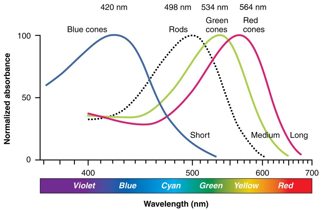
The three kinds of cone each respond best to a different part of the spectrum (Figure 6). Short-wavelength cones peak near 420 nm (violet-blue), medium-wavelength cones near 534 nm (green) and long-wavelength cones near 564 nm (yellow-green), despite being called "red" cones. Rods peak near 498 nm. The curves overlap widely, so any color excites two or three cone types. Your brain works out the color from the ratio of their activity: yellow light, for example, excites long-wavelength cones strongly and medium-wavelength cones somewhat, and almost no short-wavelength cones.
Color blindness is a reduced ability to tell some colors apart, because one kind of cone pigment is missing or altered. The most common kind is red–green: the medium- or long-wavelength pigment is missing or shifted, and reds, oranges and greens look alike. The genes for those two pigments sit on the X chromosome. A male has only one X, so one faulty copy is enough to change his color vision; a female usually has a working copy on her other X. Red–green color blindness therefore affects about 1 in 12 males of northern European ancestry but only about 1 in 200 females.
Phototransduction: how light becomes a signal
Phototransduction is the conversion of light into a change in a photoreceptor's membrane potential. It runs backward from what you might expect. In the dark, photoreceptors are depolarized and release neurotransmitter all the time. Light hyperpolarizes them and cuts their release. Figure 7 sets the two states side by side for a rod.
The pigment
A rod's outer segment is a stack of about a thousand flattened membrane discs, and each disc is packed with rhodopsin. Rhodopsin has two parts:
- Opsin, a protein that spans the disc membrane. It belongs to the same family as the G protein–coupled receptor proteins you met with chemical signaling.
- Retinal, a small light-absorbing molecule made from a vitamin in your diet, which sits in a pocket of the opsin. In the dark it has a kink in its carbon chain; this bent form is called 11-cis-retinal.
When retinal absorbs a photon, the kink straightens and it becomes all-trans-retinal. This change of shape caused by light is photoisomerization (photo- = light, iso- = same, mer- = part: the same atoms in a new arrangement). It is the only step in vision that needs light. Everything after it is chemistry. Cone pigments work the same way; each kind has retinal attached to a slightly different opsin, which sets the wavelength it absorbs best.
In the dark
- The outer segment holds a high level of cyclic GMP (cGMP), a second messenger related to cAMP.
- cGMP holds open cation channels in the outer segment membrane. Na+ and some Ca2+ flow in steadily. This inward flow is called the dark current.
- The dark current keeps the rod depolarized, at about −40 mV instead of the −70 mV typical of a neuron at rest.
- The depolarized rod releases glutamate steadily onto its bipolar cells.
In the light
- A photon straightens retinal from 11-cis to all-trans. The opsin changes shape and rhodopsin becomes active.
- Active rhodopsin switches on many molecules of a G protein called transducin.
- Transducin switches on an enzyme that breaks down cGMP, and cGMP falls.
- With less cGMP, the cation channels close, and the dark current stops.
- K+ still leaks out, so the rod hyperpolarizes toward −70 mV.
- The hyperpolarized rod releases less glutamate. The change in glutamate is the signal: bipolar cells respond to it, and the ganglion cells change their firing.
The cascade amplifies: one active rhodopsin switches on dozens of transducin molecules, and each enzyme they switch on breaks down many cGMP molecules. That is how a single photon can close enough channels to change a rod's voltage.
Bleaching, recovery and dark adaptation
After absorbing light, all-trans-retinal comes away from its opsin. The pigment has been "bleached" and cannot respond again until it is rebuilt. The loose retinal moves to the pigmented epithelium, where enzymes put the kink back, and it returns to rejoin an opsin. In bright daylight rods are saturated: nearly all their cation channels are already closed, so more light cannot change their signal, and cones do most of the work.
Step into the dark and rhodopsin is gradually rebuilt. Cone sensitivity recovers within about 10 minutes, rod sensitivity over 20 to 30 minutes or more; this slow gain is dark adaptation. The reverse, light adaptation, takes only seconds to a minute or two. Because retinal is made from a dietary vitamin, a severe shortage of that vitamin causes night blindness: too little rhodopsin for rods to work in dim light.
The visual pathway
Hold a finger up in front of you, slightly to your right. Light from it lands on the left half of each retina: the outer (temporal) half of your left eye, and the inner (nasal) half of your right eye. The visual pathway sorts signals so that everything you see to your right ends up in your left hemisphere, and everything to your left in your right hemisphere (Figure 8).
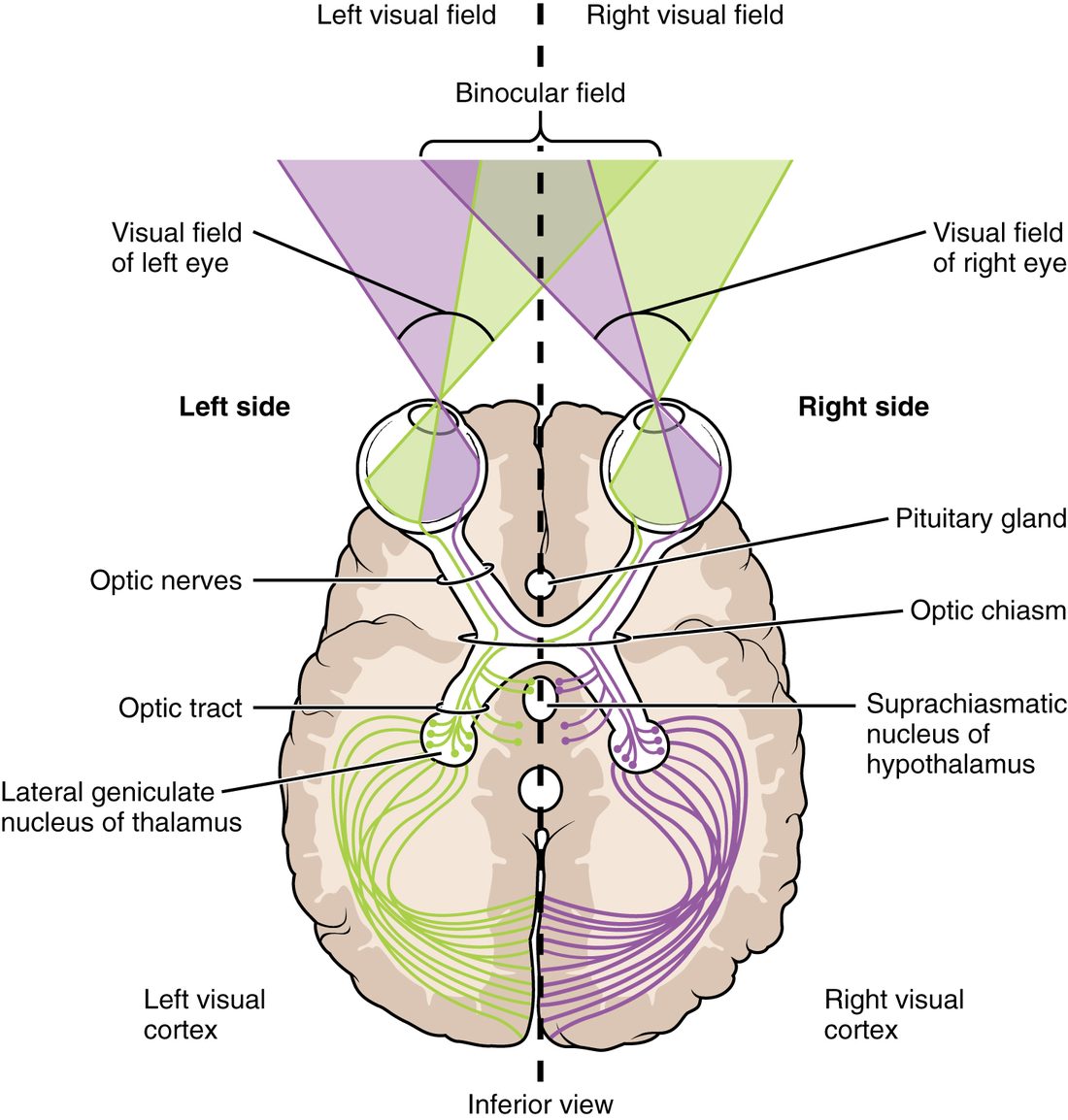
- Optic nerve. The ganglion cell axons of each eye leave at the optic disc and run back through the optic canal as the optic nerve.
- Optic chiasm. The two optic nerves meet just above the pituitary gland, in front of its stalk, at the optic chiasm (chiasm = cross, from the Greek letter chi, X). Axons from the nasal half of each retina cross to the other side here. Axons from the temporal half stay on their own side.
- Optic tract. Behind the chiasm, each optic tract carries axons from the temporal half of the same eye and the nasal half of the other eye. Those two halves see the same side of the world: the left optic tract carries everything in your right visual field, the whole area you can see without moving your eyes.
- Lateral geniculate nucleus. Most optic tract axons synapse in the lateral geniculate nucleus (genicul- = little knee, for its bent shape) of the thalamus, the relay you would expect for a sense that reaches the cortex.
- Visual cortex. Axons from the lateral geniculate nucleus fan out as the optic radiation to the primary visual cortex of the occipital lobe, which maps the opposite visual field point by point.
Some axons leave the pathway before the thalamus. At the chiasm, a few go up to the hypothalamus, where light resets the daily sleep–wake rhythm. From the optic tract, others go to the superior colliculi, which turn your eyes and head toward sudden movement, and to the midbrain area that controls the pupils.
Where the pathway is damaged, the field loss tells you
Because each part of the pathway carries a known part of the field, the pattern of loss points to the site of damage (Figure 9). A loss of half the visual field is called a hemianopia (hemi- = half, an- = without, -opia = vision).
| Site of damage | Fibers cut | Field loss | Typical cause |
|---|---|---|---|
| One optic nerve | All fibers from that eye | That eye blind; the other eye normal | Injury, inflammation of the nerve, glaucoma (gradually) |
| Middle of the optic chiasm | The crossing fibers from both nasal retinas | Outer (temporal) half of each eye's field: bitemporal hemianopia | A pituitary tumor pressing up from below |
| One optic tract, optic radiation or visual cortex | Everything carrying the opposite visual field | The opposite half of the field in both eyes: homonymous hemianopia | Stroke, tumor |
A person with bitemporal hemianopia often first notices bumping into doorframes or cars appearing suddenly from the side. A person with a right occipital stroke may miss food on the left side of the plate and bump into things on the left, until they learn to turn their head toward the blind side.
The pupillary light reflex
Shine a penlight into one eye and both pupils narrow within a second. The narrowing of the lit pupil is the direct response; the narrowing of the other one is the consensual response. This is the pupillary light reflex, a visceral reflex with this arc:
- Sensory receptor cells: photoreceptors and some light-sensitive ganglion cells in the retina.
- Afferent pathway: ganglion cell axons in the optic nerve, which leave the optic tract before the thalamus.
- Integration center: the pretectal area of the midbrain, which signals the parasympathetic nuclei of the oculomotor nerve on both sides.
- Efferent pathway: parasympathetic fibers in each oculomotor nerve, relaying in a small ganglion behind the eye.
- Effector: the circular sphincter muscle of each iris contracts, and both pupils narrow.
Two words describe pupil size. Miosis (mei- = less) is narrowing of the pupil, from parasympathetic activity: bright light, the near response, and opioid drugs cause it. Mydriasis is widening of the pupil, from sympathetic activity on the dilator fibers or loss of parasympathetic drive: dim light, fear and eye drops used to examine the retina cause it.
Because the arc has separate afferent and efferent halves, testing each eye tells you which half is damaged:
- Afferent damage (one optic nerve). Light in the damaged eye moves neither pupil, because no signal gets in. Light in the healthy eye narrows both, because both efferent paths work.
- Efferent damage (one oculomotor nerve). The pupil on that side stays wide whichever eye is lit, while the other pupil narrows normally. A wide pupil that ignores light in a patient with a head injury can mean rising pressure inside the skull is squeezing the oculomotor nerve, which is an emergency.
Beyond the primary visual cortex: two streams
The primary visual cortex detects edges, contrast, color and motion. From there, processing splits into two routes, the dorsal and ventral visual streams (Figure 10).
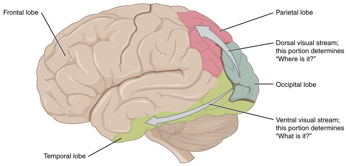
- The dorsal stream runs up into the parietal lobe. It works out where things are and how they are moving, and guides your hand and eyes toward them: the "where" or "how" stream.
- The ventral stream runs forward and down into the temporal lobe. It works out what things are, recognizing objects and faces: the "what" stream.
Damage shows the split. A person with ventral stream damage may be unable to name a key they are looking at, yet reach out and pick it up correctly. A person with dorsal stream damage may name the key at once but fumble when reaching for it.