Vision
1Why this matters
Mr. Chen, 52, has scraped both sides of his car pulling into the garage this month and keeps clipping doorframes. His glasses are new and his reading is fine. On a visual field test, he cannot see anything in the outer half of either eye's view. A scan shows a pituitary tumor pressing up from below on the one spot where fibers from both eyes cross. To see why a single small mass steals exactly those halves, you need to follow light from the front of the eye to the back of the brain.
2What this builds on
3Quick check before you start
1. Which cranial nerve moves most of the eye muscles and narrows the pupil?
- Optic nerve (II)
- Oculomotor nerve (III)
- Abducens nerve (VI)
Show the answer
The oculomotor nerve supplies four of the six eye muscles and the muscle that raises the upper lid, and carries parasympathetic fibers that narrow the pupil. The optic nerve is sensory, and the abducens supplies only the muscle that turns the eye outward.
- Optic nerve (II):
- Correct: Oculomotor nerve (III):
- Abducens nerve (VI):
2. Where is the primary visual cortex?
- In the occipital lobe
- In the postcentral gyrus
- In the temporal lobe
Show the answer
The primary visual cortex lies in the occipital lobe at the back of the brain. The postcentral gyrus holds the primary somatosensory cortex, and the temporal lobe holds the primary auditory cortex.
- Correct: In the occipital lobe:
- In the postcentral gyrus:
- In the temporal lobe:
3. A G protein–coupled receptor protein is activated. What does it do next?
- It opens as an ion channel
- It switches on a G protein, which changes the activity of an enzyme
- It moves into the nucleus
Show the answer
An activated G protein–coupled receptor protein switches on a G protein inside the cell, which switches an enzyme on or off and so changes the level of a second messenger.
- It opens as an ion channel:
- Correct: It switches on a G protein, which changes the activity of an enzyme:
- It moves into the nucleus:
4Anatomy
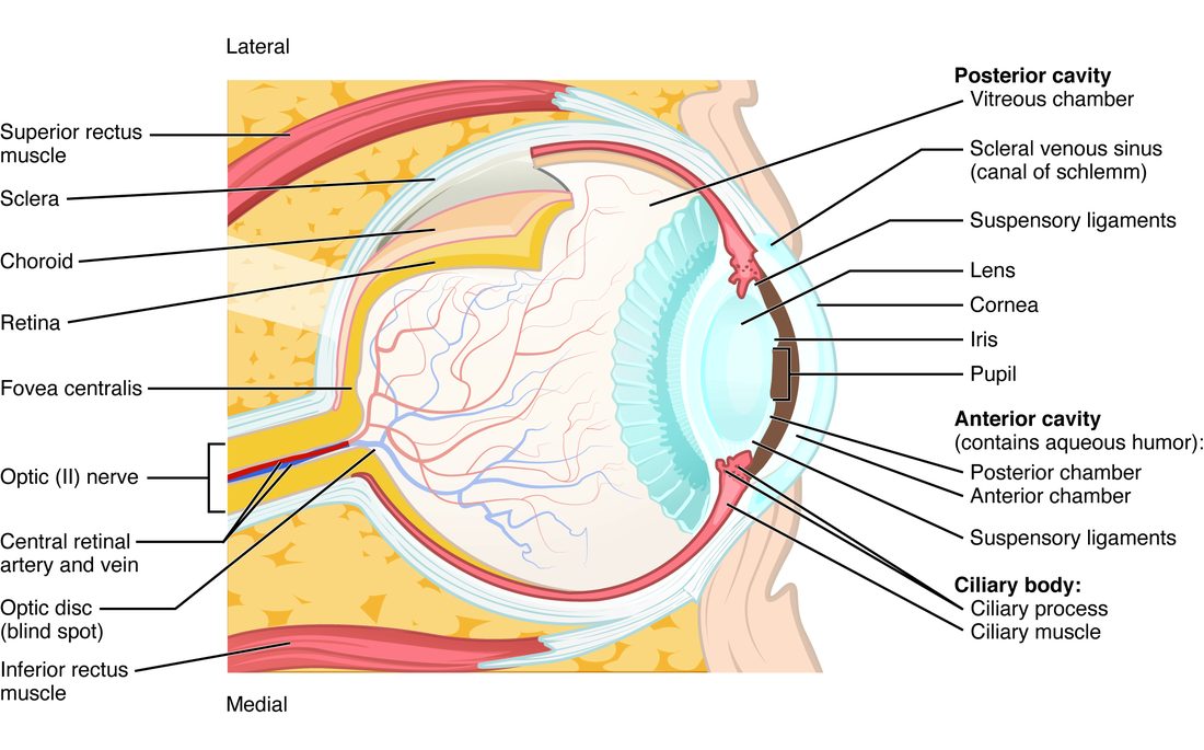
With labels hidden, select a box to reveal its label.
5How it works, step by step
- Light from an object enters the eye.The cornea and the lens refract it, and the rays from each point meet on the retina as a sharp, upside-down image.
- Light reaches the rods and cones at the back of the retina.Retinal in the pigment changes from 11-cis to all-trans (photoisomerization) and activates the pigment.
- The active pigment switches on the G protein transducin.An enzyme breaks down cGMP, the cGMP-gated cation channels close, and the photoreceptor hyperpolarizes.
- The hyperpolarized photoreceptor releases less glutamate onto bipolar cells.The bipolar cells change their output, and retinal ganglion cells change their rate of action potentials.
- Ganglion cell axons leave the eye as the optic nerve, and nasal fibers cross at the optic chiasm.Each optic tract carries the opposite visual field to the lateral geniculate nucleus of the thalamus, which relays it to the primary visual cortex.
6Core concepts
7A common mistake
The wrong idea: Light switches photoreceptors on: it depolarizes rods and cones and makes them fire.
What actually happens: It is the other way round. In the dark, cGMP holds cation channels open, so rods and cones are depolarized and release glutamate all the time. Light starts a cascade that destroys cGMP, closes those channels and hyperpolarizes the cell, which cuts its glutamate release. Rods and cones never fire action potentials; the first cells in the pathway to do so are the retinal ganglion cells.
8Check yourself
Anything you miss goes into your review queue.
1. Name the pinned structure.
- Fovea centralis
- Ciliary body
- Optic disc
- Choroid
Show the answer
The optic disc is where the ganglion cell axons leave the eye as the optic nerve and the central retinal vessels enter. It has no photoreceptors, so it makes the blind spot.
- Fovea centralis: The fovea is the small cone-only pit of sharpest vision, beside the optic disc rather than where the nerve leaves.
- Ciliary body: The ciliary body is the ring of muscle and folds around the lens at the front of the eye.
- Correct: Optic disc: Correct. The spot where the optic nerve leaves the eye is the optic disc, the blind spot.
- Choroid: The choroid is the dark, vessel-rich middle layer of the wall, not a spot on the retina.
2. You look up from a distant road sign to read a text message. What happens in your eye?
- The ciliary muscle relaxes, the zonule fibers tighten and the lens flattens
- The ciliary muscle contracts, the zonule fibers tighten and pull the lens rounder
- The cornea bulges forward and bends light more strongly
- The ciliary muscle contracts, the zonule fibers go slack and the lens rounds up
Show the answer
Near vision needs more bending. Parasympathetic fibers in the oculomotor nerve contract the ring-shaped ciliary muscle, which shrinks toward the lens. The zonule fibers go slack, and the elastic lens rounds up and refracts more.
- The ciliary muscle relaxes, the zonule fibers tighten and the lens flattens: That is what happens when you look from near to far, not far to near.
- The ciliary muscle contracts, the zonule fibers tighten and pull the lens rounder: Taut zonule fibers pull the lens flat, not round. When the ciliary muscle contracts, the fibers slacken.
- The cornea bulges forward and bends light more strongly: The cornea does most of the bending, but its shape is fixed. Only the lens changes for near vision.
- Correct: The ciliary muscle contracts, the zonule fibers go slack and the lens rounds up: Correct. Slack zonules let the elastic lens round up.
3. An eyeball is slightly too long from front to back. Where does light from a distant object focus, and what lens corrects it?
- Behind the retina; a convex lens
- In front of the retina; a convex (converging) lens
- Behind the retina; a concave lens
- In front of the retina; a concave lens
Show the answer
In a long eye (myopia), the cornea and lens bring distant light to a focus before it reaches the retina. A concave lens spreads the rays slightly first, which moves the focus back onto the retina.
- Behind the retina; a convex lens: A focus behind the retina is hyperopia, caused by an eye that is too short.
- In front of the retina; a convex (converging) lens: The focus is in front of the retina, but a convex lens would add bending and move it even farther forward.
- Behind the retina; a concave lens: A long eye puts the focus in front of the retina, not behind it.
- Correct: In front of the retina; a concave lens: Correct. Myopia: focus in front of the retina, corrected with a diverging (concave) lens.
4. Why is vision sharpest at the fovea?
- Rods there feed many ganglion cells each, which multiplies their signal
- It lies closest to the lens, so the image there is least blurred
- Each cone has its own ganglion cell, and the layers above are pushed aside
- It has the most blood vessels, so its photoreceptors receive the best oxygen supply
Show the answer
The fovea holds only tightly packed cones. Each foveal cone connects through its own bipolar cell to its own ganglion cell, so its signal reports one tiny spot, and light reaches the cones without passing through the other layers.
- Rods there feed many ganglion cells each, which multiplies their signal: The fovea has no rods, and rods converge many-to-one onto ganglion cells, which lowers acuity rather than raising it.
- It lies closest to the lens, so the image there is least blurred: The fovea is at the back of the eye, not especially close to the lens; its sharpness comes from its wiring.
- Correct: Each cone has its own ganglion cell, and the layers above are pushed aside: Correct. Low convergence and an unobstructed path give the fovea its acuity.
- It has the most blood vessels, so its photoreceptors receive the best oxygen supply: Blood vessels are kept away from the fovea; vessels in front of it would block light.
5. A rod has been in darkness. A flash of light now falls on it. Predict the change in each variable in the rod, compared with darkness.
| Variable | Change |
|---|---|
| cGMP in the outer segment | — |
| Number of open cation channels | — |
| Membrane potential (how positive the inside is) | — |
| Glutamate release onto bipolar cells | — |
Show the answer
In photoreceptors, light turns a signal off. It lowers cGMP, closes the cation channels that carried the dark current, hyperpolarizes the rod and reduces its glutamate release.
- cGMP in the outer segment: down. Active rhodopsin switches on transducin, which switches on the enzyme that breaks down cGMP.
- Number of open cation channels: down. These channels are held open by cGMP, so they close as cGMP falls.
- Membrane potential (how positive the inside is): down. With the Na+ inflow cut off while K+ still leaks out, the rod hyperpolarizes from about −40 toward −70 mV.
- Glutamate release onto bipolar cells: down. A hyperpolarized rod releases less neurotransmitter; the fall in glutamate is the light signal.
6. Mr. Chen, 52, keeps clipping doorframes and is startled by cars that seem to appear from nowhere on both sides. Testing shows he has lost the outer (temporal) half of the visual field of each eye. A scan shows a mass. Where is it most likely pressing?
- The middle of the optic chiasm
- The left optic tract
- The right optic nerve
- The occipital lobe on both sides
Show the answer
The outer half of each eye's field is seen by the nasal half of each retina. Those are exactly the axons that cross in the middle of the optic chiasm. A pituitary tumor growing up from below typically presses there, causing bitemporal hemianopia.
- Correct: The middle of the optic chiasm: Correct. Crossing fibers from both nasal retinas run through the middle of the chiasm.
- The left optic tract: A left optic tract lesion removes the right half of the field in both eyes, not the outer half of each.
- The right optic nerve: A right optic nerve lesion blinds the right eye only.
- The occipital lobe on both sides: Damage to both occipital lobes would cause far wider loss; a single mass pressing on one structure fits the chiasm.
7. Light shone into Ms. Park's left eye makes neither pupil narrow. Light shone into her right eye makes both pupils narrow. Where is the damage?
- Left oculomotor nerve
- Right oculomotor nerve
- Left optic nerve
- Right optic chiasm
Show the answer
When the right eye is lit, both pupils narrow, so both efferent paths (both oculomotor nerves and both iris sphincters) work. When the left eye is lit, nothing happens, so the signal never gets in: the afferent path from the left eye, the left optic nerve, is damaged.
- Left oculomotor nerve: A left oculomotor lesion would leave the left pupil wide whichever eye is lit, but here it narrows when the right eye is lit.
- Right oculomotor nerve: A right oculomotor lesion would stop the right pupil narrowing, but it narrows when the right eye is lit.
- Correct: Left optic nerve: Correct. Light in the damaged eye cannot start the reflex; light in the healthy eye drives both pupils.
- Right optic chiasm: The chiasm has no separate "right" half for one eye's pupil fibers, and damage there would not silence one eye's input completely.
8. After a stroke, Mr. Olsen cannot say what a cup on the table is, or recognize his daughter's face, yet he reaches out and grasps the cup smoothly. Which part of visual processing is damaged?
- The dorsal stream in the parietal lobe
- The lateral geniculate nucleus
- The optic chiasm
- The ventral stream in the temporal lobe
Show the answer
The ventral stream, running from the occipital lobe forward into the temporal lobe, identifies what things are, including faces. His ability to locate and grasp the cup shows that the dorsal stream still works.
- The dorsal stream in the parietal lobe: The dorsal stream guides reaching and locating, which he does well.
- The lateral geniculate nucleus: Damage to the lateral geniculate nucleus would cause loss of part of the visual field, not a loss of recognition with normal reaching.
- The optic chiasm: Chiasm damage causes field loss, typically the outer halves, not a loss of object recognition.
- Correct: The ventral stream in the temporal lobe: Correct. Recognizing objects and faces is the job of the ventral "what" stream.
9Summary
The accessory structures protect and move the eye: the eyelids (raised by the levator palpebrae superioris), the conjunctiva, the lacrimal gland whose tears drain through the puncta, canaliculi and nasolacrimal duct into the nose, and six extraocular muscles (lateral rectus by VI, superior oblique by IV, the rest by III). The wall has three layers: fibrous (sclera and cornea), vascular (choroid, ciliary body, iris around the pupil) and neural (retina). The lens, held by zonule fibers, separates the aqueous humor, which drains into the scleral venous sinus (poor drainage and raised pressure are the main risk factor for glaucoma, damage to the optic nerve), from the vitreous humor. The cornea does most of the refraction; accommodation rounds the lens for near vision when the ciliary muscle contracts and the zonules slacken. Myopia focuses in front of the retina, hyperopia behind it, and presbyopia is the stiffening lens of middle age. Rods (dim light, one pigment, high convergence) and cones (bright light, color, the fovea) hyperpolarize in light: photoisomerization of retinal activates the pigment, transducin lowers cGMP and the channels close. Nasal fibers cross at the optic chiasm, so each optic tract, lateral geniculate nucleus and visual cortex handles the opposite visual field, and field loss localizes damage. The pupillary light reflex narrows both pupils through the midbrain and both oculomotor nerves. Beyond the primary visual cortex, the dorsal stream finds where things are and the ventral stream what they are.