Cortical functions, memory, language and sleep
1Why this matters
Mr. Castillo, 62, is eating breakfast when his words suddenly stop coming. He understands everything his wife says and nods to answer, but when he tries to speak he manages only "Coffee... no... walk," slowly and with great effort, and he is plainly frustrated. His right hand is weak too. A blocked artery has cut off blood to the lower part of his left frontal lobe: the area just in front of the one that moves his face and hand, and part of that motor area too.
2What this builds on
3Quick check before you start
1. Which sulcus separates the frontal lobe from the parietal lobe?
- The lateral sulcus
- The central sulcus
- The parieto-occipital sulcus
Show the answer
The central sulcus runs from the top of the hemisphere down its side, with the frontal lobe in front and the parietal lobe behind. The lateral sulcus lies above the temporal lobe.
- The lateral sulcus:
- Correct: The central sulcus:
- The parieto-occipital sulcus:
2. Which structure is needed to form new memories of facts and events?
- The hippocampus
- The cerebellum
- The amygdala
Show the answer
The hippocampus, part of the limbic system in the temporal lobe, is needed to form new memories of facts and events. The cerebellum coordinates movement; the amygdala judges emotional importance.
- Correct: The hippocampus:
- The cerebellum:
- The amygdala:
3. At a chemical synapse, what makes an excitatory postsynaptic potential (EPSP)?
- Neurotransmitter binds to receptor proteins that let positive ions into the postsynaptic cell
- The action potential jumps directly across the gap
- The postsynaptic cell releases neurotransmitter back to the presynaptic cell
Show the answer
Neurotransmitter released from the presynaptic terminal binds to receptor proteins on the postsynaptic membrane. Opening channels that let positive ions in depolarizes the cell a little: an EPSP.
- Correct: Neurotransmitter binds to receptor proteins that let positive ions into the postsynaptic cell:
- The action potential jumps directly across the gap:
- The postsynaptic cell releases neurotransmitter back to the presynaptic cell:
4Anatomy
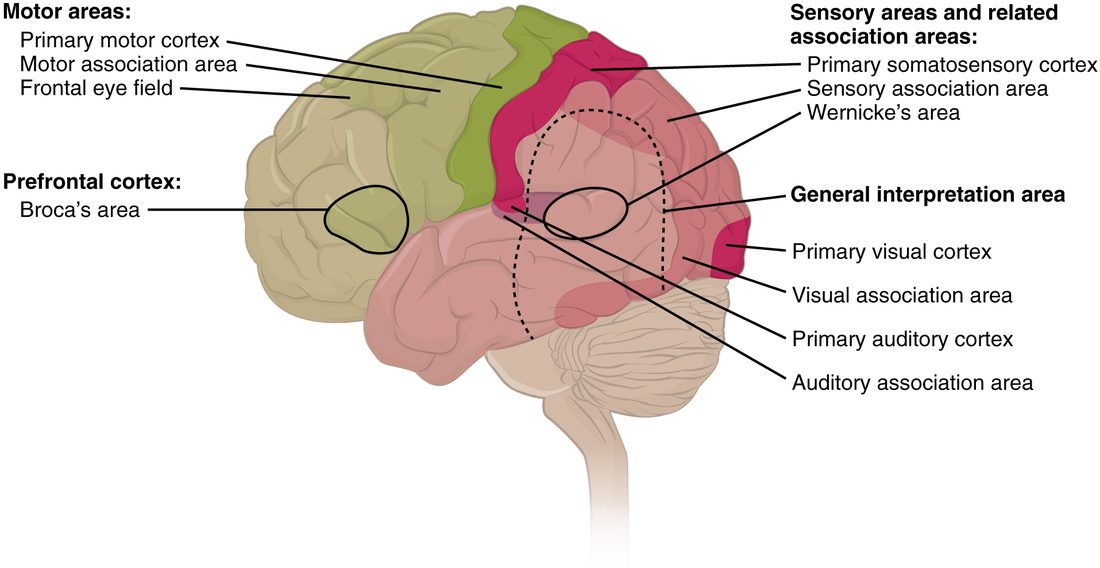
With labels hidden, select a box to reveal its label.
5How it works, step by step
- Sound waves from a spoken word reach your ears, and signals relay through the thalamus.The primary auditory cortex in the temporal lobe receives the sound.
- In the classic model, the primary auditory cortex passes the sound pattern to Wernicke's area beside it.Wernicke's area recognizes the word and its meaning.
- Wernicke's area sends the word forward along a curved band of white matter.Broca's area in the frontal lobe receives it and builds a speech plan.
- Broca's area passes the plan to the face area of the primary motor cortex next to it.The primary motor cortex drives the muscles of the lips, tongue and throat, and you say the word.
6Core concepts
7A common mistake
The wrong idea: Memories are stored in the hippocampus.
What actually happens: The hippocampus is needed to form new declarative memories and to consolidate them, but long-term memories end up stored in the cerebral cortex. Henry Molaison, who lost much of both hippocampi, could not form new memories of events, yet he kept his general knowledge from before his surgery and could still learn new skills, which depend on the basal nuclei and cerebellum.
8Check yourself
Anything you miss goes into your review queue.
1. Mr. Castillo, 62, understands what is said to him, but his own speech is slow, effortful and made of single words, and he is frustrated by it. His right arm is weak. Where is the damage most likely to be?
- Left lower frontal lobe
- Left upper temporal lobe
- Right parietal lobe
- Left occipital lobe
Show the answer
Slow, effortful speech with good understanding is expressive (Broca's) aphasia, from damage to Broca's area in the lower left frontal lobe. Broca's area lies next to the motor cortex, so right-sided weakness often comes with it.
- Correct: Left lower frontal lobe: Correct. Broca's area is in the lower left frontal lobe.
- Left upper temporal lobe: Damage to Wernicke's area in the upper left temporal lobe causes fluent speech with poor understanding, the opposite pattern.
- Right parietal lobe: Right parietal damage typically causes neglect of the left side of space, not loss of speech.
- Left occipital lobe: Left occipital damage causes loss of vision in the right half of the scene, not trouble speaking.
2. Loss of blood flow destroys the hand area of the left primary motor cortex, and nothing else. Predict the change in each.
| Variable | Change |
|---|---|
| Strength of the right hand | — |
| Strength of the left hand | — |
| Ability to feel touch on the right hand | — |
| Understanding of spoken words | — |
Show the answer
Each primary area has one job for one side of the body. Destroying the hand area of the left precentral gyrus weakens only the right hand; sensation, the other hand and language, handled by other areas, are unchanged.
- Strength of the right hand: down. The left primary motor cortex sends commands to the muscles of the right side, and its hand area drives the right hand, so the right hand weakens.
- Strength of the left hand: no change. The left hand is controlled by the right hemisphere's motor cortex, which is undamaged.
- Ability to feel touch on the right hand: no change. Touch reaches the primary somatosensory cortex in the postcentral gyrus, behind the central sulcus, which was not damaged.
- Understanding of spoken words: no change. Understanding depends on Wernicke's area in the temporal lobe, far from the hand area of the motor strip.
3. The lips occupy more of the primary somatosensory cortex than the whole back. What best explains this?
- The lips are closer to the brain, so their signals arrive stronger
- The lips have far more sensory receptors per square centimeter than the back
- The back sends most of its touch signals to the cerebellum instead of to the cerebral cortex
- The lips are used for speech, so they are mapped in Broca's area as well
Show the answer
The cortex given to a body part depends on how densely it is supplied with sensory receptors, not on its size. The lips have many more per square centimeter than the back, and every incoming signal needs cortical neurons to receive it, so the lips get a large area.
- The lips are closer to the brain, so their signals arrive stronger: Distance from the brain does not change signal strength; action potentials arrive at full size.
- Correct: The lips have far more sensory receptors per square centimeter than the back: Correct. Sensory receptor density sets how much cortex a body part gets.
- The back sends most of its touch signals to the cerebellum instead of to the cerebral cortex: Touch from the back reaches the somatosensory cortex too; it simply needs less cortex because it has fewer sensory receptors.
- The lips are used for speech, so they are mapped in Broca's area as well: The map in the somatosensory cortex is about sensation, and Broca's area does not receive touch signals.
4. After encephalitis destroys both of his hippocampi, Mr. Park cannot remember his nurse from one visit to the next. Which ability is he most likely to keep?
- Remembering what he ate this morning
- Learning the names of new staff
- Getting better at a new motor skill with daily practice
- Recalling a conversation from an hour ago
Show the answer
The hippocampi are needed to form new declarative memories, so he has anterograde amnesia for facts and events. Procedural memory for skills uses the basal nuclei and cerebellum, so he can still improve at a practiced skill, as Henry Molaison did at mirror drawing.
- Remembering what he ate this morning: This morning's meal is a new memory of an event, which needs the hippocampi to form.
- Learning the names of new staff: New names are new facts, which need the hippocampi to form.
- Correct: Getting better at a new motor skill with daily practice: Correct. Procedural learning does not depend on the hippocampus.
- Recalling a conversation from an hour ago: A conversation an hour ago is a new memory of an event, which needs the hippocampi.
5. Which describe REM sleep? Select all that apply.
- The EEG shows fast, small waves like the awake brain
- Most skeletal muscles are paralyzed, but the diaphragm keeps working
- Slow, large delta waves dominate the EEG
- Most vivid, story-like dreams happen
- REM periods are longest in the first cycle of the night
Show the answer
In REM sleep the EEG looks awake, the eyes dart, dreams are vivid, and brainstem neurons block the motor neurons to almost all skeletal muscles except the eye muscles and diaphragm. REM periods lengthen toward morning.
- Correct: The EEG shows fast, small waves like the awake brain: Yes. The REM EEG is fast and small, like being awake.
- Correct: Most skeletal muscles are paralyzed, but the diaphragm keeps working: Yes. The body is limp except the eye muscles and the diaphragm, so breathing continues.
- Slow, large delta waves dominate the EEG: No. Slow, large delta waves are the sign of N3, deep non-REM sleep.
- Correct: Most vivid, story-like dreams happen: Yes. Most vivid, story-like dreams happen in REM sleep.
- REM periods are longest in the first cycle of the night: No. REM periods are shortest early in the night and grow longer toward morning.
6. According to this graph, when does most N3 (deep) sleep happen?
- Evenly across the night
- In the first three hours
- In the last three hours
- Only during brief awakenings
Show the answer
The line reaches N3 for long stretches in the first two cycles (about 0.6 to 1.2 and 2 to 2.6 hours), briefly around 4 hours, and not at all after that. Deep sleep is concentrated early in the night.
- Evenly across the night: The graph shows N3 early and absent late, not evenly spread.
- Correct: In the first three hours: Correct. Nearly all N3 falls in the first three hours.
- In the last three hours: The last three hours contain REM and N2 but no N3.
- Only during brief awakenings: Awakenings are at the top of the graph, the opposite of N3.
7. Years ago, surgeons cut Ms. Novak's corpus callosum to stop her seizures from spreading. With her eyes closed, she is handed a key in her left hand. What happens?
- She names it at once and picks it out with either hand
- She cannot name it, but her left hand can pick it out
- She can neither name it nor pick it out with her left hand
- She names it but cannot pick it out with her left hand
Show the answer
Touch from the left hand reaches the right hemisphere. With the corpus callosum cut, it cannot be passed to the left hemisphere, which produces speech in most people. So she cannot say "key," but the right hemisphere knows the object and guides the left hand to choose it.
- She names it at once and picks it out with either hand: Touch from one hand reaches mainly the opposite hemisphere, and without the corpus callosum it cannot be shared.
- Correct: She cannot name it, but her left hand can pick it out: Correct. The right hemisphere knows the key but cannot speak it.
- She can neither name it nor pick it out with her left hand: Sensation in the left hand is normal; it reaches the right hemisphere, which can guide the hand.
- She names it but cannot pick it out with her left hand: Naming needs the left hemisphere, which produces speech in most people, and the touch cannot reach it; the right hemisphere can still guide the left hand.
8. After long-term potentiation, the same amount of glutamate released at a synapse produces a larger EPSP than before. What change in the postsynaptic cell best explains this?
- Its resting membrane potential has become more negative
- Its axon has been freshly myelinated
- It has stopped making receptor proteins for glutamate
- It has more glutamate receptor proteins in its membrane
Show the answer
Strong, repeated activity lets calcium into the postsynaptic cell, which triggers the insertion of more glutamate receptor proteins. The same glutamate now opens more channels, giving a larger EPSP.
- Its resting membrane potential has become more negative: A more negative resting potential would move the cell further from threshold. That is not what long-term potentiation changes; the EPSP grows because more glutamate receptor proteins open.
- Its axon has been freshly myelinated: Myelin speeds conduction along an axon; it does not change the size of an EPSP at a synapse on the dendrites.
- It has stopped making receptor proteins for glutamate: Fewer receptor proteins would shrink the EPSP.
- Correct: It has more glutamate receptor proteins in its membrane: Correct. More receptor proteins mean a bigger response to the same transmitter.
9Summary
Cortical areas are specialized. The primary motor cortex (precentral gyrus, area 4) sends commands to the opposite side, planned by the premotor and supplemental motor areas; the prefrontal cortex runs executive function. The primary somatosensory (postcentral gyrus), visual (occipital) and auditory (temporal) cortices receive signals first, association areas interpret them, and multimodal integration areas combine them. Body maps along the two central gyri give the most cortex to the hands, lips and tongue. The hemispheres specialize, with language usually on the left. Damage to Broca's area causes expressive aphasia, to Wernicke's area receptive aphasia, and to the link between them conduction aphasia. Working memory holds about four chunks; the hippocampus consolidates declarative memories into the cortex by strengthening synapses, while procedural memory uses the basal nuclei and cerebellum. The EEG records summed synaptic potentials, and sleep cycles every 90 minutes through N1, N2, N3 and REM.