The neurological exam
1Why this matters
Mr. Adeyemi, 68, wakes up and cannot button his shirt. His wife notices the left corner of his mouth sagging and his words slurring. In the emergency department, a nurse checks his face, arm drift, speech and reflexes in under five minutes and says: "right hemisphere, probably a stroke, last known well at 6 a.m." Every item she checked is a test of one part of the pathways you have just learned, and the pattern told her where to look.
2What this builds on
3Quick check before you start
1. Where does the lateral corticospinal tract cross the midline?
- At the pyramidal decussation in the lower medulla
- In the spinal cord at each segment
- In the internal capsule
Show the answer
About 85–90% of corticospinal axons cross at the pyramidal decussation, where the medulla meets the spinal cord, and descend as the lateral corticospinal tract.
- Correct: At the pyramidal decussation in the lower medulla:
- In the spinal cord at each segment:
- In the internal capsule:
2. Which set of findings points to lower motor neuron damage?
- Spasticity, brisk tendon jerks and a Babinski sign
- Flaccid weakness, wasting, fasciculations and lost tendon jerks
- Normal strength with an intention tremor
Show the answer
Losing the lower motor neuron cuts the final common path to the muscle: tone falls, reflexes are lost, the muscle wastes and sick neurons twitch. Spasticity, brisk jerks and a Babinski sign are upper motor neuron signs; an intention tremor with normal strength is cerebellar.
- Spasticity, brisk tendon jerks and a Babinski sign:
- Correct: Flaccid weakness, wasting, fasciculations and lost tendon jerks:
- Normal strength with an intention tremor:
3. Which pathway carries vibration and joint position sense up the spinal cord?
- The spinothalamic tract
- The lateral corticospinal tract
- The dorsal column pathway
Show the answer
Vibration, fine touch and position sense climb the dorsal columns on the same side and cross in the medulla. The spinothalamic tract carries pain and temperature, and the corticospinal tract is motor.
- The spinothalamic tract:
- The lateral corticospinal tract:
- Correct: The dorsal column pathway:
4Anatomy
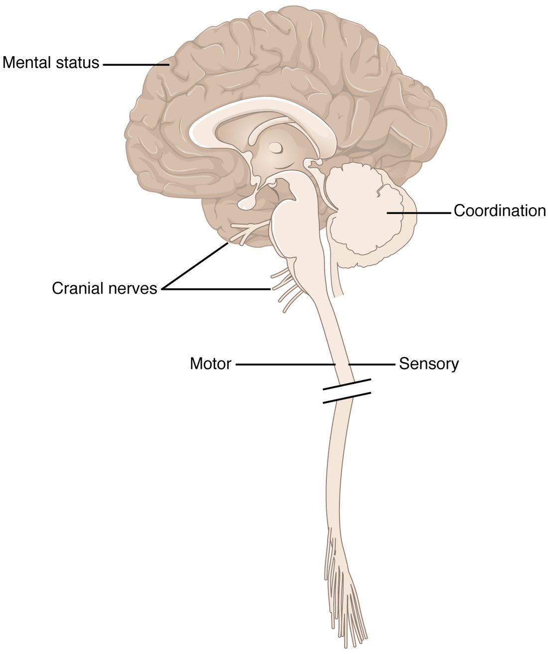
With labels hidden, select a box to reveal its label.
5How it works, step by step
- An artery supplying the right motor cortex and internal capsule is blocked by a clot.Upper motor neurons for the left face, arm and leg, and their axons, lose their blood supply and stop firing within minutes.
- Commands no longer reach the crossed corticospinal and corticobulbar pathways to the left side.The left arm drifts down and the lower left face droops, while the forehead, which also gets input from the left hemisphere, still wrinkles.
- The examiner finds upper motor neuron weakness of the face and body on the same side, with no cranial nerve problem on the other side.The damage is placed above the facial nucleus in the pons, most likely in the right cerebral hemisphere or internal capsule.
- The onset was sudden and the patient was last known to be well two hours ago.A stroke is suspected, and a brain scan is done at once to tell an ischemic stroke from a hemorrhagic one before treatment.
6Core concepts
7A common mistake
The wrong idea: Weakness on one side of the body means damage on that same side of the brain.
What actually happens: Above the pyramidal decussation, motor pathways serve the opposite side, so a weak left arm and leg with a brisk left knee jerk points to the right hemisphere, internal capsule or brainstem. Only below the decussation, in the spinal cord, does damage weaken the same side. The face helps: in the brainstem, the damaged cranial nerve is on the same side as the lesion while the body weakness is on the opposite side.
8Check yourself
Anything you miss goes into your review queue.
1. A patient opens his eyes only when you press on his fingernail, makes moaning sounds with no words, and bends his arm away from the pressure. What is his Glasgow Coma Scale score?
- 6
- 8
- 10
- 7
Show the answer
Eyes open to pressure: E2. Sounds without words: V2. Bending away from pressure (normal flexion): M4. Total 2 + 2 + 4 = 8, in the severe range.
- 6: 6 would need a motor score of 2 (extension). Bending the arm away from pressure scores 4.
- Correct: 8: Correct. E2 + V2 + M4 = 8.
- 10: 10 would need higher scores, for example eyes opening to voice and inappropriate words.
- 7: 7 would need abnormal flexion (M3) rather than bending away from the pressure.
2. A woman stands steadily with her feet together and eyes open, but sways and nearly falls as soon as she closes her eyes. She cannot tell whether her big toe is moved up or down, and she cannot feel a tuning fork on her ankles. Where is the problem?
- The cerebellar vermis
- The dorsal columns or the large sensory axons from the legs
- The lateral corticospinal tracts
- The spinothalamic tracts
Show the answer
This is a positive Romberg test. Loss of position and vibration sense means the dorsal column pathway, or the sensory axons feeding it, is damaged. With eyes open, vision makes up for the missing position sense; with eyes closed it cannot.
- The cerebellar vermis: Cerebellar damage makes a person unsteady with the eyes open as well, and it does not remove vibration or position sense.
- Correct: The dorsal columns or the large sensory axons from the legs: Correct. Lost position sense, compensated by vision, is the Romberg pattern.
- The lateral corticospinal tracts: Damage to the corticospinal tracts causes weakness with brisk reflexes, not loss of vibration and position sense.
- The spinothalamic tracts: The spinothalamic tracts carry pain and temperature, not vibration or position sense.
3. A man has weakness of the whole left side of his face, forehead included, and weakness of his right arm and leg with brisk right reflexes and a right Babinski sign. Where is the single lesion?
- The left cerebral hemisphere
- The right cerebral hemisphere
- The left side of the pons
- The cervical spinal cord on the right
Show the answer
Whole-face weakness including the forehead is a lower motor neuron sign of the left facial nerve or its nucleus, which lies in the pons. The corticospinal axons for the right body run past it and have not yet crossed. One lesion on the left side of the pons hits both. Face on one side, body on the other is the mark of a brainstem lesion.
- The left cerebral hemisphere: A left hemisphere lesion would weaken the right body and the lower right face, sparing the forehead. It would not weaken the left face.
- The right cerebral hemisphere: A right hemisphere lesion would weaken the left body, not the right.
- Correct: The left side of the pons: Correct. The left facial nucleus and the uncrossed corticospinal axons sit side by side here.
- The cervical spinal cord on the right: A cervical cord lesion would not affect the face, and a right-sided cord lesion would weaken the right body but not the left face.
4. A man can lift his arm off the bed against gravity but cannot hold it up against any push from the examiner. What strength grade is this?
- 1
- 2
- 3
- 4
Show the answer
Grade 3 is movement against gravity but not against resistance.
- 1: Grade 1 is a flicker of contraction with no movement.
- 2: Grade 2 is movement only with gravity taken away, such as sliding the arm along the bed.
- Correct: 3: Correct. Against gravity, not against resistance.
- 4: Grade 4 is movement against some resistance, which he cannot manage.
5. At lunch, a 70-year-old woman's right arm suddenly goes weak, the right corner of her mouth droops and her speech comes out jumbled. Her husband calls emergency services. What must happen before she can be given a clot-dissolving drug?
- A brain scan to rule out a hemorrhagic stroke
- A 24-hour wait to see whether the signs clear
- A full mental status exam to grade her aphasia
- A lumbar puncture to test her cerebrospinal fluid
Show the answer
Ischemic and hemorrhagic strokes can look the same at the bedside, and a clot-dissolving drug would worsen bleeding. A scan must tell them apart first, as fast as possible.
- Correct: A brain scan to rule out a hemorrhagic stroke: Correct. Only a scan can tell a blocked artery from a burst one.
- A 24-hour wait to see whether the signs clear: Waiting wastes the hours in which clot-dissolving treatment works. Nearly two million neurons can die each minute in a large stroke.
- A full mental status exam to grade her aphasia: Her language problem is already clear from the history. A detailed exam would delay treatment and would not tell bleeding from blockage.
- A lumbar puncture to test her cerebrospinal fluid: A lumbar puncture does not tell an ischemic stroke from a bleed into the brain, and it would delay treatment.
6. A man has 20 minutes of weakness and numbness in his left hand, then recovers completely. A scan shows no dead brain tissue. What is the best description, and why does it matter?
- A migraine that needs no further tests
- A completed ischemic stroke that has now recovered
- A transient ischemic attack, with a high risk of stroke in the coming days
- A hemorrhagic stroke that has already healed
Show the answer
Brief stroke-like signs that clear, with no dead tissue on the scan, define a transient ischemic attack. It is a warning: the risk of a full stroke is highest in the following days, so it is investigated and treated urgently.
- A migraine that needs no further tests: Treating it as harmless misses the warning. Sudden one-sided weakness is assumed to be vascular until proven otherwise.
- A completed ischemic stroke that has now recovered: A stroke leaves dead brain tissue on the scan. His scan shows none.
- Correct: A transient ischemic attack, with a high risk of stroke in the coming days: Correct. A TIA, which needs urgent treatment.
- A hemorrhagic stroke that has already healed: Bleeding into the brain leaves blood and damaged tissue on the scan, and it does not clear in 20 minutes.
7. A stab wound cuts the left half of the spinal cord at T8. Weeks later, predict each finding compared with before the injury.
| Variable | Change |
|---|---|
| Strength of the left leg | — |
| Knee jerk on the left | — |
| Position sense in the left foot | — |
| Pain sensation in the right foot | — |
| Pain sensation in the left foot | — |
| Strength of the arms | — |
Show the answer
A hemisection (Brown-Séquard syndrome) removes the tracts that have already crossed (corticospinal) or have not yet crossed (dorsal columns) on the same side, and the spinothalamic tract carrying pain from the opposite side.
- Strength of the left leg: down. The left lateral corticospinal tract, already crossed in the medulla, carries commands for the left leg.
- Knee jerk on the left: up. The left leg's reflex arcs survive below the cut but lose descending control, so after an early absent phase they become brisk.
- Position sense in the left foot: down. The left dorsal column carries position sense from the left side and does not cross until the medulla.
- Pain sensation in the right foot: down. Pain from the right foot crosses within a segment or two of entry and climbs in the left spinothalamic tract, which is cut.
- Pain sensation in the left foot: no change. Pain from the left foot crosses to the right spinothalamic tract, which is intact.
- Strength of the arms: no change. The motor neurons and tracts for the arms are in the cervical cord, above T8.
8. A 45-year-old has pain down the back of his left leg, a weak push-off with his left foot, an absent left ankle jerk and numbness along the outer edge of his left foot. His knee jerks and plantar reflexes are normal. Where is the lesion?
- The left S1 nerve root
- The right motor cortex
- The spinal cord at T12
- The left cerebellar hemisphere
Show the answer
Weakness of the calf, a lost ankle jerk and numbness in the S1 dermatome all belong to one root on one side: lower motor neuron and sensory findings in the territory of left S1, often from a herniated disc.
- Correct: The left S1 nerve root: Correct. Motor, reflex and sensory findings all fit one root.
- The right motor cortex: Cortex damage would cause upper motor neuron signs: brisk reflexes and a Babinski sign, not a lost ankle jerk.
- The spinal cord at T12: A cord lesion at T12 would affect both legs or produce upper motor neuron signs below it, not a single lost reflex with a dermatome of numbness.
- The left cerebellar hemisphere: Cerebellar damage causes clumsiness with normal strength and does not remove reflexes or sensation.
9Summary
The neurological exam localizes a problem by testing each part of the nervous system in turn. The mental status exam tests the cerebrum: consciousness, orientation, attention, memory, language and judgment. The Glasgow Coma Scale scores eye (1–4), verbal (1–5) and motor (1–6) responses from 3 to 15, with 8 or less severe. The cranial nerve exam tests the brainstem and its nerves, including the corneal reflex (V in, VII out). The sensory exam tests the spinothalamic tract (pinprick, temperature) and dorsal columns (vibration, position sense), finds a dermatome or sensory level, and tests the parietal cortex with stereognosis and graphesthesia. The motor exam checks bulk, tone, strength (0–5), pronator drift and reflexes, sorting upper from lower motor neuron damage. The coordination and gait exam tests the cerebellum; a positive Romberg test (steady with eyes open, falling with eyes closed) points to lost position sense. A stroke is a sudden loss of function from interrupted blood supply, ischemic or hemorrhagic, recognized by BE FAST; a TIA leaves no dead tissue. Spinal cord injury causes tetraplegia (cervical) or paraplegia (lower), and a hemisection splits motor and sensory losses between the two sides.