Motor pathways and movement control
1Why this matters
Mr. Okoro, 64, woke up unable to lift his right arm or leg. A month later his right arm is stiff and flexed, his right knee jerks briskly at the lightest tap, and when his doctor strokes the sole of his right foot, his big toe rises instead of curling down. His neighbor on the ward cannot lift her right foot either, but her leg is floppy, its muscles are wasting and small twitches ripple under the skin. Same weakness, opposite signs: one lost an upper motor neuron, the other a lower one.
2What this builds on
3Quick check before you start
1. Where is the primary motor cortex?
- The postcentral gyrus
- The precentral gyrus
- The occipital lobe
Show the answer
The primary motor cortex is the precentral gyrus, just in front of the central sulcus. The postcentral gyrus behind it is the primary somatosensory cortex.
- The postcentral gyrus:
- Correct: The precentral gyrus:
- The occipital lobe:
2. Where do the cell bodies of the motor neurons that drive skeletal muscles of the leg sit?
- In the dorsal root ganglion
- In the dorsal horn
- In the ventral horn
Show the answer
Motor neurons for skeletal muscle have their cell bodies in the ventral horn and send axons out through the ventral root. The dorsal horn and dorsal root ganglion are sensory.
- In the dorsal root ganglion:
- In the dorsal horn:
- Correct: In the ventral horn:
3. Where does the spinothalamic tract cross the midline?
- In the spinal cord, within a segment or two of entry
- In the medulla oblongata
- In the thalamus
Show the answer
Second-order spinothalamic axons cross in the spinal cord close to where the signal entered. The dorsal column pathway is the one that crosses in the medulla.
- Correct: In the spinal cord, within a segment or two of entry:
- In the medulla oblongata:
- In the thalamus:
4Anatomy
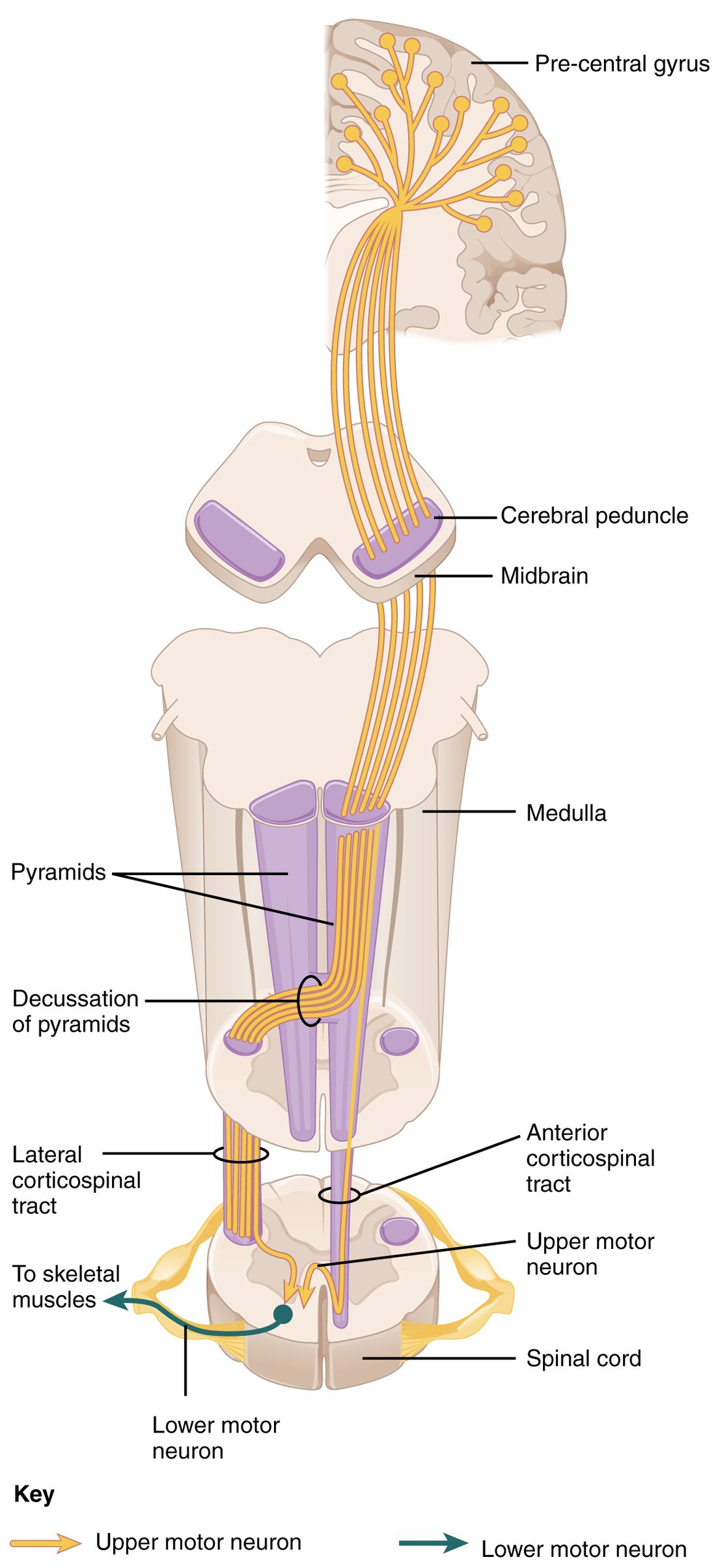
With labels hidden, select a box to reveal its label.
5How it works, step by step
- An upper motor neuron in the left precentral gyrus fires an action potential.The action potential travels down its axon through the internal capsule, the cerebral peduncle, the pons and the left pyramid of the medulla.
- The axon reaches the pyramidal decussation at the bottom of the medulla.It crosses the midline and descends in the right lateral corticospinal tract.
- At its target segment, the axon ends in the right ventral horn and releases glutamate.A lower motor neuron there depolarizes to threshold and fires.
- The lower motor neuron's action potential travels out through the ventral root and a spinal nerve.It releases acetylcholine at the neuromuscular junction, and the muscle fibers of its motor unit on the right side of the body contract.
- While the movement runs, the cerebellum compares the command with proprioceptor signals, and the basal nuclei keep competing movements braked.Corrections through the thalamus to the motor cortex make the movement accurate and smooth.
6Core concepts
7A common mistake
The wrong idea: Any damage to the motor system makes muscles weak and floppy.
What actually happens: Only lower motor neuron damage makes a limb floppy for good, because the final common path to the muscle is gone. Upper motor neuron damage leaves the lower motor neurons and their spinal circuits intact but frees them from the brain's control, so after an early floppy phase the limb becomes stiff (spastic), with brisk tendon jerks and a Babinski sign. The signs point to which neuron is damaged.
8Check yourself
Anything you miss goes into your review queue.
1. Put the corticospinal pathway for moving the right hand in order, starting in the brain.
- Upper motor neuron in the left precentral gyrus
- Internal capsule
- Pyramid of the medulla oblongata
- Pyramidal decussation
- Right lateral corticospinal tract
- Lower motor neuron in the right ventral horn
Show the answer
The upper motor neuron's axon leaves the left precentral gyrus, funnels through the internal capsule, runs in the cerebral peduncle, forms part of the pyramid on the front of the medulla, crosses at the pyramidal decussation, descends in the right lateral corticospinal tract and synapses on a lower motor neuron in the right ventral horn of the cervical enlargement.
- Correct order: 1. Upper motor neuron in the left precentral gyrus 2. Internal capsule 3. Pyramid of the medulla oblongata 4. Pyramidal decussation 5. Right lateral corticospinal tract 6. Lower motor neuron in the right ventral horn
2. Blood flow is cut off to a small area of the left internal capsule that carries corticospinal axons. Which side of the body becomes weak?
- The left side of the body
- The right side of the body
- Both sides equally
- Neither side
Show the answer
The internal capsule lies above the pyramidal decussation. The corticospinal axons there have not crossed yet, and they are on their way to cross in the lower medulla and control the right side. Losing them weakens the right side.
- The left side of the body: Same-side weakness follows damage below the pyramidal decussation, in the spinal cord. The internal capsule is above it.
- Correct: The right side of the body: Correct. Damage above the decussation weakens the opposite side.
- Both sides equally: Each internal capsule carries axons for the opposite side only. Weakness on both sides would need damage on both sides.
- Neither side: The axons of the upper motor neurons pass through the internal capsule. Cutting them stops the signal, even though their cell bodies in the cortex survive.
3. A limb is weak and floppy, its tendon jerks are absent, its muscles have wasted over six weeks and small twitches ripple under the skin. Where is the damage most likely to be?
- The opposite motor cortex
- The internal capsule
- The lateral corticospinal tract
- The ventral horn or ventral root
Show the answer
Flaccidity, lost tendon jerks, marked atrophy and fasciculations are lower motor neuron signs. The lower motor neuron's cell body is in the ventral horn, and its axon leaves through the ventral root.
- The opposite motor cortex: Motor cortex damage is upper motor neuron damage. After the first days it produces stiffness and brisk tendon jerks, not wasting with twitches.
- The internal capsule: The internal capsule carries upper motor neuron axons. Damage there causes spastic weakness of the opposite side.
- The lateral corticospinal tract: The lateral corticospinal tract is made of upper motor neuron axons. Cutting it leaves the lower motor neurons healthy, so there are no fasciculations and little wasting.
- Correct: The ventral horn or ventral root: Correct. These are the signs of a lower motor neuron lesion.
4. Patient A cannot move the lower right side of her face, but she wrinkles her forehead normally on both sides. Patient B cannot move any part of the right side of his face, forehead included. Which statement explains the difference?
- A: right facial nerve; B: left motor cortex
- A and B: right facial nerve, worse in B
- A: left motor cortex or its axons; B: right facial nerve
- A: the right motor cortex or its axons; B: the left facial nerve
Show the answer
The forehead part of the facial nucleus gets corticobulbar input from both hemispheres, while the lower face part gets it mainly from the opposite hemisphere. Upper motor neuron damage on the left therefore spares the right forehead and weakens only the lower right face. The facial nerve is the lower motor neuron for the whole half of the face, so damage to it weakens everything on that side.
- A: right facial nerve; B: left motor cortex: This is the reverse. Facial nerve damage weakens the forehead too, and cortex damage spares it.
- A and B: right facial nerve, worse in B: Sparing of the forehead is not a matter of severity. Any facial nerve damage that weakens the lower face also reaches the forehead, because one nerve supplies both.
- Correct: A: left motor cortex or its axons; B: right facial nerve: Correct. A has an upper motor neuron lesion on the left; B has a lower motor neuron lesion on the right.
- A: the right motor cortex or its axons; B: the left facial nerve: The corticobulbar input to the lower face comes from the opposite hemisphere, so right-sided face weakness points to the left cortex, and it points to the right facial nerve, not the left.
5. A 9-month-old baby's big toe extends upward when the outer edge of her sole is stroked. What does this mean?
- Damage to her corticospinal tract above the lumbar cord
- Damage to the lower motor neurons that supply her toes
- A normal finding at her age
- Weakness of the muscles that curl her toes downward
Show the answer
In infants the corticospinal axons are not yet fully myelinated, so the upward big toe is normal. It changes to the adult downward response by about 1 to 2 years of age.
- Damage to her corticospinal tract above the lumbar cord: In an adult this would be the Babinski sign of corticospinal damage, but in a 9-month-old it is expected.
- Damage to the lower motor neurons that supply her toes: Lower motor neuron damage would weaken the toe muscles or remove the response altogether, not produce an upward toe.
- Correct: A normal finding at her age: Correct. The upward big toe is normal until the corticospinal tract matures.
- Weakness of the muscles that curl her toes downward: An upward toe is an active extension, not a failure of the flexors. It reflects the maturity of the descending control, not muscle strength.
6. In Parkinson's disease, the dopamine neurons of the substantia nigra die. Predict each change in the basal nuclei loop.
| Variable | Change |
|---|---|
| Activity of the direct (go) pathway | — |
| Activity of the indirect (stop) pathway | — |
| Inhibition of the thalamus by the globus pallidus output | — |
| Ease of starting a movement | — |
Show the answer
Dopamine favors movement by boosting the direct pathway and damping the indirect one. Without it, the brake from the globus pallidus on the thalamus presses harder, and movement becomes slow and hard to begin.
- Activity of the direct (go) pathway: down. Dopamine normally excites the direct-pathway striatal neurons, so losing it weakens them.
- Activity of the indirect (stop) pathway: up. Dopamine normally inhibits the indirect-pathway striatal neurons, so losing it frees them.
- Inhibition of the thalamus by the globus pallidus output: up. Less direct-pathway inhibition and more indirect-pathway excitation both make the globus pallidus output fire more.
- Ease of starting a movement: down. A more inhibited thalamus excites the motor cortex less, so movements are slow, small and hard to start.
7. This sequence describes how the direct pathway of the basal nuclei lets a chosen movement go ahead. Which step is wrong?
- The motor cortex excites direct-pathway neurons of the striatum
- The striatal neurons inhibit the output neurons of the globus pallidus
- The globus pallidus output now inhibits the thalamus more strongly
- The thalamus excites the motor cortex, and the movement goes ahead
Show the answer
The globus pallidus output is itself inhibitory and fires all the time. When the striatum inhibits it, it fires less, so the thalamus is inhibited less, not more. Removing that brake frees the thalamus to excite the cortex.
- The motor cortex excites direct-pathway neurons of the striatum: This step is right. Cortical input to the striatum is excitatory.
- The striatal neurons inhibit the output neurons of the globus pallidus: This step is right. Direct-pathway striatal neurons are inhibitory and act on the globus pallidus output.
- Correct: The globus pallidus output now inhibits the thalamus more strongly: This is the error. An inhibited globus pallidus inhibits the thalamus less: the brake is released.
- The thalamus excites the motor cortex, and the movement goes ahead: This step is right. A released thalamus excites the motor cortex.
9Summary
Voluntary movement uses two neurons. The upper motor neuron sits in the motor cortex or brainstem and stays in the CNS; the lower motor neuron sits in the ventral horn or a cranial nerve nucleus and is the final common path to skeletal muscle. The corticospinal (pyramidal) tract runs from the cortex through the internal capsule, cerebral peduncles, pons and pyramids; about 85–90% crosses at the pyramidal decussation in the lower medulla and descends as the lateral corticospinal tract to the limbs, while the anterior corticospinal tract serves the trunk. The corticobulbar tract drives the cranial nerve nuclei, mostly from both sides, except the lower face. Brainstem tracts (vestibulospinal, reticulospinal, tectospinal, rubrospinal) control posture and tone. The basal nuclei select movements through a direct (go) and an indirect (stop) pathway, and dopamine favors go. The cerebellum's three divisions correct balance, posture and skilled movement through its three peduncles, and its damage shows on the same side. Lower motor neuron damage causes flaccid weakness, lost tendon jerks, atrophy and fasciculations; upper motor neuron damage causes spastic weakness, hyperreflexia and a Babinski sign.