The neurological exam is a set of bedside tests, each built to check one part of the nervous system, so that a clinician can work out where a problem is before any scan is taken. This page goes through the components of the neurological exam in the usual order (mental status, cranial nerves, sensory, motor with reflexes, and coordination and gait), shows how the Glasgow Coma Scale scores consciousness, how to recognize a stroke, what a spinal cord injury does at each level, and how the findings are put together to place a lesion.
What the exam is for
A woman arrives with a weak right hand. The weakness could come from her left cerebral cortex, her internal capsule, her brainstem, her cervical spinal cord, a C8 nerve root, her ulnar nerve or the hand muscles themselves. Each of those places leaves a different pattern of findings, and the exam is designed to read the pattern.
The neurological exam is that structured set of tests. It asks two questions, in this order:
- Where is the problem? This is localization. The exam answers it, using what you have learned about pathways: which side they serve, where they cross and which segment or nerve they use.
- What is the problem? The history answers it, mostly from the time course: seconds (loss of blood flow), hours to days (infection, inflammation), months (a slowly growing mass or a degenerative disease).
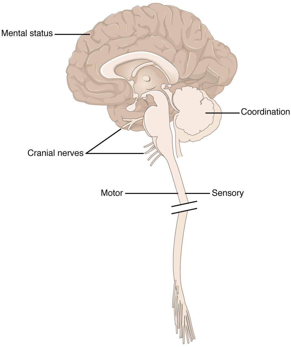
The exam has five parts, and each one tests particular regions of the nervous system (Figure 1):
- the mental status exam tests the cerebrum;
- the cranial nerve exam tests the brainstem and its nerves;
- the sensory exam tests the ascending pathways, from nerve endings to the cortex;
- the motor exam, which includes the reflexes, tests the descending pathways, the lower motor neurons and the muscles;
- the coordination exam, with the gait exam, tests the cerebellum and the systems that feed it.
A full exam takes a long time. A screening exam checks a few items from each part in a few minutes, and the clinician goes into detail wherever something is abnormal.
The mental status exam
The mental status exam tests the cerebrum and the systems that keep you awake. It runs from the most basic function to the most complex:
- Level of consciousness. Alert, drowsy but rousable by voice, rousable only by pressure, or unresponsive. Staying awake depends on the reticular activating system of the brainstem acting on both hemispheres, so a drop in consciousness means either brainstem trouble or trouble affecting both hemispheres at once.
- Orientation. Does the person know who they are, where they are, the date and why they are here? A person who knows all four is "oriented times four".
- Attention. Can they repeat a string of digits, or count back from 100 by sevens?
- Memory. Can they recall three words after five minutes? That tests forming new long-term memories, which needs the hippocampus.
- Language. Fluency, comprehension, naming objects and repeating a sentence. You met the patterns in cortical functions: effortful, halting speech with good comprehension points to Broca's area (expressive aphasia); fluent speech with poor comprehension points to Wernicke's area (receptive aphasia). In most people both lie in the left hemisphere.
- Judgment and planning. Tested with a problem or a hypothetical situation. These executive functions depend on the prefrontal cortex.
The Glasgow Coma Scale
When a person is too unwell for a full mental status exam, for example after a head injury, their level of consciousness is scored with the Glasgow Coma Scale (GCS). It rates the best response in three categories and adds the scores:
| Score | Eye opening (E) | Verbal response (V) | Motor response (M) |
|---|---|---|---|
| 6 | – | – | Obeys commands |
| 5 | – | Oriented | Localizes: moves a hand toward the site of pressure |
| 4 | Spontaneously | Confused | Normal flexion: bends the arm away from pressure |
| 3 | To sound (voice) | Inappropriate words | Abnormal flexion: slow, stiff bending of the arm at the elbow and wrist |
| 2 | To pressure (pain) | Incomprehensible sounds | Extension: straightens and turns in the arm |
| 1 | None | None | None |
The total runs from 3 (no response at all) to 15 (fully awake and oriented). Totals are grouped as mild (13 to 15), moderate (9 to 12) and severe (8 or less). A score of 8 or less is the usual working definition of coma. Clinicians report the parts as well as the total, because the same total can hide very different patients.
Worked example: scoring the GCS
Problem. After a fall, a man lies with his eyes closed. They open when you press firmly on his fingernail, but not when you call his name. He mutters "stop, go away" but cannot say where he is. When you press on the top of his shoulder, his hand comes up to push yours away. What is his GCS?
- Eyes. No opening to voice, opening to pressure: E2.
- Verbal. Real words, but not a conversation and not answers to the questions asked: inappropriate words, V3.
- Motor. His hand goes to the site of the pressure: he localizes, M5.
- Add. 2 + 3 + 5 = 10.
Answer. GCS 10 (E2 V3 M5), a moderate impairment. Had he merely bent his arm away from the pressure, his motor score would be 4 and his total 9. Had his hand not moved at all, it would be 1 and his total 6, in the severe range.
The cranial nerve exam
You met every cranial nerve test in the cranial nerves topic. In the exam they are run in number order, and each finding is compared across the two sides:
| Nerve | Quick test |
|---|---|
| I olfactory | Identify a familiar smell with each nostril (often skipped) |
| II optic | Read letters on a chart with each eye; check side vision; look at the back of the eye with a light |
| II and III | Shine a light into one eye and watch both pupils narrow |
| III, IV, VI | Follow a finger in an H shape with both eyes; look for a drooping eyelid (III) |
| V trigeminal | Light touch on the forehead, cheek and jaw; clench the jaw |
| VII facial | Raise the eyebrows, squeeze the eyes shut, smile, puff out the cheeks |
| VIII vestibulocochlear | Hear a finger rub or a whisper at each ear |
| IX, X | Say "ah" and watch the back of the roof of the mouth rise evenly; gag |
| XI accessory | Shrug the shoulders and turn the head against resistance |
| XII hypoglossal | Stick out the tongue; it points toward a weak side |
One test has its own name. The corneal reflex is the blink test: a wisp of cotton touched to the clear front surface of the eye makes both eyes blink. The touch travels in on the ophthalmic branch of the trigeminal nerve (V), crosses in the pons, and the blink goes out on both facial nerves (VII). It is a reflex arc through the brainstem, which is why it is also checked in a person who cannot cooperate: once sedating drugs and a very low body temperature are ruled out, an absent corneal reflex in a deeply unresponsive patient points to failure of the brainstem around the pons.
Cranial nerve findings are powerful for localization because each nerve joins the brainstem at a known level. A cranial nerve problem on one side together with weakness of the opposite side of the body points to the brainstem, as you will see below.
The sensory exam
The sensory exam tests each ascending pathway separately, because the two main pathways cross at different levels:
- Spinothalamic tract: pain, tested with a clean pin (pinprick), and temperature, tested with a cool metal object.
- Dorsal column pathway: vibration, tested with a buzzing tuning fork on a bony point such as the big toe joint, and joint position sense (proprioception): with eyes closed, is the toe being moved up or down?
- Light touch travels in both, so it is a general screen.
Each modality is tested on both sides and from the feet upward. Comparing the findings with the dermatomes tells you whether the loss follows one nerve root, one peripheral nerve, a "stocking" pattern in both feet (typical of disease of many peripheral nerves) or a sensory level, a line across the trunk below which sensation is lost, which means the spinal cord is damaged at about that segment.
Cortical sensation
If the basic sensations are intact, the clinician can test the parietal cortex, which turns raw touch into meaning. With eyes closed:
- Stereognosis (stereo- = solid, gnosis = knowledge): name an object, such as a coin or a key, placed in the hand.
- Graphesthesia (graph- = write, esthesia = sensation): name a number traced on the palm with a fingertip.
- Two-point discrimination: tell whether one point or two are touching the fingertip.
Failing these tests with normal pinprick, vibration and position sense points to the parietal lobe on the opposite side. If the basic sensations are already lost, these tests cannot be interpreted, because the information never reached the cortex.
The motor exam
The motor exam applies what you learned about upper and lower motor neurons. It has five parts:
- Inspection. Muscle bulk (wasting), fasciculations and abnormal movements such as a tremor.
- Tone. The clinician moves the relaxed limb: normal, reduced (flaccidity) or increased (spasticity, or the rigidity of Parkinson's disease).
- Strength. Each muscle group is graded on a scale of 0 to 5.
- Pronator drift. A sensitive test for mild upper motor neuron weakness in the arm.
- Reflexes. Deep tendon reflexes graded 0 to 4+, and the plantar reflex.
| Strength grade | Meaning |
|---|---|
| 0 | No contraction at all |
| 1 | A flicker of contraction, but no movement |
| 2 | Moves the joint only with gravity taken away (sliding along the bed) |
| 3 | Moves against gravity, but not against any resistance |
| 4 | Moves against some resistance, but weaker than normal |
| 5 | Normal strength |
For pronator drift, the person holds both arms straight out in front, palms up, and closes their eyes. A healthy person holds the position. With mild upper motor neuron weakness, one forearm slowly turns palm-down (pronates) and the arm drifts downward. It works because upper motor neuron damage weakens the supinators and extensors of the arm more than the pronators and flexors, and closing the eyes removes the visual correction.
The findings are then sorted with the table you met in motor pathways: weakness with raised tone, brisk reflexes and a Babinski sign points to the upper motor neuron; weakness with wasting, fasciculations, low tone and lost reflexes points to the lower motor neuron.
The coordination and gait exam
The coordination exam tests the cerebellum, in a person strong enough to move normally:
- Finger-to-nose: touch your nose, then the examiner's moving finger, back and forth. Overshooting (dysmetria) and a tremor that grows near the target point to the cerebellar hemisphere on the same side.
- Heel-to-shin: lying down, run one heel down the other shin, the same test for the leg.
- Rapid alternating movements: slap the thigh with the palm, then the back of the hand, as fast as you can. Clumsy, irregular rhythm is another cerebellar sign.
The gait exam watches the person walk normally, then heel-to-toe along a line (tandem walking), then on the heels and on the toes. Each abnormal gait points somewhere: a wide-based, staggering gait (ataxia) to the cerebellum, especially the vermis; a stiff leg swung out in an arc to upper motor neuron damage on that side; small shuffling steps to Parkinson's disease; a foot that slaps down or catches its toe to weakness of the muscles that lift the foot.
The Romberg test
Balance while standing uses three inputs: vision, the balance organs deep in the skull, and position sense from the legs, carried by the dorsal columns. In the Romberg test, the person stands with feet together, first with eyes open, then with eyes closed.
- Steady with eyes open, sways or falls with eyes closed: the test is positive. Position sense from the legs is lost, and vision had been making up for it. This points to the dorsal columns or the large sensory axons of the peripheral nerves.
- Unsteady even with eyes open: vision cannot make up for the problem. This is typical of cerebellar damage, and the Romberg test is not "positive" in the strict sense.
Recognizing a stroke
A man at a restaurant suddenly cannot hold his fork in his right hand. The right corner of his mouth droops, and when he tries to speak, the words come out wrong. Nothing hurt. This is what a stroke looks like.
A stroke, or cerebrovascular accident (CVA), is the sudden loss of function in part of the brain when its blood supply is interrupted. Neurons have almost no energy stores, so within minutes of losing blood flow they stop working, and they start to die soon after. A typical large ischemic stroke kills nearly two million neurons a minute, which is why the rule is "time is brain". There are two kinds:
| Ischemic stroke | Hemorrhagic stroke | |
|---|---|---|
| Cause | A blocked artery: a clot forms in it or travels to it | A burst artery bleeds into or around the brain |
| Share of strokes | About 85–87% | About 13–15% |
| Typical onset | Sudden loss of function, often painless | Sudden loss of function, often with a severe headache, vomiting and falling consciousness |
| How the damage happens | Neurons downstream of the block run out of oxygen and glucose | Loss of flow downstream, plus the pressure of the blood mass on the brain |
| Emergency treatment | Dissolving or removing the clot, within hours | Controlling the bleeding and the pressure; clot-dissolving drugs would make it worse |
Because the treatments are opposite, a brain scan must tell the two apart before any clot-dissolving drug is given. The signs alone cannot.
The warning signs
The public is taught the signs as BE FAST:
- Balance: sudden loss of balance or coordination.
- Eyes: sudden loss of vision in one or both eyes, or double vision.
- Face: one side of the face droops; ask the person to smile.
- Arm: one arm is weak; ask them to raise both arms and watch for one drifting down.
- Speech: slurred, strange or absent speech.
- Time: note when the person was last known to be well, and call emergency services at once.
The signs localize like any other finding. The restaurant patient has weakness of the right hand and lower right face, with sparing of the forehead, plus an aphasia. That places the damage in the left hemisphere, in the motor cortex for the face and hand and in the language areas next to it: one artery supplies that whole region.
Transient ischemic attack
A transient ischemic attack (TIA) is a brief episode of stroke-like signs caused by a temporary loss of blood flow, with no dead brain tissue on a scan. The signs usually last minutes, and most clear within an hour. A TIA is a warning: the risk of a full stroke is highest in the next few days, so it is treated as an emergency even though the signs have gone.
Spinal cord injury
A spinal cord injury cuts some or all of the tracts at one level and destroys the gray matter of the segments at that level. Below it, the cord's own circuits survive but lose their connection to the brain. Everything depends on the level:
- The neurological level is the lowest segment with normal sensation and strength on both sides.
- A complete injury leaves no movement or sensation at all in the lowest sacral segments. An incomplete injury spares some, and the chance of recovery is much better.
| Tetraplegia (quadriplegia) | Paraplegia | |
|---|---|---|
| Meaning of the word | tetra- (Greek) or quadri- (Latin) = four; -plegia = paralysis | para- = beside; -plegia = paralysis |
| Level of injury | Cervical cord | Thoracic, lumbar or sacral cord |
| Limbs affected | All four, plus the trunk | Both legs, plus the trunk below the level |
| Hands | Weak or paralyzed | Normal |
| Breathing | At C3 or above, the diaphragm fails and the person needs a ventilator; at C4 and C5 it works partly or fully, though breathing support may be needed at first; below that, weak chest muscles still weaken breathing | The diaphragm works normally; a high thoracic injury weakens the intercostal and abdominal muscles |
The diaphragm is the key to cervical injuries. Its phrenic nerves come from C3, C4 and C5, and students remember this as "C3, 4 and 5 keep the diaphragm alive". An injury above C3 stops breathing at once.
Right after a sudden injury, the cord below it goes quiet for days to weeks: the limbs are floppy and the reflexes absent. Then reflexes return and become brisk, with spasticity and Babinski signs, the upper motor neuron picture you met in motor pathways. At the level of the injury itself, the destroyed ventral horn cells leave a narrow band of lower motor neuron weakness.
Hemisection: damage to one half of the cord
You worked this pattern out in the sensory and motor pathways topics. When one half of the cord is cut, a hemisection (hemi- = half), also called Brown-Séquard syndrome, the tracts that have already crossed and those that have not yet crossed are both lost:
- Same side, below the injury: upper motor neuron weakness (the lateral corticospinal tract crossed in the medulla) and loss of vibration and position sense (the dorsal columns cross in the medulla).
- Opposite side, from a segment or two below: loss of pain and temperature (the spinothalamic tract crossed on entry).
Putting it together: localizing a lesion
Localization follows a fixed order of questions. Figure 2 shows the main branches.
Worked example: three patients, three places
Patient A. Sudden weakness of the left arm and leg, with brisk left reflexes and a left Babinski sign. The lower left face droops; the forehead moves normally.
- Brisk reflexes and a Babinski sign: upper motor neuron.
- Face and body weak on the same side, lower face only: the damage is above the facial nucleus in the pons, in the path of the corticobulbar and corticospinal axons before either has crossed.
- Left body, so the damage is on the right: the right hemisphere or right internal capsule. The sudden onset suggests a stroke.
Patient B. The whole right side of the face is weak, forehead included, and the left arm and leg are weak with brisk reflexes.
- Whole-face weakness including the forehead: the right facial nerve or its nucleus, a lower motor neuron finding.
- Upper motor neuron weakness of the opposite side of the body: the corticospinal axons, which have not yet crossed at the level of the pons.
- One place damages both: the right side of the pons, where the facial nucleus sits beside the descending corticospinal axons. Findings on opposite sides of face and body are the signature of a brainstem lesion.
Patient C. Both legs are stiff and weak, with brisk knee and ankle jerks and two Babinski signs. The arms and face are normal. Pinprick is lost below the level of the navel.
- Upper motor neuron signs in both legs only: the damage is below the arms' segments, so below the cervical enlargement.
- A sensory level at the navel matches the T10 dermatome.
- The spinal cord at about T10: a paraplegia.