How do taste and smell work? Both are chemical senses: their sensory receptors respond to molecules that dissolve and bind to them. Taste works through taste buds in your mouth, and smell through a small patch of nerve tissue high in your nose. This page explains the structures of each, how a molecule becomes an electrical signal, the five taste qualities, the path each signal takes to the brain, and why a blocked nose makes food taste flat. It also corrects a famous error: the "tongue map."
Two chemical senses
Hold your nose and put a jelly bean in your mouth. You can tell it is sweet, but you cannot tell whether it is cherry or lemon. Let go of your nose, and the flavor arrives at once. Two senses were at work, and only one of them was blocked.
- Gustation (gust- = taste) is the sense of taste. It detects a small set of taste qualities in what you put in your mouth.
- Olfaction (olfact- = smell) is the sense of smell. It detects airborne molecules that reach the top of your nose, including molecules that rise from food in your mouth up the back of your throat.
Both use chemoreceptors, the sensory receptors that respond to particular chemicals. In both, a molecule has to dissolve in a watery film first: saliva for taste, mucus for smell. Both are special senses, so their signals travel in cranial nerves. Figure 1 shows the structures side by side.
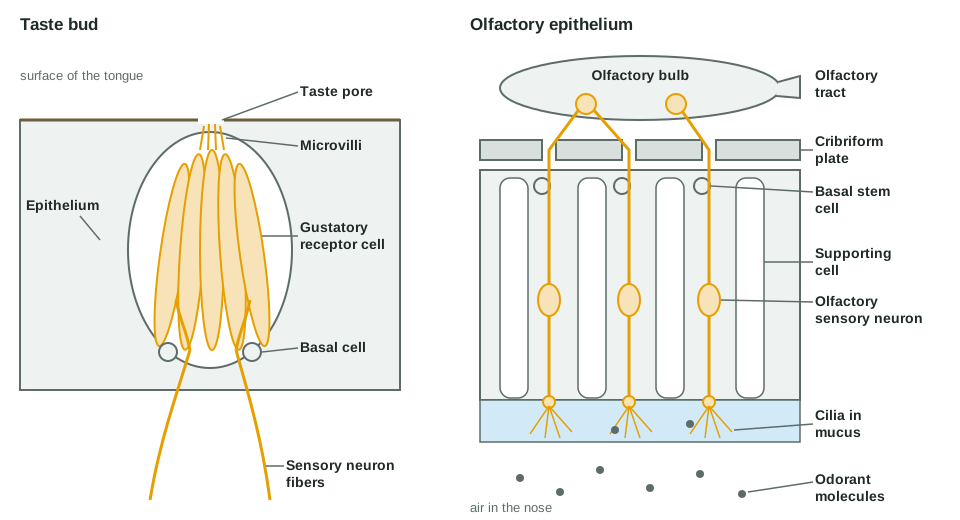
Taste buds and gustatory receptor cells
The bumps you can see on your tongue are not taste buds. Taste buds are microscopic, and most sit in the walls of some of those bumps. You have a few thousand of them, mostly on the tongue, with smaller numbers on the back of the roof of the mouth, the throat and the epiglottis.
A taste bud is an onion-shaped cluster of about 50 to 100 cells set into the epithelium. Its main parts are:
- Gustatory receptor cells are the taste cells. They are not neurons. They are specialized epithelial cells, so they are sensory receptor cells in the sense of the last topics: they detect the stimulus and pass the signal to a sensory neuron at a synapse.
- Each gustatory receptor cell has microvilli at its tip that poke into the taste pore, a small opening in the surface. Dissolved chemicals, called tastants, reach the cell there.
- Basal cells at the base of the bud are stem cells. Taste cells wear out and are replaced every week or two on average, and the basal cells make the new ones. That is why a burnt tongue recovers its taste within days.
The five taste qualities
A taste quality is one kind of taste, a submodality in the language of the last topic. Current evidence supports five. Each has its own chemical trigger and its own way into the cell.
| Quality | Typical trigger | How it enters the cell's signal | Everyday example |
|---|---|---|---|
| Salty | Sodium ions (Na+) | Na+ flows in through sodium channels and depolarizes the cell directly | Potato chips |
| Sour | Acids, which release hydrogen ions (H+) | H+ flows in through a proton channel and depolarizes the cell directly | Lemon juice |
| Sweet | Sugars, and artificial sweeteners | Binds a G protein–coupled receptor protein; a second messenger does the rest | Honey |
| Umami | Glutamate, an amino acid | Binds a G protein–coupled receptor protein; a second messenger does the rest | Broth, aged cheese, ripe tomato |
| Bitter | Many unrelated compounds, including many plant toxins and caffeine | Binds one of about 25 G protein–coupled receptor proteins; a second messenger does the rest | Black coffee |
Umami (from a Japanese word for "savory, delicious") is the savory taste of glutamate. It is the taste that monosodium glutamate adds to food.
From tastant to signal
Salty and sour work through ion channels. Positive ions flow straight into the cell, and the cell depolarizes. Sweet, umami and bitter work through receptor proteins coupled to G proteins, the same kind of receptor protein you met with second messengers. Binding sets off a second messenger cascade inside the cell, calcium inside the cell rises, and the cell releases neurotransmitter. Either way, the taste cell depolarizes: this change in voltage is the cell's receptor potential.
A gustatory receptor cell has no axon, so it cannot send a signal to the brain itself. A larger receptor potential makes it release more neurotransmitter onto the endings of a sensory neuron. More tastant, more release, and the sensory neuron fires action potentials faster. Sweet, bitter and umami cells release mainly ATP as their neurotransmitter.
The tongue map is a myth
You may have seen a drawing of the tongue with "sweet" at the tip, "sour" and "salty" on the sides and "bitter" at the back. It is wrong. Every region of the tongue that has taste buds can detect all five qualities. A single taste bud usually holds cells for several qualities.
The map came from a misreading of a 1901 study that found small differences in how sensitive each region was. A later book redrew those small differences as separate zones, and the drawing spread. The real differences are slight. For example, the back of the tongue is somewhat more sensitive to bitter, and a strong bitter taste there readily triggers gagging. You can test it yourself: put a little salt on the tip of your tongue, where the map says only sweet is sensed. You will taste salt at once.
The gustatory pathway
Taste signals from different parts of the mouth travel in three cranial nerves:
- The facial nerve (VII) carries taste from the front two thirds of the tongue.
- The glossopharyngeal nerve (IX) carries taste from the back third of the tongue.
- The vagus nerve (X) carries taste from the throat and epiglottis.
All three deliver taste to one nucleus in the medulla oblongata. From there, signals go two ways. Some go up to the thalamus and then to the gustatory cortex, in the insula and the nearby part of the frontal lobe, where taste becomes a perception. Others stay in the brainstem and drive reflexes: saliva flows when you taste food, and a bitter or spoiled taste can trigger gagging.
The olfactory epithelium
Smell starts in the olfactory epithelium, a patch of tissue about the size of a postage stamp on each side of the roof of your nose, just under the cribriform plate of the ethmoid bone. Look at the right half of Figure 1. It holds three kinds of cells:
- Olfactory sensory neurons are the sensory receptors for smell. Unlike taste cells, they are neurons themselves. Each is a bipolar neuron. Its dendrite runs down to the surface and ends in a knob with a tuft of long, non-moving cilia that lie in the mucus. Its unmyelinated axon runs up through a hole in the cribriform plate.
- Supporting cells surround and support the neurons.
- Basal stem cells divide to replace olfactory sensory neurons, which live roughly one to two months. They are among the very few neurons in your body that are replaced throughout life.
Glands under the epithelium keep it coated in mucus. An odorant is any molecule you can smell. To be smelled, an odorant has to float in the air, reach the top of your nose, and dissolve in that mucus. Sniffing pulls more air up to the epithelium, which is why you sniff to catch a faint smell.
How an odorant becomes a signal
The cilia of olfactory sensory neurons carry odorant receptor proteins. Humans have about 400 kinds. Each olfactory sensory neuron makes just one kind. Here is what happens when an odorant arrives:
- The odorant dissolves in the mucus and binds an odorant receptor protein on a cilium.
- The receptor protein activates a G protein. The G protein switches on an enzyme that makes cAMP, the second messenger.
- cAMP opens ligand-gated cation channels in the cilium. Na+ and Ca2+ flow in.
- The Ca2+ opens chloride channels. These neurons hold unusually high chloride inside, so Cl− flows out, which depolarizes the cell further.
- This depolarization is the neuron's receptor potential. It spreads to the axon, and if it reaches threshold, action potentials fire.
The cascade amplifies the signal: each activated enzyme makes many cAMP molecules, and the chloride channels add a second, larger current on top of the first. In the olfactory bulb, thousands of neurons of the same type then feed the same few small clusters of synapses, which add their weak signals together. Signal amplification and this pooling are why a trace of a strong odorant can be enough.
Telling thousands of odors apart with 400 receptor proteins
If you have only about 400 kinds of odorant receptor protein, how do you tell apart far more odors than that? Each odorant binds several kinds of receptor protein, some strongly and some weakly. Each receptor protein binds several odorants. So each odor activates its own pattern across the 400 kinds, like a chord played on a keyboard. Your brain reads the pattern, not a single key.
The olfactory pathway
The axons of olfactory sensory neurons pass through the cribriform plate in small bundles. Together they form the olfactory nerve (I). They end in the olfactory bulb, the swelling at the front end of each olfactory tract on the underside of the frontal lobe.
In the bulb, all the axons from neurons that make the same receptor protein converge on the same few small clusters of synapses. So the bulb sorts incoming signals by receptor type before passing them on. From the bulb, the olfactory tract carries signals backward.
Smell is unusual in two ways:
- It reaches the cortex without first stopping in the thalamus. Every other sense relays in the thalamus before reaching the cortex. Olfactory signals go straight to the olfactory cortex on the inner surface of the temporal lobe. Only later do some pass through the thalamus to the frontal lobe, where smell combines with taste into flavor.
- It connects directly with the limbic system. Olfactory signals reach the amygdala and areas next to the hippocampus in one or two synapses. That tight link is why a smell can bring back a memory, and the feelings that went with it, faster than a photograph can.
Flavor: taste plus smell
Back to the jelly bean. When you chew, odorants rise from your mouth up the back of your throat to the olfactory epithelium. Your brain combines these with taste signals, texture and temperature, and you perceive one flavor, which feels as if it comes from your mouth. Most of what you call "taste" is actually smell. That is why food tastes flat when a cold blocks your nose: your taste buds work normally, but odorants cannot reach the epithelium.
Both senses show sensory adaptation. Smell adapts especially fast: within minutes you stop noticing the smell of your own home or perfume, while a visitor notices it at once.
When smell fails: anosmia
Anosmia (an- = without, osm- = smell) is the loss of the sense of smell. Common causes follow from the anatomy:
- Head injury. A blow, especially to the back of the head, can jolt the brain against the skull. The thin axons passing through the cribriform plate can tear.
- Viral illness. Colds, influenza and COVID-19 can damage the supporting cells and the epithelium. Smell often returns over weeks to months as basal stem cells rebuild the epithelium.
- Blockage. Swelling or growths in the nose stop odorants from reaching the epithelium.
- Age and brain disease. Smell declines with age, and loss of smell can be an early sign of some brain diseases, such as Parkinson's disease.
People with anosmia usually say they have lost their sense of taste. Testing shows they can still tell sweet from salty: what they have lost is the smell part of flavor. Losing smell is also a safety risk, because a person cannot smell smoke, a gas leak or spoiled food.
Summary
Gustation and olfaction are chemical senses. Taste buds hold gustatory receptor cells, epithelial cells whose microvilli reach into the taste pore and which pass signals to sensory neurons; basal cells replace them. The five taste qualities are salty and sour, which enter through ion channels, and sweet, umami and bitter, which act through G protein–coupled receptor proteins. All five are detected wherever there are taste buds: the tongue map is a myth. Taste travels in cranial nerves VII, IX and X to the medulla, then through the thalamus to the insula. Olfactory sensory neurons are bipolar neurons in the olfactory epithelium, replaced by basal stem cells. An odorant binds a receptor protein on the cilia, cAMP opens cation channels, and chloride leaving depolarizes the cell further. Axons pass through the cribriform plate to the olfactory bulb and olfactory tract, reaching the cortex and limbic system without a first stop in the thalamus. Flavor is mostly smell, and anosmia is often felt as a loss of taste.