Spinal nerves and plexuses
1Why this matters
Marcus, 44, a roofer, lifted a bundle of shingles and felt a pop in his lower back. Now a burning pain runs from his right buttock down the back of his thigh and calf to the outer edge of his foot, and his little toe feels numb. His thigh muscles work, but he struggles to rise onto the toes of his right foot. One pinched nerve root, S1, explains every part of that picture, and by the end of this topic you will be able to say why.
2What this builds on
3Quick check before you start
1. Which root of a spinal cord segment carries sensory axons into the cord?
- The ventral root
- The dorsal root
- Both roots equally
Show the answer
The dorsal root carries sensory axons in, and its ganglion holds their cell bodies. The ventral root carries motor axons out from the ventral horn.
- The ventral root:
- Correct: The dorsal root:
- Both roots equally:
2. Which kind of connective tissue forms a tough wrapping made mostly of collagen fibers running in many directions?
- Dense irregular connective tissue
- Areolar tissue
- Adipose tissue
Show the answer
Dense irregular connective tissue is packed with collagen fibers running in several directions, so it resists pulling from any side. It wraps organs such as nerves and forms the dermis.
- Correct: Dense irregular connective tissue:
- Areolar tissue:
- Adipose tissue:
3. In the peripheral nervous system, what is a nerve?
- A single long neuron
- A cluster of neuron cell bodies
- A bundle of axons wrapped in connective tissue
Show the answer
A nerve is a bundle of many axons with connective tissue around them. A cluster of cell bodies outside the CNS is a ganglion.
- A single long neuron:
- A cluster of neuron cell bodies:
- Correct: A bundle of axons wrapped in connective tissue:
4Anatomy
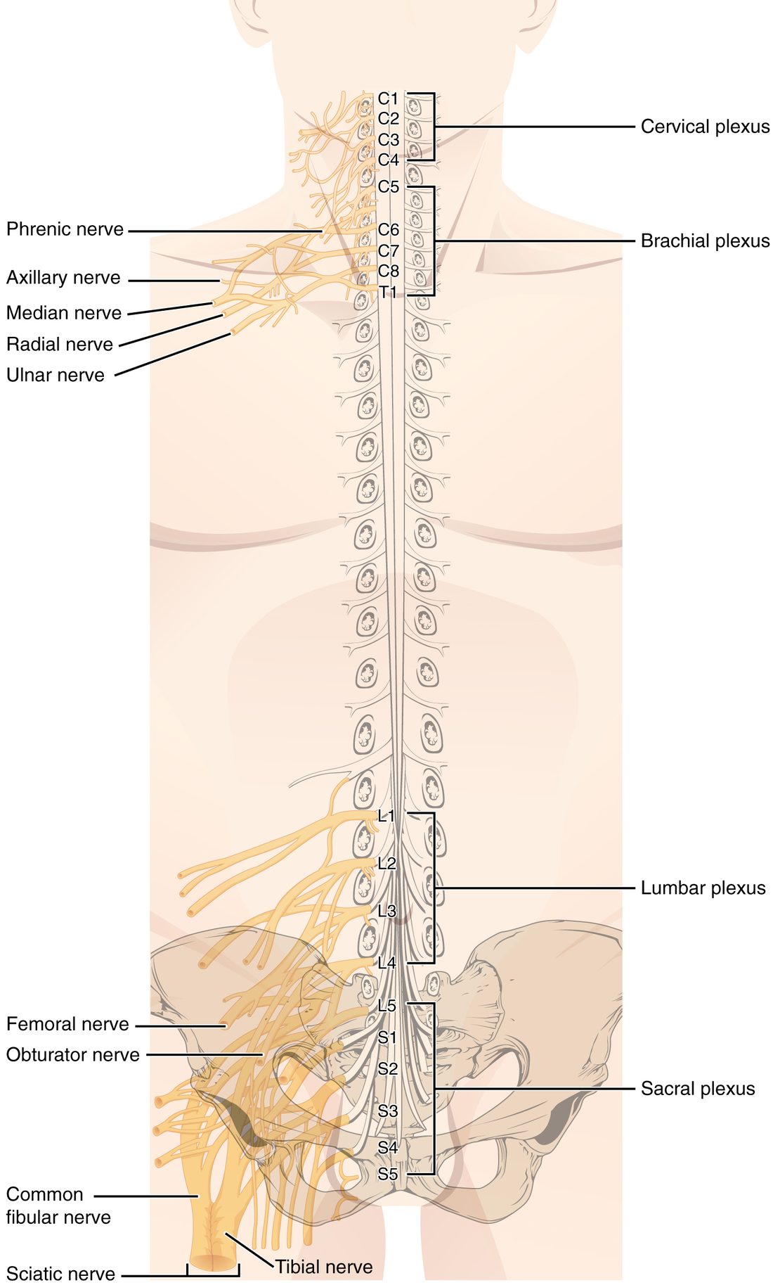
With labels hidden, select a box to reveal its label.
5How it works, step by step
- A pinprick touches the skin on the outer edge of your right foot, in the S1 dermatome.Sensory endings there fire action potentials in sensory axons that run in small skin branches joining the tibial nerve.
- The action potentials travel up the tibial nerve and the sciatic nerve.They reach the sacral plexus, where the S1 axons sort back into the S1 ventral ramus.
- The ventral ramus joins the S1 spinal nerve inside the sacrum's openings.In the spinal nerve, the sensory axons separate from the motor axons and enter the dorsal root.
- The signal passes the cell bodies in the S1 dorsal root ganglion and continues along the dorsal root.It enters the dorsal horn of the S1 segment of the cord, where it is passed on toward the brain.
- A herniated disc presses on the S1 root before it joins the plexus.Signals from that dermatome are blocked or distorted, so the outer foot feels numb and pain is felt along the whole path the S1 axons serve, the pattern called sciatica.
6Core concepts
7A common mistake
The wrong idea: Each dorsal root supplies its dermatome alone, so cutting one dorsal root leaves a sharply bordered numb band.
What actually happens: Neighboring dermatomes overlap: each patch of skin is supplied mainly by one spinal nerve and partly by the nerves above and below. Cutting one dorsal root only reduces sensation in its dermatome. A band of complete numbness appears only when two or three neighboring roots are damaged. The sharp borders on dermatome maps are a drawing convention.
8Check yourself
Anything you miss goes into your review queue.
1. Name the pinned plexus.
- Cervical plexus
- Brachial plexus
- Lumbar plexus
- Sacral plexus
Show the answer
The plexus formed by the ventral rami of C5 to T1, at the level of the shoulder, is the brachial plexus. Its nerves supply the upper limb.
- Cervical plexus: The cervical plexus is higher, formed by C1 to C4. It supplies the neck and the diaphragm, not the arm.
- Correct: Brachial plexus: Correct. C5 to T1 form the brachial plexus.
- Lumbar plexus: The lumbar plexus comes from L1 to L4 in the lower back and supplies the front of the thigh.
- Sacral plexus: The sacral plexus sits in the pelvis, from L4 to S4, and supplies the back of the thigh, the leg and the foot.
2. Name the pinned nerve.
- Femoral nerve
- Common fibular nerve
- Sciatic nerve
- Obturator nerve
Show the answer
The femoral nerve (L2 to L4) leaves the lumbar plexus, passes into the front of the thigh and supplies the quadriceps femoris.
- Correct: Femoral nerve: Correct. This is the femoral nerve.
- Common fibular nerve: The common fibular nerve is a branch of the sciatic nerve behind the knee, far lower than this.
- Sciatic nerve: The sciatic nerve runs down the back of the thigh from the sacral plexus, not across the front.
- Obturator nerve: The obturator nerve also comes from L2 to L4, but on this figure it is the branch just below the pinned one, heading to the medial thigh.
3. A surgeon cuts across a nerve and sees several round bundles, each with its own sleeve. Which layer forms the sleeve around each bundle?
- Epineurium
- Endoneurium
- Perineurium
- Myelin sheath
Show the answer
Each bundle is a fascicle, and the perineurium wraps each fascicle. Its cells are joined by tight junctions and control what passes from the blood into the fascicle.
- Epineurium: The epineurium wraps the whole nerve and holds the fascicles together; it is the outer layer, not the sleeve around each bundle.
- Endoneurium: The endoneurium wraps single axons inside a fascicle, not whole bundles.
- Correct: Perineurium: Correct. The perineurium wraps each fascicle.
- Myelin sheath: Myelin wraps single axons and is made by Schwann cells; it is not a connective tissue layer around a bundle.
4. After a fracture of the middle of the humerus, a patient cannot extend his wrist or fingers; his hand hangs limply when he holds his arm out, palm down. Which nerve is most likely injured?
- Median nerve
- Radial nerve
- Ulnar nerve
- Musculocutaneous nerve
Show the answer
The radial nerve spirals around the shaft of the humerus and supplies the wrist and finger extensors. Damage there causes wrist drop.
- Median nerve: The median nerve supplies most of the flexors of the wrist and fingers and the thenar muscles. Damage weakens gripping and the thumb, not extension.
- Correct: Radial nerve: Correct. Wrist drop after a mid-shaft humerus fracture points to the radial nerve.
- Ulnar nerve: The ulnar nerve supplies most of the small muscles inside the hand. Damage weakens finger spreading and can curl the ring and little fingers, but wrist extension is spared.
- Musculocutaneous nerve: The musculocutaneous nerve supplies the elbow flexors at the front of the arm, not the wrist extensors.
5. A soccer player is kicked hard on the outer side of the knee, just below the joint. Afterward his toes drag when he walks, and he lifts his knee high with each step. Which nerve was injured?
- Femoral nerve
- Tibial nerve
- Obturator nerve
- Common fibular nerve
Show the answer
The common fibular nerve wraps around the neck of the fibula just under the skin and supplies the dorsiflexors at the front of the leg. Damage causes foot drop.
- Femoral nerve: The femoral nerve supplies the quadriceps; damage makes the knee buckle, not the toes drag.
- Tibial nerve: The tibial nerve runs deep in the back of the leg to the plantar flexors, so it is not easily hit at the side of the knee, and its loss would weaken pushing off rather than lifting the foot.
- Obturator nerve: The obturator nerve supplies the adductors of the medial thigh, not the muscles that lift the foot.
- Correct: Common fibular nerve: Correct. A blow at the fibular neck injures the common fibular nerve, causing foot drop.
6. In the figure, which spinal nerve supplies the skin at marker 6, the navel?
- T4
- T10
- L1
- T12
Show the answer
The navel lies in the T10 dermatome. The thoracic dermatomes run as horizontal bands, T4 at the nipple line and T10 at the navel.
- T4: T4 is the nipple line, marker 5, well above the navel.
- Correct: T10: Correct. The navel is the T10 landmark.
- L1: L1 supplies the groin, marker 7, below the navel.
- T12: T12 lies below the navel, just above the groin.
7. A surgeon cuts one thoracic dorsal root, T8, to treat pain. Afterward, the T8 band of skin feels slightly dull but is not numb. What best explains this?
- Neighboring dermatomes overlap, so T7 and T9 still supply part of the T8 band
- The T8 ventral root carries enough sensory axons to take over
- The dorsal root ganglion was left intact, so its neurons still send signals into the cord
- Sensory axons regrow through the cut root within hours
Show the answer
Each patch of skin is supplied mainly by one spinal nerve and partly by the ones above and below. With only T8 cut, T7 and T9 still carry some signals from the band, so sensation is reduced but not lost.
- Correct: Neighboring dermatomes overlap, so T7 and T9 still supply part of the T8 band: Correct. Overlap from neighboring dermatomes keeps partial sensation.
- The T8 ventral root carries enough sensory axons to take over: The ventral root carries motor axons out; it cannot bring sensory signals in to replace a cut dorsal root.
- The dorsal root ganglion was left intact, so its neurons still send signals into the cord: The ganglion holds the cell bodies, but their central axons run through the cut root to the cord, so the ganglion alone cannot deliver the signal.
- Sensory axons regrow through the cut root within hours: Regrowth, where it happens, takes weeks to months at about a millimeter a day, and cut dorsal roots regrow into the cord poorly.
9Summary
A nerve is a bundle of axons wrapped in endoneurium (each axon), perineurium (each fascicle) and epineurium (the whole nerve). Each spinal nerve forms where a sensory dorsal root and a motor ventral root join in the intervertebral foramen, so all 31 pairs (8 cervical, 12 thoracic, 5 lumbar, 5 sacral, 1 coccygeal) are mixed nerves. Each splits into a dorsal ramus, for the deep back muscles and the skin of the back, and a larger ventral ramus, for the front and sides of the trunk and the limbs. The thoracic ventral rami run alone as intercostal nerves; the others weave into the cervical (C1–C4), brachial (C5–T1), lumbar (L1–L4) and sacral (L4–S4) plexuses. Key nerves: phrenic (diaphragm), axillary (deltoid), radial (extensors; wrist drop), median (carpal tunnel), ulnar (hand muscles; funny bone), femoral (quadriceps), sciatic, dividing into tibial (plantar flexion) and fibular (dorsiflexion; foot drop). A dermatome is the skin strip supplied by one spinal nerve; neighboring dermatomes overlap.