Hearing and equilibrium
1Why this matters
Aiden, 6, has had a cold for a week, and now his left ear feels "full" and his teacher says he ignores her when she speaks from his left. His doctor presses a humming tuning fork to the middle of his forehead, and Aiden says the sound is louder in his bad ear. That surprises his parents, but it tells the doctor exactly where the problem is: not in the cochlea or the nerve, but in the small air space behind the eardrum.
2What this builds on
3Quick check before you start
1. What opens a mechanically gated ion channel?
- A change in membrane voltage
- A physical force that stretches or deforms the membrane
- A chemical messenger binding to it
Show the answer
Mechanically gated channels open when the membrane or a structure attached to the channel is stretched or pushed. Voltage-gated channels respond to voltage, and ligand-gated channels to a bound chemical.
- A change in membrane voltage:
- Correct: A physical force that stretches or deforms the membrane:
- A chemical messenger binding to it:
2. In which skull bone do the middle and inner ear lie?
- Temporal bone
- Occipital bone
- Sphenoid bone
Show the answer
The middle and inner ear are hollowed out of the temporal bone, whose external acoustic meatus is the bony part of the ear canal.
- Correct: Temporal bone:
- Occipital bone:
- Sphenoid bone:
3. Which of these describes receptor potentials?
- An action potential in a sensory neuron's axon
- The resting potential of a sensory cell
- A graded change in a sensory cell's membrane potential caused by a stimulus
Show the answer
A stimulus opens or closes ion channels in a sensory receptor cell, producing a graded change in its membrane potential. Its size depends on the strength of the stimulus.
- An action potential in a sensory neuron's axon:
- The resting potential of a sensory cell:
- Correct: A graded change in a sensory cell's membrane potential caused by a stimulus:
4Anatomy
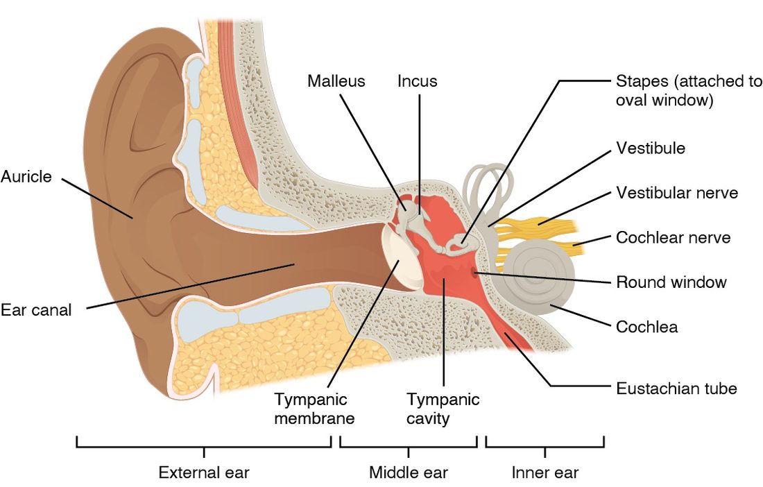
With labels hidden, select a box to reveal its label.
5How it works, step by step
- Sound waves travel down the ear canal and vibrate the tympanic membrane.The malleus, incus and stapes carry the vibration across the middle ear, and the stapes pushes in and out of the oval window with about 20 times the pressure.
- The stapes sets up pressure waves in the perilymph of the scala vestibuli.The basilar membrane vibrates most at the place tuned to the sound's frequency: near the base for high pitches, near the apex for low.
- The basilar membrane moves up and down under the tectorial membrane.The stereocilia of the hair cells bend, and tip links pull mechanically gated channels open.
- K+ from the endolymph flows into the hair cell.The hair cell depolarizes, Ca2+ enters, and the cell releases more glutamate onto its sensory neuron.
- The sensory neurons of the cochlear nerve fire faster.Signals pass through the cochlear nuclei, superior olivary nuclei, inferior colliculus and medial geniculate nucleus to the primary auditory cortex, where you perceive the sound.
6Core concepts
7A common mistake
The wrong idea: Each ear sends its signals only to the opposite side of the brain, so damage to one auditory cortex makes the opposite ear deaf.
What actually happens: Auditory fibers cross at several levels, starting in the brainstem, so each ear reaches the auditory cortex on both sides. Damage to one auditory cortex does not deafen either ear. Deafness in one ear points to that ear, its cochlear nerve or its cochlear nuclei.
8Check yourself
Anything you miss goes into your review queue.
1. Name the pinned structure.
- Malleus
- Stapes
- Incus
- Tympanic membrane
Show the answer
The incus is the middle ossicle. It links the malleus, attached to the eardrum, to the stapes in the oval window.
- Malleus: The malleus is the ossicle attached to the inside of the eardrum, the first in the chain.
- Stapes: The stapes is the last ossicle, whose footplate sits in the oval window.
- Correct: Incus: Correct. The middle bone of the chain is the incus.
- Tympanic membrane: The tympanic membrane is the sheet stretched across the end of the ear canal, not a bone.
2. Put the steps in order, from a sound entering the ear to the hair cells being stimulated.
- Sound waves vibrate the tympanic membrane
- The malleus, incus and stapes carry the vibration across the middle ear
- The stapes rocks in the oval window
- Pressure waves travel through the perilymph of the scala vestibuli
- The basilar membrane vibrates
- The hair cells' stereocilia bend
Show the answer
The eardrum vibrates, the ossicles pass the vibration to the oval window, the stapes sets up waves in the perilymph, the basilar membrane vibrates, and the tectorial membrane slides across the hair cells and bends their stereocilia.
- Correct order: 1. Sound waves vibrate the tympanic membrane 2. The malleus, incus and stapes carry the vibration across the middle ear 3. The stapes rocks in the oval window 4. Pressure waves travel through the perilymph of the scala vestibuli 5. The basilar membrane vibrates 6. The hair cells' stereocilia bend
3. A smoke alarm gives a high-pitched 4,000 Hz tone. Where on the basilar membrane is the vibration largest?
- Near the apex, where the membrane is wide and floppy
- Evenly along the whole length of the membrane
- Near the base, where the membrane is narrow and stiff
- At the round window, where the membrane ends
Show the answer
High frequencies vibrate the narrow, stiff base of the basilar membrane most, close to the oval window. Low frequencies travel farther and peak near the wide, floppy apex. Which hair cells are stimulated tells the brain the pitch.
- Near the apex, where the membrane is wide and floppy: The wide, floppy apex vibrates most for low frequencies, not high ones.
- Evenly along the whole length of the membrane: Each frequency makes the traveling wave peak at one place; the membrane does not vibrate equally along its length.
- Correct: Near the base, where the membrane is narrow and stiff: Correct. High pitch is coded near the base.
- At the round window, where the membrane ends: The round window is where the scala tympani ends at the middle ear; the basilar membrane lies along the cochlear duct.
4. The stereocilia of a hair cell are pushed toward the tallest stereocilium. What happens to the hair cell?
- Tip links slacken, channels close and the cell hyperpolarizes
- Tip links stretch, channels open, K+ flows in and the cell depolarizes
- The hair cell fires an action potential along its axon to the brainstem
- Tip links stretch, channels open, K+ flows out and the cell hyperpolarizes
Show the answer
Bending toward the tallest stereocilium stretches the tip links, which pull mechanically gated channels open. K+ from the endolymph flows in and depolarizes the cell, which then releases more glutamate onto its sensory neuron.
- Tip links slacken, channels close and the cell hyperpolarizes: This is what happens when the bundle bends away from the tallest stereocilium.
- Correct: Tip links stretch, channels open, K+ flows in and the cell depolarizes: Correct. Toward the tallest: channels open and the cell depolarizes.
- The hair cell fires an action potential along its axon to the brainstem: Hair cells have no axon. They release neurotransmitter onto sensory neurons, which fire the action potentials.
- Tip links stretch, channels open, K+ flows out and the cell hyperpolarizes: K+ flows in, not out, because the endolymph around the stereocilia is rich in K+ and positively charged.
5. Ms. Abara, 34, has muffled hearing in her right ear. In the Weber test she hears the tuning fork louder in her right ear. In the Rinne test on the right, she hears the fork longer on the mastoid process than beside the ear canal. What is the most likely diagnosis?
- Sensorineural hearing loss in the right ear
- Conductive hearing loss in the right ear
- Sensorineural hearing loss in the left ear
- Conductive hearing loss in the left ear
Show the answer
Bone conduction beating air conduction on the right means sound is blocked on its way through the right external or middle ear: a conductive loss. The Weber test fits, because a conductive loss makes the fork louder in the affected ear.
- Sensorineural hearing loss in the right ear: A right sensorineural loss would send the Weber sound to the left ear, and air conduction would still beat bone on the right.
- Correct: Conductive hearing loss in the right ear: Correct. Weber to the right plus bone better than air on the right means a right conductive loss.
- Sensorineural hearing loss in the left ear: A left sensorineural loss would also send the Weber sound right, but it would not make bone beat air in the right Rinne test.
- Conductive hearing loss in the left ear: A left conductive loss would send the Weber sound to the left ear.
6. A stroke destroys the primary auditory cortex on the left side only. What happens to the patient's hearing?
- He is deaf in the right ear
- He is deaf in the left ear
- Neither ear goes deaf, since each ear reaches both hemispheres
- He loses the ability to hear high-pitched sounds in both of his ears
Show the answer
Auditory fibers cross at several levels, beginning at the superior olivary nuclei, so each ear reaches both auditory cortices. Losing one cortex does not deafen either ear, although locating sounds and understanding speech can suffer.
- He is deaf in the right ear: Unlike touch, hearing from each ear reaches both hemispheres, so one-sided cortical damage does not deafen the opposite ear.
- He is deaf in the left ear: The left ear also sends signals to the intact right cortex.
- Correct: Neither ear goes deaf, since each ear reaches both hemispheres: Correct. Bilateral pathways protect against deafness from one-sided cortical damage.
- He loses the ability to hear high-pitched sounds in both of his ears: Frequency is mapped across each auditory cortex; losing one side does not remove high pitches from both ears.
7. A child spins in a chair for a minute and then stops suddenly. She feels as if she is spinning the other way. Why?
- Otoliths have come loose from the maculae
- The endolymph keeps flowing and bends the cupula the opposite way
- The cupula is heavy and is pulled downhill by gravity when she stops
- The vestibulocochlear nerve keeps firing from fatigue
Show the answer
During steady spinning, the endolymph catches up with the canal and the cupula returns upright. When she stops, the fluid's inertia keeps it flowing, which bends the cupula the opposite way and signals a turn in the other direction.
- Otoliths have come loose from the maculae: Loose otoliths cause brief positional dizziness, not the normal after-spin sensation everyone feels.
- Correct: The endolymph keeps flowing and bends the cupula the opposite way: Correct. The lagging fluid now overshoots and bends the cupula the other way.
- The cupula is heavy and is pulled downhill by gravity when she stops: The cupula has the same density as endolymph and no crystals, so gravity does not pull it.
- The vestibulocochlear nerve keeps firing from fatigue: Nerve fatigue would reduce firing, not create a clear sense of turning in a particular direction.
8. You turn your head quickly to the right while reading a sign. Through the vestibulo-ocular reflex, what do your eyes do?
- Both eyes turn right, ahead of the head
- Only the right eye turns left
- Both eyes stay fixed in their sockets and the image slides
- Both eyes turn left by the same amount
Show the answer
The semicircular canals signal the head turn to the vestibular nuclei, which drive the eye muscles to turn both eyes the same amount in the opposite direction. The image stays still on the retinas and the sign stays sharp.
- Both eyes turn right, ahead of the head: Turning the eyes with the head would make the image slide across the retinas, the opposite of what the reflex does.
- Only the right eye turns left: The reflex moves both eyes together, keeping them aligned.
- Both eyes stay fixed in their sockets and the image slides: Without the reflex the image would slide and blur; the reflex prevents this.
- Correct: Both eyes turn left by the same amount: Correct. The eyes counter-rotate to hold the image steady.
9Summary
The external ear (auricle, ear canal, tympanic membrane) collects sound. The middle ear's ossicles (malleus, incus, stapes) carry it to the oval window and raise its pressure about 20-fold; the auditory tube equalizes air pressure. The inner ear holds the cochlea, the vestibule and the semicircular canals. In the cochlea, the scala vestibuli and scala tympani hold perilymph and the cochlear duct holds K+-rich endolymph; the spiral organ (organ of Corti) sits on the basilar membrane under the tectorial membrane. Bending stereocilia toward the tallest opens mechanically gated channels, K+ flows in and the hair cell releases more glutamate; bending away does the opposite. Pitch is coded by place (high frequencies at the base, low at the apex), loudness by firing rate and recruitment. The auditory pathway runs through the cochlear nuclei, superior olivary nuclei, inferior colliculus and medial geniculate nucleus to the auditory cortex, reaching both hemispheres. Conductive loss (outer or middle ear) sends the Weber sound to the bad ear and makes bone beat air in the Rinne test; sensorineural loss (cochlea or nerve) sends the Weber sound to the good ear with air still better than bone. The utricle and saccule use otoliths on a gel membrane to sense tilt and straight-line movement; the semicircular canals use a cupula moved by lagging endolymph to sense turning, and drive the vestibulo-ocular reflex.