Sympathetic and parasympathetic divisions
1Why this matters
Mr. Okafor, 61, a long-time smoker, notices in the mirror that his right pupil is smaller than his left and his right eyelid droops a little. On a hot day, only the left side of his face sweats. A scan finds a tumor at the very top of his right lung, pressing on a chain of nerve ganglia beside his upper thoracic spine. To see why a lung tumor changes his eye and his sweating, you need the wiring of the two autonomic divisions.
2What this builds on
3Quick check before you start
1. Which horn of the spinal cord's gray matter holds the cell bodies of autonomic motor neurons in the thoracic cord?
- Dorsal horn
- Ventral horn
- Lateral horn
Show the answer
The small lateral horn, present from T1 to L2, holds autonomic motor cell bodies. The dorsal horn is sensory and the ventral horn holds somatic motor neurons.
- Dorsal horn:
- Ventral horn:
- Correct: Lateral horn:
2. In the peripheral nervous system, what is a ganglion?
- A cluster of neuron cell bodies outside the CNS
- A bundle of axons inside the CNS
- A cluster of neuron cell bodies inside the CNS
Show the answer
A ganglion is a cluster of neuron cell bodies in the PNS. Inside the CNS the same kind of cluster is called a nucleus, and a bundle of axons is a tract.
- Correct: A cluster of neuron cell bodies outside the CNS:
- A bundle of axons inside the CNS:
- A cluster of neuron cell bodies inside the CNS:
3. Which cranial nerve is the only one that reaches the organs of the chest and abdomen?
- Glossopharyngeal nerve (IX)
- Vagus nerve (X)
- Accessory nerve (XI)
Show the answer
The vagus nerve (wandering nerve) leaves the brainstem and runs down to the heart, lungs and digestive organs.
- Glossopharyngeal nerve (IX):
- Correct: Vagus nerve (X):
- Accessory nerve (XI):
4Anatomy
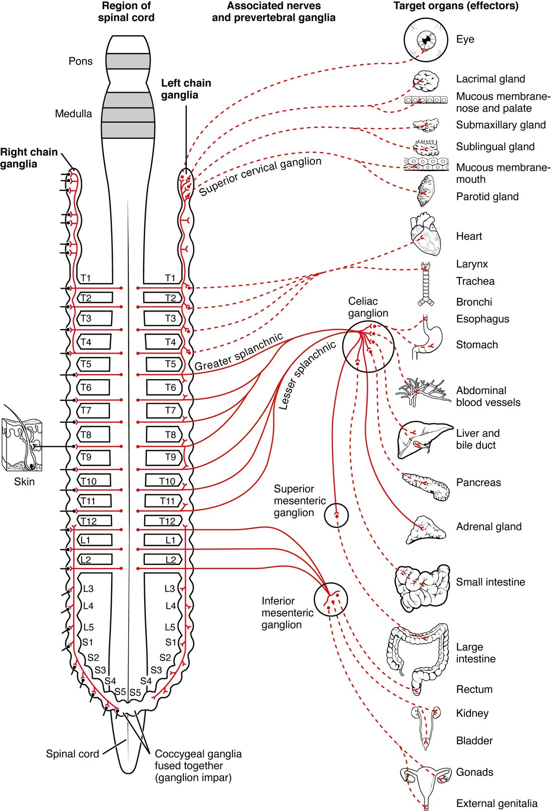
With labels hidden, select a box to reveal its label.
5How it works, step by step
- A sympathetic preganglionic neuron in the lateral horn of T1–L2 fires.Its myelinated axon leaves in the ventral root and turns into the sympathetic chain through a white ramus communicans.
- Inside the chain, the preganglionic axon branches up and down and synapses on many postganglionic neurons.Postganglionic neurons at many levels fire at once.
- Their unmyelinated axons leave through gray rami into every spinal nerve, or through sympathetic nerves to the head and chest.Sweat glands, skin blood vessels, the heart and the eye all receive the signal together.
- Other preganglionic axons pass through the chain in splanchnic nerves, some reaching the adrenal medulla directly.Chromaffin cells release epinephrine into the blood, which carries the signal to every tissue and prolongs it for minutes.
6Core concepts
7A common mistake
The wrong idea: The sympathetic division is the part of the ANS that comes from the spinal cord, and the parasympathetic division is the part that comes from the brain.
What actually happens: Both divisions start in the CNS and use two neurons. The sympathetic division leaves only from the thoracic and upper lumbar cord (T1–L2). The parasympathetic division leaves from the brainstem, in cranial nerves III, VII, IX and X, and also from the sacral cord (S2–S4), which supplies the bladder, rectum and reproductive organs.
8Check yourself
Anything you miss goes into your review queue.
1. Where are the cell bodies of sympathetic preganglionic neurons?
- In the brainstem and in sacral segments S2–S4
- In the lateral horn of the spinal cord, segments T1–L2
- In the chain ganglia beside the vertebral column
- In the ventral horn at every spinal segment
Show the answer
Sympathetic preganglionic cell bodies sit in the lateral horn of the thoracic and upper lumbar cord, T1–L2. That is why the division is called thoracolumbar.
- In the brainstem and in sacral segments S2–S4: The brainstem and S2–S4 are the parasympathetic (craniosacral) origins.
- Correct: In the lateral horn of the spinal cord, segments T1–L2: Correct. The lateral horn of T1–L2 holds every sympathetic preganglionic cell body.
- In the chain ganglia beside the vertebral column: The chain ganglia hold the cell bodies of postganglionic neurons. Preganglionic cell bodies are inside the CNS.
- In the ventral horn at every spinal segment: The ventral horn holds somatic motor neurons that supply skeletal muscle, and it runs the whole cord. Sympathetic output starts only at T1–L2.
2. Which cranial nerves carry parasympathetic preganglionic fibers?
- III, VII, IX and X
- V, VII, IX and X
- III, IV, VI, VII and X
- I, II, VIII and X
Show the answer
The oculomotor (III), facial (VII), glossopharyngeal (IX) and vagus (X) nerves carry the cranial parasympathetic outflow. The rest of the parasympathetic outflow leaves from S2–S4.
- Correct: III, VII, IX and X: Correct. Remember them as 3, 7, 9 and 10.
- V, VII, IX and X: The trigeminal nerve (V) is sensory to the face and motor to the chewing muscles. It carries no parasympathetic preganglionic fibers of its own.
- III, IV, VI, VII and X: Nerves IV and VI are purely somatic motor nerves to eye muscles. Of the eye-movement nerves, only nerve III carries parasympathetic fibers.
- I, II, VIII and X: Nerves I, II and VIII are sensory nerves for smell, vision, and hearing and balance. They carry no motor output at all.
3. Select every feature of the parasympathetic division.
- Preganglionic fibers leave in cranial nerves and from S2–S4
- Ganglia sit near or inside the target organ
- Postganglionic fibers reach the skin through gray rami communicantes
- Preganglionic fibers are long and postganglionic fibers are short
- A preganglionic neuron branches to many chain ganglia
- The vagus nerve carries most of its preganglionic fibers
Show the answer
The parasympathetic division is craniosacral, uses terminal or intramural ganglia in or beside its targets, so its preganglionic fibers are long and postganglionic fibers short, and about three quarters of its fibers travel in the vagus nerve. It has no chain and no gray rami.
- Correct: Preganglionic fibers leave in cranial nerves and from S2–S4: Correct. Nerves III, VII, IX and X, plus S2–S4.
- Correct: Ganglia sit near or inside the target organ: Correct. These are the terminal and intramural ganglia.
- Postganglionic fibers reach the skin through gray rami communicantes: Gray rami carry sympathetic postganglionic fibers. Parasympathetic fibers do not reach the skin.
- Correct: Preganglionic fibers are long and postganglionic fibers are short: Correct. The ganglion sits at the target, so the first neuron covers almost the whole distance.
- A preganglionic neuron branches to many chain ganglia: Branching through the chain is the sympathetic pattern. Parasympathetic preganglionic neurons contact only a few postganglionic neurons, and there is no parasympathetic chain.
- Correct: The vagus nerve carries most of its preganglionic fibers: Correct. The vagus carries about three quarters of all parasympathetic preganglionic fibers.
4. A fiber is traced in the gray ramus that joins spinal nerve C6. What kind of fiber is it most likely to be?
- A sympathetic preganglionic fiber entering the chain
- A parasympathetic postganglionic fiber heading to the arm
- A somatic motor fiber heading to an arm muscle
- An unmyelinated sympathetic postganglionic fiber returning from the chain
Show the answer
Gray rami carry unmyelinated sympathetic postganglionic fibers from the chain back into spinal nerves. Their preganglionic neurons sit in the upper thoracic cord; the fibers climbed the chain to a cervical ganglion before synapsing.
- A sympathetic preganglionic fiber entering the chain: Preganglionic fibers enter the chain through white rami, and there are no white rami at C6 because no sympathetic fibers leave the cord there.
- A parasympathetic postganglionic fiber heading to the arm: The parasympathetic division does not supply the limbs and has no fibers in the gray rami.
- A somatic motor fiber heading to an arm muscle: Somatic motor fibers leave in the ventral root and stay in the spinal nerve. They do not pass through the rami communicantes.
- Correct: An unmyelinated sympathetic postganglionic fiber returning from the chain: Correct. A gray ramus at any level carries postganglionic sympathetic fibers back into the spinal nerve.
5. Mr. Okafor, 61, has a tumor at the very top of his right lung, beside the upper thoracic vertebrae. His right pupil is smaller than his left, his right upper eyelid droops slightly, and the right side of his face does not sweat. Which structure has the tumor most likely damaged?
- The right oculomotor nerve
- The right sympathetic chain
- The right vagus nerve
- The right facial nerve
Show the answer
The sympathetic supply to the whole head leaves the cord at T1–T2 and climbs the chain to the superior cervical ganglion. A tumor at the lung apex can compress the chain there. The eye muscle that widens the pupil, a small smooth muscle in the eyelid and the facial sweat glands all lose their sympathetic input, so the pupil is small, the lid droops and the skin stays dry.
- The right oculomotor nerve: Oculomotor damage would remove the parasympathetic fibers that narrow the pupil, so the pupil would be large, not small. It would also paralyze several eye movements.
- Correct: The right sympathetic chain: Correct. Losing sympathetic input to the head produces a small pupil, a slightly drooping lid and loss of facial sweating on that side.
- The right vagus nerve: The vagus nerve supplies the organs of the chest and abdomen. It does not supply the pupil, the eyelid or the sweat glands of the face.
- The right facial nerve: The facial nerve's parasympathetic fibers go to the tear and saliva glands, and its motor fibers to the muscles of facial expression. Facial sweat glands get only sympathetic fibers.
6. Complete the pathway: lateral horn → white ramus → through the chain → splanchnic nerve → ____ → epinephrine released into the blood.
- Chromaffin cells of the adrenal medulla
- Postganglionic neurons in the celiac ganglion
- A gray ramus communicans
- Cells of the outer layer of the adrenal gland
Show the answer
Preganglionic fibers synapse directly on chromaffin cells in the adrenal medulla, which release epinephrine and some norepinephrine into the blood.
- Correct: Chromaffin cells of the adrenal medulla: Correct. Chromaffin cells stand in for postganglionic neurons.
- Postganglionic neurons in the celiac ganglion: Fibers to the adrenal medulla pass through the celiac ganglion without synapsing. If they synapsed there, the second neuron would release its transmitter onto an organ, not into the blood.
- A gray ramus communicans: Gray rami carry postganglionic fibers back to spinal nerves, toward the skin and limbs. They are not on the route to the adrenal gland.
- Cells of the outer layer of the adrenal gland: The outer layer of the adrenal gland is controlled by hormones, not by sympathetic fibers. Epinephrine comes from the medulla.
7. After a transplant, a heart has no nerve connections at all. Unlike a skeletal muscle that loses its nerve, it keeps contracting. Which feature of autonomic motor pathways does this illustrate?
- Autonomic effectors have their own activity, which the nerves speed up or slow down
- Autonomic signals travel through two neurons, so one cut neuron is replaced by the other
- Autonomic nerves regrow within hours after being cut
- Heart muscle is controlled by somatic motor neurons that are not cut in surgery
Show the answer
Skeletal muscle contracts only when a somatic motor neuron excites it, so cutting the nerve paralyzes it. Cardiac muscle, smooth muscle and many glands have their own built-in activity. Autonomic nerves adjust that activity up or down rather than starting it.
- Correct: Autonomic effectors have their own activity, which the nerves speed up or slow down: Correct. The heart beats without nerves; its autonomic supply only changes the rate and strength.
- Autonomic signals travel through two neurons, so one cut neuron is replaced by the other: The two neurons are in series, not in parallel. Cutting either one breaks the pathway.
- Autonomic nerves regrow within hours after being cut: Cut peripheral axons regrow slowly, over weeks to months, and after a transplant the heart stays largely without nerves for a long time.
- Heart muscle is controlled by somatic motor neurons that are not cut in surgery: Cardiac muscle is an autonomic effector. No somatic motor neurons supply it.
8. Select every target that receives sympathetic fibers but no parasympathetic fibers.
- Sweat glands in the skin
- Arrector pili muscles
- The adrenal medulla
- The heart
- Smooth muscle in most blood vessel walls
- The iris
Show the answer
Parasympathetic fibers do not travel in the gray rami or spinal nerves, so they never reach the skin or most blood vessels, and they do not supply the adrenal medulla. The heart and the iris get fibers from both divisions.
- Correct: Sweat glands in the skin: Correct. Sweat glands get sympathetic fibers through the gray rami.
- Correct: Arrector pili muscles: Correct. The arrector pili muscles are supplied only by sympathetic fibers.
- Correct: The adrenal medulla: Correct. Only sympathetic preganglionic fibers reach the adrenal medulla.
- The heart: The heart gets sympathetic fibers from the upper thoracic chain and parasympathetic fibers from the vagus.
- Correct: Smooth muscle in most blood vessel walls: Correct. Most blood vessels receive only sympathetic fibers.
- The iris: The iris gets parasympathetic fibers from nerve III, which narrow the pupil, and sympathetic fibers, which widen it.
9Summary
Autonomic motor pathways use two neurons: a preganglionic neuron in the CNS and a postganglionic neuron in an autonomic ganglion, whose unmyelinated axon releases transmitter from varicosities onto cardiac muscle, smooth muscle or glands. The sympathetic (thoracolumbar) division leaves the lateral horn at T1–L2 through white rami, synapses in the chain ganglia beside the spine or passes through splanchnic nerves to the celiac, superior mesenteric and inferior mesenteric ganglia, and returns fibers to every spinal nerve through gray rami. Its short preganglionic fibers branch widely, and some end on chromaffin cells of the adrenal medulla, which release epinephrine into the blood. The parasympathetic (craniosacral) division leaves in cranial nerves III, VII, IX and X and from S2–S4, with long preganglionic fibers running to terminal or intramural ganglia at the organ, so its effects stay local.