Autonomic control and visceral reflexes
1Why this matters
Ms. Farah, 24, gets up quickly from a hospital bed after a long night of vomiting. Her vision grays out, and she grabs the rail. A nurse notes her heart rate jump from 80 to 115 within a few seconds, and the dizziness passes. Nothing she chose to do raised that heart rate. A reflex in her brainstem shifted the balance between her two autonomic divisions, and this page shows how.
2What this builds on
3Quick check before you start
1. Which part of the brain links the nervous and endocrine systems and helps regulate body temperature, hunger and thirst?
- Cerebellum
- Hypothalamus
- Thalamus
Show the answer
The hypothalamus, below the thalamus, sets body temperature, hunger, thirst and water balance, and controls the autonomic nervous system.
- Cerebellum:
- Correct: Hypothalamus:
- Thalamus:
2. What is the difference between a somatic reflex and a visceral (autonomic) reflex?
- A somatic reflex acts on skeletal muscle; a visceral reflex acts on cardiac muscle, smooth muscle or glands
- A somatic reflex has no integration center; a visceral reflex has one
- A somatic reflex is always conscious; a visceral reflex is always spinal
Show the answer
Both kinds of reflex have a sensory receptor, an afferent path, an integration center, an efferent path and an effector. They differ in the effector: skeletal muscle for somatic, cardiac or smooth muscle and glands for visceral.
- Correct: A somatic reflex acts on skeletal muscle; a visceral reflex acts on cardiac muscle, smooth muscle or glands:
- A somatic reflex has no integration center; a visceral reflex has one:
- A somatic reflex is always conscious; a visceral reflex is always spinal:
3. Which receptor protein does acetylcholine from the vagus nerve bind on the heart?
- Beta-1
- Nicotinic
- Muscarinic
Show the answer
Parasympathetic postganglionic neurons release acetylcholine onto muscarinic receptor proteins, which slow the heart by opening potassium channels.
- Beta-1:
- Nicotinic:
- Correct: Muscarinic:
4Anatomy
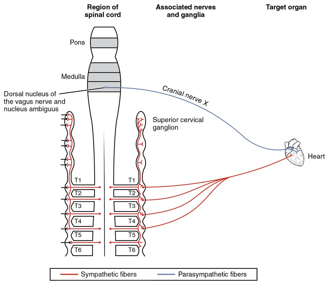
With labels hidden, select a box to reveal its label.
5How it works, step by step
- Standing up quickly pools blood in the legs, and pressure in the large arteries dips.Stretch-sensitive sensory receptors in the artery walls fire less, and fewer signals reach the medulla oblongata.
- The medulla oblongata receives less stretch input.It lowers vagal tone to the heart and raises sympathetic tone to the heart and vessels.
- Less acetylcholine on muscarinic receptor proteins and more norepinephrine on beta-1 and alpha-1 receptor proteins act on the heart and vessels.The heart beats faster and harder, and vessels in the skin, gut, kidneys and resting muscle narrow.
- The heart pumps more and the vessels resist flow more.Arterial pressure rises back toward normal, the stretch-sensitive sensory receptors fire at their usual rate again, and autonomic tone returns to its resting balance.
6Core concepts
7A common mistake
The wrong idea: The autonomic nervous system works on its own, independently of the brain.
What actually happens: Autonomic means self-governing, but the ANS is run by the CNS. Its preganglionic neurons sit in the brainstem and spinal cord; brainstem centers integrate autonomic reflexes; and the hypothalamus, fed by the limbic system and cortex, sets the overall balance. That is why fear speeds your heart and embarrassment makes you blush.
8Check yourself
Anything you miss goes into your review queue.
1. In a resting adult, atropine raises the heart rate by about 30 beats a minute, but a beta blocker lowers it by only a few beats. What does this show?
- At rest, vagal tone on the heart is much stronger than its sympathetic tone
- The heart carries no beta-1 receptor proteins while the body is at rest
- Atropine directly stimulates beta-1 receptor proteins
- The sympathetic division is silent at rest
Show the answer
Removing a tone reveals how much it was doing. Blocking the vagus nerve's effect frees the heart from a large brake, so the rate jumps. Blocking beta-1 removes only a small sympathetic push, so the rate falls a little.
- Correct: At rest, vagal tone on the heart is much stronger than its sympathetic tone: Correct. Vagal tone dominates the resting heart.
- The heart carries no beta-1 receptor proteins while the body is at rest: The heart carries beta-1 receptor proteins all the time; exercise and fright act through them. At rest they are simply receiving little norepinephrine.
- Atropine directly stimulates beta-1 receptor proteins: Atropine blocks muscarinic receptor proteins. It has no action on beta-1 receptor proteins.
- The sympathetic division is silent at rest: The sympathetic division keeps a low resting tone, which is why a beta blocker lowers the rate at all.
2. A patient is given an injection of phenylephrine, an alpha-1 agonist, into a vein. Predict each variable, first as the direct effect of the drug, before any reflex acts, and then about a minute later, once autonomic reflexes have acted. Compare each with the value before the injection.
| Variable | Change |
|---|---|
| Diameter of skin blood vessels, direct drug effect | — |
| Blood pressure, direct drug effect | — |
| Heart rate, direct drug effect | — |
| Vagal output to the heart, a minute later | — |
| Heart rate, a minute later | — |
| Blood pressure, a minute later | — |
Show the answer
Immediately, phenylephrine narrows vessels through alpha-1 receptor proteins and blood pressure rises, with no direct effect on the heart. The blood pressure reflex then increases vagal tone and lowers sympathetic tone, so the heart rate falls. Negative feedback corrects only part of the rise, so pressure stays somewhat high while the drug acts.
- Diameter of skin blood vessels, direct drug effect: down. Phenylephrine activates alpha-1 receptor proteins on vessel smooth muscle, which contracts and narrows the vessels.
- Blood pressure, direct drug effect: up. Narrower vessels throughout the body raise the pressure the heart must push against, so arterial pressure rises.
- Heart rate, direct drug effect: no change. Phenylephrine does not act on beta-1 receptor proteins, so it does not directly change the heart rate.
- Vagal output to the heart, a minute later: up. Stretch-sensitive sensory receptors in the walls of the large arteries fire more as pressure rises. The medulla oblongata responds by increasing vagal output and reducing sympathetic output.
- Heart rate, a minute later: down. More vagal tone, acting through muscarinic receptor proteins on the heart, slows the heart below its starting rate.
- Blood pressure, a minute later: up. The reflex slows the heart and trims sympathetic tone, but the drug is still holding alpha-1 receptor proteins active, so pressure is pulled back only part of the way and stays above its starting value.
3. The small vessels in the skin of your fingers have no parasympathetic supply. How does your nervous system widen them when you warm up?
- It sends acetylcholine to them through the vagus nerve
- It lowers sympathetic tone, so the vessels constrict less
- It raises sympathetic tone, which widens them through alpha-1 receptor proteins
- It cannot widen them; only local heat can
Show the answer
Vasomotor nerves keep finger skin vessels partly constricted at rest. Firing less lets the smooth muscle relax, and the vessels widen. One division with a resting tone gives two-way control.
- It sends acetylcholine to them through the vagus nerve: The vagus nerve does not supply skin vessels, and most blood vessels have no parasympathetic fibers at all.
- Correct: It lowers sympathetic tone, so the vessels constrict less: Correct. Withdrawing sympathetic tone widens the vessels.
- It raises sympathetic tone, which widens them through alpha-1 receptor proteins: Alpha-1 receptor proteins make vessel smooth muscle contract. More sympathetic tone narrows skin vessels.
- It cannot widen them; only local heat can: The hypothalamus widens these vessels through the nervous system, by withdrawing sympathetic tone. Local warmth helps, but it is not the only route.
4. Ms. Tran's right oculomotor nerve is compressed after a head injury. A penlight is shone into her right eye. What happens to her pupils?
- Both pupils narrow normally
- Neither pupil narrows
- The right pupil narrows, but the left pupil stays wide
- The left pupil narrows but the right does not
Show the answer
Her right retina and optic nerve still send the light signal to the midbrain, which drives both Edinger–Westphal nuclei. The left oculomotor nerve is intact, so the left pupil narrows. The right oculomotor nerve cannot carry the parasympathetic output, so the right pupil stays wide.
- Both pupils narrow normally: The right pupil needs the right oculomotor nerve to narrow, and that nerve is compressed.
- Neither pupil narrows: The sensory side works and the midbrain sends the signal to both sides, so the left pupil still responds.
- The right pupil narrows, but the left pupil stays wide: The right pupil's efferent pathway is the damaged one, so the right pupil is the one that fails.
- Correct: The left pupil narrows but the right does not: Correct. The input is intact and the midbrain crosses the signal, but only the left efferent pathway works.
5. Select every effect of strong sympathetic activation.
- Blood vessels in the skin narrow
- The liver releases glucose into the blood
- The pupils narrow
- Sweating increases
- Gut movement speeds up
- The small airways widen
Show the answer
Sympathetic activation narrows skin vessels (alpha-1), widens the airways (beta-2, mostly epinephrine), releases glucose from the liver (beta-2), and drives sweating (muscarinic, through cholinergic sympathetic fibers). It widens the pupils and slows the gut.
- Correct: Blood vessels in the skin narrow: Correct. Norepinephrine from the vasomotor nerves acts on alpha-1 receptor proteins and contracts the smooth muscle of skin vessels.
- Correct: The liver releases glucose into the blood: Correct. Epinephrine on beta-2 receptor proteins makes the liver break down glycogen.
- The pupils narrow: Narrowing the pupils is a parasympathetic effect. Sympathetic activity widens them through alpha-1 receptor proteins.
- Correct: Sweating increases: Correct. Sympathetic cholinergic fibers drive the eccrine sweat glands.
- Gut movement speeds up: Speeding the gut is a parasympathetic effect. Sympathetic activity slows it.
- Correct: The small airways widen: Correct. Epinephrine relaxes airway smooth muscle through beta-2 receptor proteins.
6. Which brain region is the main integrator of autonomic function, linking emotions, body temperature and water balance to autonomic output?
- Cerebellum
- Hypothalamus
- Primary motor cortex
- Thalamus
Show the answer
The hypothalamus compares temperature, water balance and energy stores with their set points, receives emotional input from the limbic system, and sends commands to the brainstem and spinal cord autonomic centers.
- Cerebellum: The cerebellum coordinates movement. It is not the integrator of autonomic output.
- Correct: Hypothalamus: Correct. The hypothalamus sits at the top of the autonomic control hierarchy below the cerebrum.
- Primary motor cortex: The primary motor cortex controls voluntary movement through somatic motor pathways.
- Thalamus: The thalamus relays sensory signals to the cortex. It does not integrate autonomic output.
7. Mr. Sato's spinal cord was completely cut at C5 an hour ago. His blood pressure is low, his heart rate is 48, and his feet are warm and pink. Which explanation fits all three findings?
- His vagus nerve was cut, so vagal tone is lost
- His adrenal medulla is releasing too much epinephrine
- He has lost sympathetic tone while vagal output continues
- His skeletal muscles are paralyzed, so they need less of the blood flow
Show the answer
Brain control of all sympathetic output runs down the cord to T1–L2, below the cut. Without sympathetic tone, vessels widen (low pressure, warm pink skin) and the heart loses its sympathetic push. The vagus nerve leaves the brainstem above the cut, so vagal tone continues unopposed and slows the heart.
- His vagus nerve was cut, so vagal tone is lost: The vagus nerve leaves the brainstem and never enters the spinal cord, so a cord injury does not cut it. Losing vagal tone would speed the heart.
- His adrenal medulla is releasing too much epinephrine: Epinephrine would speed the heart and constrict skin vessels, the opposite of these findings. The adrenal medulla also depends on T-segment preganglionic neurons cut off from the brain.
- Correct: He has lost sympathetic tone while vagal output continues: Correct. Lost sympathetic tone plus intact vagal tone explains all three findings.
- His skeletal muscles are paralyzed, so they need less of the blood flow: Paralysis does not explain a slow heart or warm feet. The findings come from lost sympathetic tone.
8. A resting patient is given a drug that blocks the nicotinic receptor proteins in every autonomic ganglion. What happens to his heart rate?
- It falls, because sympathetic input to the heart is removed
- It stays the same, because the two effects cancel exactly
- It rises, because removing both tones takes away the larger vagal brake
- It stops, because the heart needs autonomic input to beat
Show the answer
Blocking every ganglion silences both divisions. On the resting heart, vagal tone is the larger influence, so removing both leaves a net loss of braking: the heart rises toward its own rate of about 100 beats a minute.
- It falls, because sympathetic input to the heart is removed: Sympathetic input is removed, but the larger vagal tone is removed too, so the net effect is a rise.
- It stays the same, because the two effects cancel exactly: The two tones are not equal at rest; vagal tone dominates.
- Correct: It rises, because removing both tones takes away the larger vagal brake: Correct. The net loss is of the vagal brake.
- It stops, because the heart needs autonomic input to beat: The heart beats on its own; autonomic nerves only adjust its rate.
9Summary
Most organs of the chest and abdomen have dual innervation: both divisions reach them, usually with opposite effects (heart rate, pupil, gut, bladder) and sometimes cooperatively (saliva). Both divisions fire at a resting autonomic tone, so one division can turn an effector up or down. Vagal tone dominates the resting heart, holding it well below its own rate of about 100; sympathetic tone in the vasomotor nerves keeps most blood vessels, which have no parasympathetic fibers, partly constricted. Sympathetic activation raises heart rate and force, shifts blood from skin and gut to muscle, widens airways and pupils, releases glucose and drives sweating, often as a mass discharge prolonged by adrenal epinephrine. Parasympathetic activation slows the heart, narrows the pupils and speeds the gut, locally and briefly. Visceral reflexes such as the pupillary light reflex and the blood pressure reflex use a two-neuron efferent limb, and control runs from the spinal cord up through brainstem centers to the hypothalamus, which receives emotional input from the limbic system.