Most of your organs get nerve fibers from both autonomic divisions, and both divisions fire all the time. That is dual innervation and autonomic tone, and together they let your nervous system turn an organ up or down smoothly instead of switching it on and off. This page explains both ideas, lists what sympathetic and parasympathetic activation each do across the body, follows two autonomic reflexes step by step, and shows how the hypothalamus and brainstem run the whole system.
Dual innervation
Your heart has two sets of autonomic nerves (Figure 1). Sympathetic fibers from the upper thoracic cord (T1–T4) climb into the chain, synapse there, and run to the heart as the cardiac accelerator nerves. Parasympathetic fibers leave the medulla oblongata in the vagus nerve and synapse in terminal ganglia on the heart itself.
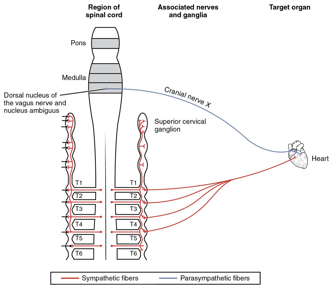
An organ that receives fibers from both divisions has dual innervation (dual = two; in- = into, nerv- = nerve). Most organs of the chest and abdomen do. The two divisions usually act in opposite directions on the same effector:
- Heart rate: sympathetic speeds it, parasympathetic slows it.
- Pupil: sympathetic widens it, parasympathetic narrows it. Here each division drives a different muscle: the radial dilator and the circular muscle of the iris.
- Stomach and intestines: sympathetic slows their movement and secretion, parasympathetic speeds them.
- Bladder: sympathetic relaxes its wall and tightens its outlet, parasympathetic contracts the wall and relaxes the outlet.
Not every pairing is a tug of war. For the glands that make saliva, both divisions increase secretion: parasympathetic activity brings a large volume of watery saliva, and sympathetic activity a small volume of thick, protein-rich saliva. That is why your mouth feels dry and sticky when you are nervous.
And some effectors have only sympathetic fibers: most blood vessels, the sweat glands, the arrector pili muscles and the adrenal medulla. These have no opposing division, so how do they get turned down? The answer is tone.
Autonomic tone
Autonomic neurons are never fully silent. At rest, both divisions fire at a low, steady rate. That background activity is autonomic tone (tonus = tension). Because there is always some tone, a division can change an effector in both directions: fire faster and the effect grows, fire slower and it shrinks.
Vagal tone on the heart
A heart with every nerve blocked, as after a transplant, beats about 100 times a minute. Yet your resting heart rate is closer to 60–80. The difference is vagal tone: steady parasympathetic firing in the vagus nerve that holds the heart below its own rate. At rest, vagal tone outweighs sympathetic tone on the heart. That is why atropine, which blocks the vagus nerve's effect, often raises a resting heart rate by 30 or more beats a minute, while a beta blocker lowers it only a little.
Sympathetic tone on blood vessels
The smooth muscle in most vessel walls is supplied by sympathetic fibers called vasomotor nerves (vas- = vessel, motor = mover). Their steady firing, sympathetic tone, keeps the vessels partly constricted all the time. More firing narrows them further; less firing lets them widen. One division, with tone, gives full two-way control with no opposing nerve at all (Figure 2).
| Vagal tone | Sympathetic tone | |
|---|---|---|
| Division | Parasympathetic | Sympathetic |
| Best example | The heart's rate at rest | The diameter of blood vessels |
| Nerve | Vagus nerve | Vasomotor nerves (and cardiac accelerator nerves to the heart) |
| Resting effect | Holds the heart about 20–40 beats a minute below its own rate | Keeps most vessels partly constricted |
| If the tone is removed | Heart rate rises (atropine, heart transplant) | Vessels widen and blood pressure falls (alpha-1 blocker, ganglion blocker, spinal cord injury) |
| If the tone increases | Heart rate falls | Vessels narrow and blood pressure rises |
Effects of sympathetic activation
Picture a car swerving toward you. Before you have even named the danger, your heart is pounding. Strong sympathetic activation produces a coordinated whole-body pattern, the fight-or-flight response, through three routes at once: postganglionic neurons releasing norepinephrine, cholinergic fibers to the sweat glands, and epinephrine from the adrenal medulla. The effects, with the receptor proteins you met in the last topic:
- Heart: faster rate and stronger contractions (beta-1), through the cardiac accelerator nerves and circulating epinephrine.
- Blood vessels: vessels in the skin, stomach, intestines and kidneys constrict (alpha-1); vessels in working skeletal muscle dilate, mainly because chemicals released by the working muscle override sympathetic constriction, with some help from epinephrine on beta-2. Blood is shifted toward the muscles, and blood pressure rises.
- Airways: smooth muscle relaxes and the airways widen (beta-2, mostly epinephrine).
- Eyes: pupils widen (alpha-1).
- Fuel: the liver breaks glycogen down and releases glucose into the blood (beta-2), and fat cells release fatty acids.
- Skin: sweating increases (muscarinic, cholinergic sympathetic fibers) and hair stands on end (alpha-1).
- Gut and bladder: movement and secretion slow, and sphincters tighten (alpha-1), so emptying is put off.
- Brain: alertness rises.
Two features set the sympathetic pattern apart. It can act as a mass discharge, many organs at once, because of the chain's wide divergence and the adrenal medulla. And the adrenal epinephrine makes its effects outlast the nerve activity by minutes. The sympathetic division can also act selectively: on a hot day, it widens the skin's vessels and drives sweating without widening your pupils or stopping your gut.
Effects of parasympathetic activation
After a big meal on a quiet evening, parasympathetic activity dominates. Its effects are the rest-and-digest pattern, mostly through muscarinic receptor proteins:
- Heart: slower rate.
- Eyes: pupils narrow (miosis), and the ciliary muscle contracts to focus the lens for near vision.
- Glands of the head: tears and large volumes of watery saliva.
- Stomach and intestines: more movement and more secretion; sphincters relax.
- Bladder and rectum: the walls contract and the sphincters relax, allowing emptying.
- Airways: smooth muscle contracts, narrowing the airways, and mucus secretion increases.
Parasympathetic effects are local and short. The terminal ganglia sit on single organs, each preganglionic neuron reaches few postganglionic neurons, and acetylcholine is destroyed within milliseconds. So your stomach can be stimulated without your pupils narrowing.
| Sympathetic activation | Parasympathetic activation | |
|---|---|---|
| Overall pattern | Fight or flight: exercise, danger, stress | Rest and digest: quiet, after meals |
| Heart rate and force | Up and stronger | Down |
| Blood vessels | Skin, gut, kidneys and resting muscle constrict; working muscle dilates | No effect on most vessels (no fibers) |
| Airways | Widen | Narrow, more mucus |
| Pupils | Widen | Narrow |
| Gut movement and secretion | Down | Up |
| Bladder | Wall relaxes, outlet tightens | Wall contracts, outlet relaxes |
| Glucose released by the liver | Up | No major effect |
| Sweating | Up | No effect (no fibers) |
| Spread | Widespread, many organs together | Local, organ by organ |
| Duration | Longer, extended by adrenal epinephrine | Brief |
Autonomic reflexes
You met the visceral (autonomic) reflex in the reflexes topic. It uses the same five parts as any reflex arc, with one difference on the way out (Figure 3):
- A sensory receptor in an organ or blood vessel detects a change (stretch, chemicals, light, temperature).
- A visceral sensory neuron carries the signal into the CNS, often in the vagus or glossopharyngeal nerve, or a spinal nerve.
- An integration center in the spinal cord or brainstem processes it.
- The efferent side is the two-neuron chain: preganglionic neuron, ganglion, postganglionic neuron.
- An effector (cardiac muscle, smooth muscle or a gland) responds.
The pupillary light reflex
Shine a penlight into one eye, and both pupils narrow within a second. You met this reflex with the eye; here is its autonomic wiring (Figure 4):
- Light strikes the retina, and retinal ganglion cells fire.
- Signals travel along the optic nerve to a small center in the midbrain (the pretectal nucleus), on both sides.
- Each pretectal nucleus signals the parasympathetic nucleus of the oculomotor nerve (the Edinger–Westphal nucleus) on both sides.
- Preganglionic fibers run in both oculomotor nerves (III) to the ciliary ganglia.
- Short postganglionic fibers release acetylcholine onto muscarinic receptor proteins of the circular iris muscle.
- The circular muscle contracts, and both pupils narrow.
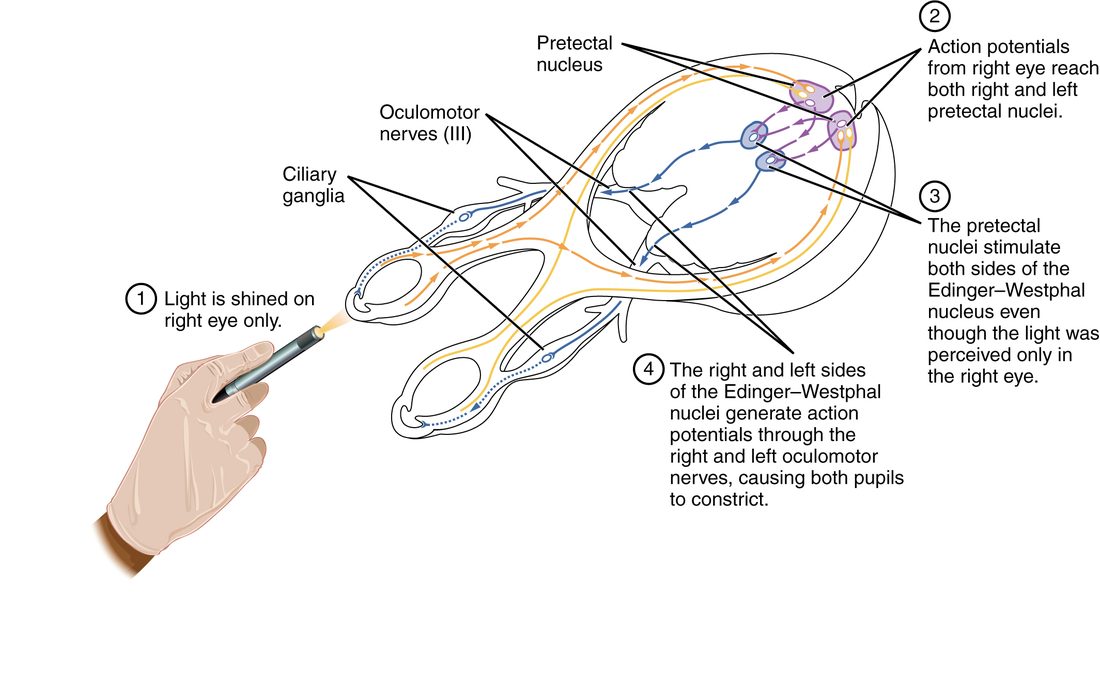
Because the midbrain crosses the signal to both sides, the unlit pupil narrows too. In dim light, the opposite happens: parasympathetic output falls and sympathetic output from the upper thoracic cord, relayed through the superior cervical ganglion, contracts the dilator muscle. Doctors test this reflex after head injuries because it checks the optic nerve, the midbrain and the oculomotor nerve in one quick look.
The blood pressure reflex
Stand up quickly from lying down. Blood pools in your legs, and for a moment less returns to your heart, so the pressure in your large arteries dips. Within a few heartbeats a reflex corrects it:
- Stretch-sensitive sensory receptors in the walls of the large arteries near your heart and in your neck are stretched less, so they fire less.
- Fewer signals reach the medulla oblongata through the glossopharyngeal and vagus nerves.
- The medulla oblongata reduces vagal output and increases sympathetic output.
- Heart rate and force rise (beta-1), and vasomotor nerves constrict small arteries in the skin, gut, kidneys and resting skeletal muscle, and the veins (alpha-1).
- Arterial pressure rises back toward normal.
A rise in pressure runs the same loop the other way: more stretch, more vagal output, less sympathetic tone, a slower heart and wider vessels. This is negative feedback carried entirely by autonomic tone, and you will study it in detail, with its sensory receptors named, in the blood pressure topic.
Central control of the ANS
The ANS is not independent of the brain. Its control runs in a hierarchy, from reflexes that need only the spinal cord up to emotions that start in the cerebrum (Figure 5).
- Spinal cord. The preganglionic neurons themselves, and simple visceral reflexes. The reflexes that empty the bladder and rectum are integrated in the sacral cord; the brain normally adds or removes permission.
- Brainstem. The main autonomic reflex centers. The medulla oblongata receives visceral sensory input from nerves IX and X, and its centers adjust heart rate, vessel diameter and breathing, and run coughing, swallowing and vomiting. The dorsal motor nucleus of the vagus sends parasympathetic output to the chest and abdomen. The midbrain runs the pupillary reflexes.
- Hypothalamus. The master integrator. It compares body temperature, water balance and energy stores with their set points and sends commands down tracts to the brainstem and spinal cord. Warm hypothalamic neurons drive skin vessel widening and sweating; cold ones drive skin vessel narrowing and shivering.
- Limbic system and cerebral cortex. Emotions and thoughts reach the hypothalamus from the limbic system, especially the amygdala. Fear starts a sympathetic surge; embarrassment widens facial vessels and you blush; the sight of blood can trigger a sudden burst of vagal activity with widened vessels, and some people faint.
You cannot usually command your heart rate the way you command your fingers. But you can change it indirectly: slow breathing, relaxation or imagining a frightening scene all act through the limbic system and hypothalamus, which is the basis of biofeedback training.
Putting it together: a sudden fright
A dog lunges at you from behind a fence. Follow the response through time:
- Within a second: the amygdala and hypothalamus drive the brainstem and spinal cord. Vagal output to the heart drops and sympathetic output jumps. Heart rate climbs (beta-1), the pupils widen (alpha-1), and skin vessels constrict so you go pale (alpha-1).
- Within a few seconds: sweat breaks out on your palms (muscarinic), and preganglionic fibers to the adrenal medulla release epinephrine into the blood.
- Over the next minute: epinephrine reaches the airways (beta-2, wider), the liver (beta-2, glucose released) and skeletal muscle vessels (beta-2, wider). Your gut stops churning.
- After the dog is gone: sympathetic firing falls back to its resting tone within seconds, and vagal tone slows the heart. But epinephrine keeps circulating for a few minutes, so your heart keeps pounding and your hands stay shaky after the danger has passed.
Common mix-ups
- "The ANS is independent of the brain." Autonomic means self-governing, but the hypothalamus and brainstem control it continuously, and emotions reach it through the limbic system.
- "A division is either on or off." Both divisions fire at a resting tone. Most changes come from turning one up and the other down.
- "Every organ gets both divisions." Most blood vessels, sweat glands, arrector pili muscles and the adrenal medulla get only sympathetic fibers, controlled by changing sympathetic tone.