Autonomic neurotransmitters and receptors
1Why this matters
Mr. Diallo, 70, is found at home with a pulse of 38 and is faint. The paramedic gives him atropine, and within a minute his heart rate climbs to 75. Twenty minutes later, his mouth is dry, his pupils are wide and his skin is hot. One drug, one kind of receptor protein, and effects in four organs. To predict all of them, you need to know which autonomic transmitter acts on which receptor protein, organ by organ.
2What this builds on
3Quick check before you start
1. Which receptor protein does acetylcholine bind at the neuromuscular junction?
- Muscarinic receptor protein
- Nicotinic receptor protein
- Beta receptor protein
Show the answer
At the motor end plate, acetylcholine binds nicotinic receptor proteins, which are ligand-gated channels that let sodium in.
- Muscarinic receptor protein:
- Correct: Nicotinic receptor protein:
- Beta receptor protein:
2. What is the difference between an ionotropic and a metabotropic receptor protein?
- An ionotropic one is itself an ion channel; a metabotropic one acts through a G protein and second messengers
- An ionotropic one binds hormones; a metabotropic one binds neurotransmitters
- An ionotropic one is always inhibitory; a metabotropic one is always excitatory
Show the answer
Ionotropic receptor proteins open a channel within themselves as soon as the transmitter binds, so they act fast. Metabotropic receptor proteins activate a G protein, which changes channels or second messengers more slowly.
- Correct: An ionotropic one is itself an ion channel; a metabotropic one acts through a G protein and second messengers:
- An ionotropic one binds hormones; a metabotropic one binds neurotransmitters:
- An ionotropic one is always inhibitory; a metabotropic one is always excitatory:
3. Where do sympathetic postganglionic neurons have their cell bodies?
- In the lateral horn of T1–L2
- In chain and collateral ganglia
- In terminal ganglia inside the organ wall
Show the answer
Sympathetic postganglionic cell bodies sit in the chain ganglia beside the spine or the collateral ganglia in front of it. The lateral horn holds the preganglionic neurons.
- In the lateral horn of T1–L2:
- Correct: In chain and collateral ganglia:
- In terminal ganglia inside the organ wall:
4Anatomy
With labels hidden, select a box to reveal its label.
5How it works, step by step
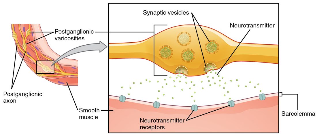
- A sympathetic postganglionic neuron fires, and action potentials reach its varicosities.Calcium enters the varicosities and norepinephrine is released onto nearby smooth muscle and heart cells.
- Norepinephrine binds whatever adrenergic receptor protein each target cell carries.Alpha-1 receptor proteins raise calcium in vessel smooth muscle; beta-1 receptor proteins raise cyclic AMP in the heart.
- Raised calcium in vessel smooth muscle and raised cyclic AMP in heart cells change how those cells work.Skin vessels constrict, while the heart beats faster and more forcefully.
- Norepinephrine also binds alpha-2 receptor proteins on the varicosity itself, and is taken back up.Further release is turned down and the signal ends within about a second, unless circulating epinephrine keeps the receptor proteins active.
6Core concepts
7A common mistake
The wrong idea: The sympathetic division always stimulates organs and the parasympathetic division always inhibits them.
What actually happens: Each division excites some organs and inhibits others, because the response is set by the receptor protein on the target, not by the division. Parasympathetic acetylcholine on muscarinic receptor proteins speeds up the gut and narrows the airways. Sympathetic epinephrine on beta-2 receptor proteins relaxes airway smooth muscle, and norepinephrine on alpha-1 receptor proteins contracts vessel smooth muscle.
8Check yourself
Anything you miss goes into your review queue.
1. Which autonomic neurons release acetylcholine?
- Only parasympathetic neurons, both preganglionic and postganglionic
- All preganglionic neurons, all parasympathetic postganglionic neurons, and sympathetic postganglionic neurons to sweat glands
- All postganglionic neurons of both the sympathetic and parasympathetic divisions
- Only the preganglionic neurons that end on the adrenal medulla
Show the answer
Every preganglionic neuron in both divisions is cholinergic, as is every parasympathetic postganglionic neuron. The one group of cholinergic sympathetic postganglionic neurons supplies the eccrine sweat glands.
- Only parasympathetic neurons, both preganglionic and postganglionic: Sympathetic preganglionic neurons also release acetylcholine, onto nicotinic receptor proteins in the chain and collateral ganglia.
- Correct: All preganglionic neurons, all parasympathetic postganglionic neurons, and sympathetic postganglionic neurons to sweat glands: Correct. These are the cholinergic neurons of the ANS.
- All postganglionic neurons of both the sympathetic and parasympathetic divisions: Most sympathetic postganglionic neurons are adrenergic and release norepinephrine.
- Only the preganglionic neurons that end on the adrenal medulla: Preganglionic neurons to the adrenal medulla are cholinergic, but so are all other preganglionic neurons and all parasympathetic postganglionic neurons.
2. On a hot day, sympathetic activity makes you sweat. Which transmitter and receptor protein act on the eccrine sweat glands?
- Norepinephrine on alpha-1 adrenergic receptor proteins
- Epinephrine on beta-2 receptor proteins
- Acetylcholine on muscarinic receptor proteins
- Acetylcholine on nicotinic receptor proteins
Show the answer
The sympathetic postganglionic neurons to eccrine sweat glands are an exception: they are cholinergic. Their acetylcholine binds muscarinic receptor proteins on the gland cells.
- Norepinephrine on alpha-1 adrenergic receptor proteins: Most sympathetic targets do receive norepinephrine, but the eccrine sweat glands are the classic exception.
- Epinephrine on beta-2 receptor proteins: Circulating epinephrine has only a small effect on eccrine sweating. The nerve supply, using acetylcholine, drives it.
- Correct: Acetylcholine on muscarinic receptor proteins: Correct. A sympathetic response carried by acetylcholine on muscarinic receptor proteins.
- Acetylcholine on nicotinic receptor proteins: Nicotinic receptor proteins are in the ganglia and adrenal medulla, not on the sweat gland cells. That is why atropine, a muscarinic blocker, stops sweating.
3. A patient starts taking metoprolol, a beta blocker that acts mainly on beta-1 receptor proteins. Predict the change in each variable at rest, compared with before the drug.
| Variable | Change |
|---|---|
| Heart rate | — |
| Force of each heart contraction | — |
| Blood pressure | — |
| Sweating | — |
| Pupil diameter | — |
Show the answer
A beta-1 blocker removes the sympathetic push on the heart only. Heart rate and force fall, so blood pressure falls. Targets that respond through alpha or muscarinic receptor proteins, such as the sweat glands and the pupil, are unchanged.
- Heart rate: down. Norepinephrine and epinephrine can no longer bind the beta-1 receptor proteins in the heart's pacemaker tissue, so the cells reach threshold more slowly.
- Force of each heart contraction: down. Blocked beta-1 receptor proteins on heart muscle cells mean less cyclic AMP and less calcium entry, so each contraction is weaker.
- Blood pressure: down. A slower, weaker heart pumps less blood into the arteries each minute, so arterial pressure falls.
- Sweating: no change. Eccrine sweat glands respond to acetylcholine on muscarinic receptor proteins, which a beta-1 blocker does not touch.
- Pupil diameter: no change. The pupil is set by alpha-1 receptor proteins on the dilator muscle and muscarinic receptor proteins on the circular muscle, neither of which is blocked.
4. A farmworker is poisoned by an organophosphate insecticide, which blocks acetylcholinesterase. He is drooling and sweating, his pupils are pinpoint, his heart rate is slow, his airways are filling with secretions, and his muscles twitch and grow weak. He is given atropine. Which problem will atropine not fix?
- The heart rate of 42 beats per minute
- The drooling
- The airway secretions
- The muscle twitching and weakness
Show the answer
Acetylcholine builds up at all cholinergic synapses. Atropine blocks only muscarinic receptor proteins, so it reverses the parasympathetic-type effects and the sweating. The twitching and weakness come from acetylcholine acting on nicotinic receptor proteins at the neuromuscular junction, which atropine does not block.
- The heart rate of 42 beats per minute: Atropine blocks the muscarinic receptor proteins through which acetylcholine slows the heart, so the rate rises.
- The drooling: Saliva secretion is driven by acetylcholine on muscarinic receptor proteins, which atropine blocks.
- The airway secretions: Airway glands secrete in response to acetylcholine on muscarinic receptor proteins, which atropine blocks. Drying these secretions is the main goal of atropine treatment.
- Correct: The muscle twitching and weakness: Correct. Skeletal muscle responds through nicotinic receptor proteins, which atropine leaves untouched.
5. During a sprint, why do the small airways widen more once the adrenal medulla is releasing its hormone than with sympathetic nerve activity alone?
- Epinephrine binds beta-2 receptor proteins far better than norepinephrine
- Epinephrine blocks muscarinic receptor proteins in the airways
- The adrenal medulla releases acetylcholine onto the airways
- Norepinephrine from nerves activates alpha-1 receptor proteins, which widen the airways
Show the answer
Airway smooth muscle relaxes through beta-2 receptor proteins. Norepinephrine binds beta-2 weakly, and few varicosities reach these muscle cells. Epinephrine arriving in the blood binds beta-2 strongly, so the airways widen more.
- Correct: Epinephrine binds beta-2 receptor proteins far better than norepinephrine: Correct. Beta-2 responses depend mainly on circulating epinephrine.
- Epinephrine blocks muscarinic receptor proteins in the airways: Epinephrine does not bind muscarinic receptor proteins. It acts on alpha and beta adrenergic receptor proteins.
- The adrenal medulla releases acetylcholine onto the airways: The adrenal medulla releases epinephrine and some norepinephrine, not acetylcholine.
- Norepinephrine from nerves activates alpha-1 receptor proteins, which widen the airways: Alpha-1 receptor proteins make smooth muscle contract, which would narrow airways, not widen them.
6. A student described how the vagus nerve slows the heart. One step is wrong. Which one?
- A vagal preganglionic neuron releases acetylcholine in a terminal ganglion in the heart wall
- Acetylcholine binds nicotinic receptor proteins on the postganglionic neuron
- The postganglionic neuron releases norepinephrine onto the heart's pacemaker tissue
- The transmitter binds muscarinic receptor proteins, and potassium channels open
- The cells hyperpolarize and take longer to reach threshold
Show the answer
Parasympathetic postganglionic neurons are cholinergic: they release acetylcholine, which binds muscarinic receptor proteins. Norepinephrine is the sympathetic transmitter, and it would speed the heart.
- A vagal preganglionic neuron releases acetylcholine in a terminal ganglion in the heart wall: This step is right. Vagal preganglionic fibers end in terminal ganglia in or on the heart.
- Acetylcholine binds nicotinic receptor proteins on the postganglionic neuron: This step is right. Ganglionic transmission in both divisions uses nicotinic receptor proteins.
- Correct: The postganglionic neuron releases norepinephrine onto the heart's pacemaker tissue: This is the error. The transmitter here is acetylcholine.
- The transmitter binds muscarinic receptor proteins, and potassium channels open: This step is right. Muscarinic receptor proteins on the pacemaker tissue open potassium channels.
- The cells hyperpolarize and take longer to reach threshold: This step is right. Starting further from threshold, the cells fire later, so the rate falls.
7. Mr. Adeyemi has a severe allergic reaction: his blood pressure is falling and his airways are closing. The paramedic injects epinephrine. Which receptor protein is responsible for reopening his airways?
- Alpha-1
- Beta-2
- Beta-1
- Muscarinic
Show the answer
Epinephrine binds beta-2 receptor proteins on airway smooth muscle, raising cyclic AMP and relaxing the muscle, so the airways reopen. At the same time, alpha-1 constricts leaky vessels and raises blood pressure, and beta-1 strengthens the heart.
- Alpha-1: Alpha-1 activation constricts blood vessels and helps restore pressure. It does not relax airway smooth muscle.
- Correct: Beta-2: Correct. Beta-2 relaxes airway smooth muscle.
- Beta-1: Beta-1 speeds and strengthens the heart. It does not act on the airways.
- Muscarinic: Epinephrine does not bind muscarinic receptor proteins, and activating them would narrow the airways.
8. Norepinephrine makes the smooth muscle of skin vessels contract, but epinephrine makes the smooth muscle of skeletal muscle vessels relax. What is the best explanation?
- Epinephrine and norepinephrine carry opposite instructions
- Skeletal muscle vessels receive parasympathetic fibers that override the signal
- The vessels carry different receptor proteins: alpha-1 or beta-2
- Skin vessels lack receptor proteins altogether
Show the answer
The response belongs to the receptor protein, not the transmitter. Skin vessels are dominated by alpha-1 receptor proteins, which raise calcium and contract smooth muscle. Skeletal muscle vessels carry many beta-2 receptor proteins, which raise cyclic AMP and relax it, and epinephrine binds beta-2 far better than norepinephrine does.
- Epinephrine and norepinephrine carry opposite instructions: Both hormones bind the same receptor proteins, alpha and beta. Neither carries an instruction of its own.
- Skeletal muscle vessels receive parasympathetic fibers that override the signal: Most blood vessels, including those in skeletal muscle, receive no parasympathetic fibers.
- Correct: The vessels carry different receptor proteins: alpha-1 or beta-2: Correct. Alpha-1 means contract; beta-2 means relax.
- Skin vessels lack receptor proteins altogether: Skin vessels carry plenty of alpha-1 receptor proteins; that is how they constrict.
9Summary
All preganglionic neurons and all parasympathetic postganglionic neurons are cholinergic, releasing acetylcholine; most sympathetic postganglionic neurons are adrenergic, releasing norepinephrine, except those to eccrine sweat glands; the adrenal medulla adds epinephrine to the blood. Acetylcholine acts on nicotinic receptor proteins (ligand-gated channels, fast and excitatory) in every ganglion and the adrenal medulla, and on muscarinic receptor proteins (G protein–coupled, excitatory or inhibitory) on parasympathetic targets and sweat glands. Norepinephrine and epinephrine act on G protein–coupled adrenergic receptor proteins: alpha-1 contracts smooth muscle, alpha-2 turns down norepinephrine release, beta-1 speeds and strengthens the heart, and beta-2, reached mainly by epinephrine, relaxes smooth muscle and releases glucose from the liver. Agonists mimic and antagonists block these receptor proteins, which is how beta blockers, albuterol, phenylephrine, atropine and nicotine produce their effects.