Insulin and glucagon keep your blood glucose inside a narrow band, fed or fasting, by pushing in opposite directions. This page starts with the pancreatic islets that make both hormones, then gives the three words for storing and releasing glucose, follows insulin and glucagon from the cell that releases each one to the tissues that answer it, and puts them together as the blood glucose feedback loop. It ends with what happens when the loop fails: diabetes mellitus on the high side, hypoglycemia on the low side.
A gland inside a gland: the pancreatic islets
Eat a bowl of rice and, within minutes, glucose from it is entering your blood. Your blood holds only about 4 to 5 grams of glucose in total, about a teaspoon. One meal can add 50 grams or more. Something has to move that glucose out of the blood and into storage quickly, and something else has to release it again between meals. Both signals come from the same small organ.
Your pancreas lies behind your stomach, across the back of your upper abdomen. Most of it is an exocrine gland: clusters of cells make digestive enzymes and send them down a duct into the gut. Scattered through that tissue are about one to three million small islands of endocrine cells, the pancreatic islets (also called the islets of Langerhans, after the student who first described them). Together they make up only 1 to 2 percent of the pancreas. Each islet is threaded with capillaries, and its hormones go straight into that blood (Figure 1).
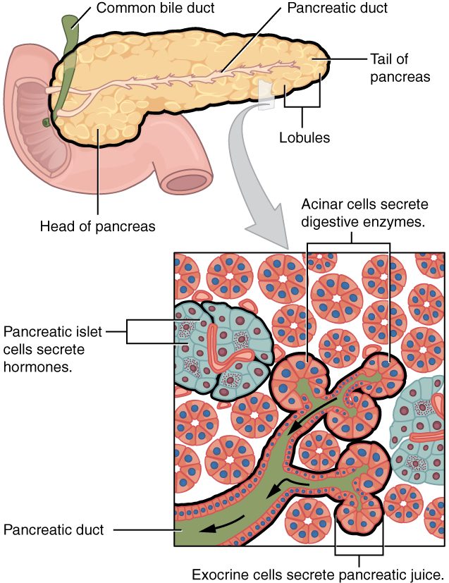
An islet holds four main types of hormone-making cell:
| Cell type | Share of the islet (human) | Hormone | Main effect |
|---|---|---|---|
| Beta cell | About 50–60% | Insulin | Lowers blood glucose |
| Alpha cell | About 30–40% | Glucagon | Raises blood glucose |
| Delta cell | About 5–10% | Somatostatin | Holds back the release of both insulin and glucagon, as a local (paracrine) signal |
| PP cell | A few percent | Pancreatic polypeptide | Slows the pancreas's digestive secretions; its full role in people is still being worked out |
The cells also signal each other. Insulin and somatostatin released inside the islet damp down the alpha cells next door. That local brake matters later, in diabetes.
Three words for storing and releasing glucose
Before following the hormones, you need three terms. Each is built from parts you can decode.
- Glycogenesis (glyc/o = sugar, -gen = producing, -genesis = making): building glycogen, the branched chain of glucose units you met with carbohydrates. Liver and skeletal muscle cells do this after a meal.
- Glycogenolysis (glycogen + -lysis = breaking apart): breaking glycogen back down into glucose.
- Gluconeogenesis (gluc/o = glucose, neo- = new, -genesis = making): making new glucose from molecules that are not carbohydrates, mainly lactate, glycerol from fats, and amino acids. The liver does most of it; the kidneys add some during a long fast.
Where the glycogen sits matters. Your liver stores about 100 grams, and it can release that glucose into the blood. Your skeletal muscles store more, roughly 400 grams, but a muscle cell lacks the enzyme that frees glucose to leave the cell, so muscle glycogen fuels only that muscle. Between meals, then, blood glucose comes from liver glycogenolysis first. Liver glycogen runs low within about a day of fasting, and from then on gluconeogenesis supplies most of the glucose. You will see these pathways step by step in carbohydrate metabolism; here you need only the names and where they happen.
Insulin: the signal of plenty
How a beta cell senses glucose
A beta cell is its own sensor. It has no separate sensory receptor and no nerve telling it glucose is high. The steps, shown in Figure 2:
- Blood glucose rises, and glucose enters the beta cell by facilitated diffusion through glucose carrier proteins. More glucose outside means more glucose inside.
- The cell breaks the glucose down in cellular respiration, and its ATP level rises.
- ATP binds and closes ATP-sensitive potassium channels in the plasma membrane. Fewer potassium ions leak out, so the membrane depolarizes.
- Depolarization opens voltage-gated calcium channels. Calcium ions flow in.
- Calcium triggers exocytosis of vesicles packed with insulin, which enters the islet's capillaries.
Other inputs adjust the output. Amino acids from a protein meal raise insulin release. Hormones from the gut, released as food arrives, raise it before blood glucose has climbed much. The vagus nerve raises it during a meal. Sympathetic activity and epinephrine lower it, through alpha-2 receptor proteins on beta cells.
What insulin does
Insulin (insul- = island, -in = substance: the hormone of the islets) is a small protein hormone, two peptide chains held together by disulfide bonds. It is water-soluble, so it acts on a receptor protein in the plasma membrane of its target cells. That receptor protein is itself an enzyme that adds phosphate groups to proteins inside the cell, starting a phosphorylation cascade like the ones you met with water-soluble hormones. The result, tissue by tissue:
- Skeletal muscle and adipose tissue: glucose gets in. These cells keep most of their glucose carriers, called GLUT4, parked in vesicles inside the cell. Insulin moves those vesicles to the plasma membrane. The carriers now let glucose in by facilitated diffusion, down its gradient. With no insulin, the door stays mostly shut.
- Liver: glucose is kept and stored. Liver cells let glucose in and out freely through carriers that insulin does not move. Insulin instead switches on glycogenesis and switches off glycogenolysis and gluconeogenesis, so the liver takes glucose up and stops releasing it.
- Muscle: glycogen and protein. Muscle cells build glycogen from the glucose they take in, and take up amino acids for protein synthesis.
- Adipose tissue: fat is stored. Fat cells build triglycerides and stop breaking down the ones they hold.
- Most cells: potassium moves in. Insulin speeds the sodium–potassium pump, so potassium moves from the plasma into cells. That is why clinicians give insulin, with glucose, to lower a dangerously high blood potassium.
Put together, insulin is a building hormone, promoting the building side of metabolism. Its net effect on the blood is that glucose, amino acids and fatty acids leave it and go into storage.
Not every tissue needs insulin to take up glucose. Your brain uses about 120 grams of glucose a day, and its neurons take it in through carriers that are always in the membrane. So the brain's glucose supply does not depend on insulin; but, as you will see, it depends entirely on how much glucose is in the blood.
Glucagon: the signal of scarcity
A few hours after you eat, glucose is still leaving your blood: your brain alone takes about 5 grams an hour. Something has to put it back. As blood glucose falls, alpha cells release glucagon (gluc- = glucose, -agon = to lead: it leads glucose out of storage). Alpha cells release more glucagon when:
- blood glucose falls. Less insulin reaching them from the beta cells next door also takes the brake off.
- amino acids rise after a protein meal. The glucagon offsets the insulin that amino acids also trigger, so a meal of pure protein does not drop your glucose.
- sympathetic nerves fire or epinephrine circulates, as in exercise or fear.
Glucagon is a peptide, water-soluble like insulin. Its main target by far is the liver. It binds G protein–coupled receptor proteins on liver cells, cyclic AMP rises, and protein kinase A switches on glycogenolysis and gluconeogenesis and switches off glycogenesis. Glucose pours out of the liver into the blood. Skeletal muscle has almost no glucagon receptor proteins, which fits with muscle glycogen staying in the muscle.
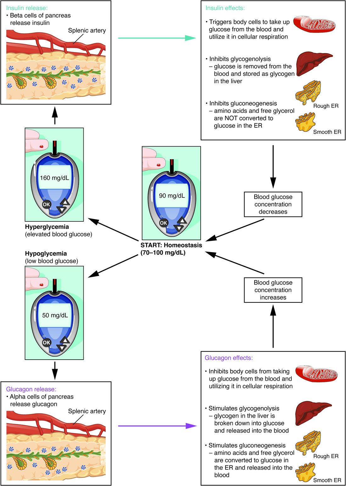
Figure 3 sets the two hormones against each other.
| Insulin | Glucagon | |
|---|---|---|
| Made by | Beta cells of the pancreatic islets | Alpha cells of the pancreatic islets |
| Chemical class | Protein (water-soluble) | Peptide (water-soluble) |
| Main stimulus | High blood glucose; also amino acids, gut hormones, the vagus nerve | Low blood glucose; also amino acids, sympathetic activity and epinephrine |
| Main targets | Liver, skeletal muscle, adipose tissue | Liver |
| Glycogen | Glycogenesis on, glycogenolysis off | Glycogenolysis on, glycogenesis off |
| Gluconeogenesis | Off | On |
| Glucose uptake by muscle and fat | Up (GLUT4 moved to the membrane) | No direct effect |
| Fat stores | Built up; breakdown blocked | Broken down when insulin is low |
| Effect on blood glucose | Down | Up |
| When it is high | After a meal | Between meals, fasting, exercise |
The two hormones are antagonistic: each opposes the other's effect on the same variable. The level of blood glucose at any moment reflects the balance between them, more than either one alone.
The blood glucose feedback loop
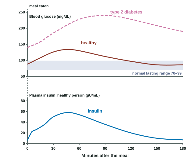
Now put the pieces into the loop you know from homeostasis. Blood glucose is the concentration of glucose in your plasma. Fasting, it sits at about 70 to 99 mg/dL (3.9 to 5.5 mmol/L). After a meal it usually peaks below 140 mg/dL and returns within two to three hours.
Worked example: converting glucose units
Problem. A glucose meter in Canada reads 7.0 mmol/L. What is that in mg/dL, the unit used in the United States?
- Find the conversion factor. One mole of glucose weighs 180 grams, so 1 mmol weighs 180 mg. There are 10 deciliters in a liter, so 1 mmol/L = 180 mg per 10 dL = 18 mg/dL.
- Multiply. 7.0 mmol/L × 18 = 126 mg/dL.
- Interpret. 126 mg/dL is above the normal fasting range. If this was a fasting reading, repeated on another day, it would meet the diagnostic cutoff for diabetes.
Answer. 126 mg/dL. To go the other way, divide mg/dL by 18.
The loop has two arms.
| Slot | Glucose high (after a meal) | Glucose low (fasting) |
|---|---|---|
| Stimulus | Blood glucose rises | Blood glucose falls |
| Sensor | Beta cells | Alpha cells (and glucose-sensing neurons in the hypothalamus) |
| Control center | Beta cells | Alpha cells |
| Signal | Insulin in the blood | Glucagon in the blood |
| Effectors | Liver, skeletal muscle, adipose tissue | Liver |
| Response | Glucose moves into cells and into storage; blood glucose falls | Liver releases glucose; blood glucose rises |
Both arms are negative feedback. As glucose falls back after a meal, the beta cells' ATP falls, their potassium channels reopen, and insulin release drops. The response has removed its own stimulus. Notice that the islet cells are both sensor and control center. Endocrine loops often work this way, with no nerve pathway at all.
Figure 4 shows the loop running in real time. Insulin starts to rise within minutes of the meal, climbs with glucose, and falls as glucose returns. The slight dip of glucose below its starting value around two and a half hours is insulin's effect outlasting the stimulus for a short while.
The pancreas is not the only player on the low side. When glucose keeps falling, the sympathetic system and epinephrine from the adrenal medulla join in, raising glucose release from the liver and cutting insulin release. Over hours, growth hormone and a stress hormone from the adrenal glands (next topic) raise it further. Only insulin lowers blood glucose. That lopsided arrangement explains why too little insulin is so damaging.
Diabetes mellitus
What it is
Diabetes mellitus (diabetes = passing through, a siphon; mellitus = honeyed) is a group of diseases in which blood glucose stays too high, because the body makes too little insulin, responds too little to it, or both. The name records what physicians noticed for centuries: large volumes of urine that tasted sweet. A chronically high blood glucose is hyperglycemia (hyper- = above, glyc- = sugar, -emia = blood condition).
Diabetes is diagnosed by any of these, confirmed on a second test: fasting glucose of 126 mg/dL or more; glucose of 200 mg/dL or more two hours after a standard glucose drink; an A1C of 6.5% or more (A1C measures how much glucose has stuck to a protein inside red blood cells, which reflects the average glucose over the past two to three months); or a random glucose of 200 mg/dL or more with the classic symptoms.
Type 1 and type 2
Type 1 diabetes is loss of insulin. The person's own immune system destroys the beta cells. Once most are gone, the body makes almost no insulin, and the person needs insulin injections or a pump to live.
Type 2 diabetes starts with insulin resistance: muscle, fat and liver cells respond less to a normal amount of insulin. Muscle moves fewer GLUT4 carriers to its membrane, and the liver keeps releasing glucose even when insulin is high. At first the beta cells make up for it by releasing more insulin, and glucose stays normal. Over years, in people whose beta cells cannot keep up, insulin output falls behind and glucose rises. Excess body fat, especially fat inside the abdomen, and physical inactivity drive insulin resistance; genes decide whose beta cells fail.
| Type 1 diabetes | Type 2 diabetes | |
|---|---|---|
| Share of cases | About 5–10% | About 90–95% |
| Main defect | Beta cells destroyed by the immune system | Insulin resistance, then beta cells that cannot keep up |
| Insulin level | Very low or absent | Normal or high early; falls later |
| Usual onset | Often in childhood, but at any age; often sudden, over weeks | Usually in adults, increasingly in young people; gradual, over years |
| Body build | Any; often weight loss before diagnosis | Usually excess body fat |
| Risk of acid blood from fat breakdown | High | Low |
| Treatment | Insulin always | Activity, diet and weight loss; drugs such as metformin; insulin if needed |
From high glucose to the classic symptoms
Follow a person with untreated diabetes:
- With too little insulin action, muscle and fat take up little glucose, and the liver keeps releasing it. Blood glucose rises.
- Your kidneys filter glucose from the blood all the time and normally take all of it back.
- Glucose left in the kidney's fluid holds water there by osmosis. Urine volume rises: polyuria (poly- = much, ur- = urine).
- Losing so much water raises the concentration of the plasma and lowers blood volume. Thirst rises: polydipsia (dips- = thirst).
- Calories leave in the urine as glucose, and cells short of glucose break down fat and muscle. The person loses weight and feels hungry despite eating (polyphagia, phag- = eat).
Acid blood without insulin
In type 1 diabetes without insulin, the loss goes further. With no insulin, nothing blocks fat breakdown, and glucagon is high because the alpha cells have lost the beta cells' brake.
These fat-derived acids pour out of the liver faster than other tissues can burn them, and they acidify the blood. The result is high glucose, acid blood, severe dehydration from polyuria, deep rapid breathing that blows off carbon dioxide, and a fruity smell on the breath. It is an emergency. People with type 2 diabetes rarely develop it, because even a little insulin is enough to hold back fat breakdown.
Long-term damage
Years of hyperglycemia damage blood vessels. Small vessels suffer first: in the retina (vision loss), the kidneys (kidney failure) and the nerves (numbness and pain, starting in the feet). Large arteries narrow faster, so heart attack and stroke are two to four times more common. Keeping glucose near normal lowers every one of these risks.
Hypoglycemia
Hypoglycemia (hypo- = below) is a blood glucose below 70 mg/dL. Below 54 mg/dL it is clinically important whatever the symptoms. It happens mostly in people treated with insulin or with drugs such as sulfonylureas: too much dose, a skipped meal or unplanned exercise.
Your body defends against a fall in a fixed order. At about 80 mg/dL, beta cells cut insulin release. At about 65 to 70 mg/dL, alpha cells raise glucagon and the adrenal medulla raises epinephrine. If glucose keeps falling, symptoms appear in two groups:
- From sympathetic activation and epinephrine: shakiness, a pounding fast heart, anxiety, sweating (through the sympathetic cholinergic fibers to sweat glands) and hunger. These are the warning signs.
- From the brain running short of glucose: trouble concentrating, confusion, slurred speech, odd behavior that can be mistaken for drunkenness, then seizures and loss of consciousness. Neurons store almost no glycogen and burn almost no fatty acids, so they depend on glucose from the blood minute by minute.
A conscious person eats or drinks about 15 grams of fast sugar (juice, glucose tablets) and rechecks after 15 minutes. An unconscious person must not be given anything by mouth; glucagon by injection or nasal spray, or glucose into a vein, brings them round by making the liver release glucose or by supplying it directly.
Summary
The pancreatic islets are the endocrine part of the pancreas. Their beta cells release insulin and their alpha cells release glucagon; delta cells release somatostatin, which damps both. A beta cell senses glucose directly: glucose breakdown raises ATP, ATP closes potassium channels, the cell depolarizes, calcium enters and insulin is released. Insulin moves GLUT4 carriers into the membranes of muscle and fat cells, drives glycogenesis in liver and muscle, blocks glycogenolysis and gluconeogenesis, and stores fat, so blood glucose falls. Glucagon acts mainly on the liver, switching on glycogenolysis and gluconeogenesis, so blood glucose rises. Together they form a negative feedback loop that holds fasting glucose at about 70 to 99 mg/dL. In diabetes mellitus, too little insulin (type 1) or too little response to it with failing beta cells (type 2) leaves glucose high, causing polyuria and polydipsia and, over years, vessel damage. In hypoglycemia, the brain is starved of glucose; glucagon and epinephrine are the first defenses.