The adrenal cortex hormones aldosterone and cortisol control two very different things: aldosterone sets how much sodium and potassium your kidneys keep, and cortisol shifts your fuel use and damps inflammation. This page shows how each adrenal gland is really two glands, cortex and medulla, walks through the three layers of the cortex and the steroid each one makes, introduces the renin–angiotensin–aldosterone system and the pituitary axis that drives cortisol, ties them into the stress response, and ends with what goes wrong when the cortex makes too little or too much.
Two glands in one
Sit an adrenal gland on your palm and it weighs about 4 to 5 grams, less than a teaspoon of sugar. You have two, one capping the upper end of each kidney (ad- = near, ren- = kidney). Slice one open and you see two distinct tissues (Figure 1):
- The adrenal cortex (cortex = bark, outer layer) makes up about 80 to 90 percent of the gland. It is true glandular epithelium, and it makes steroid hormones.
- The adrenal medulla (medulla = inner part), which you met with the sympathetic division, is the core. Its chromaffin cells are modified postganglionic sympathetic neurons, and they release epinephrine and some norepinephrine when preganglionic fibers stimulate them.
The two parts develop from different embryonic tissues, make different kinds of hormone and answer different signals. They share a capsule and a blood supply, and, as you will see, that shared blood lets one talk to the other.
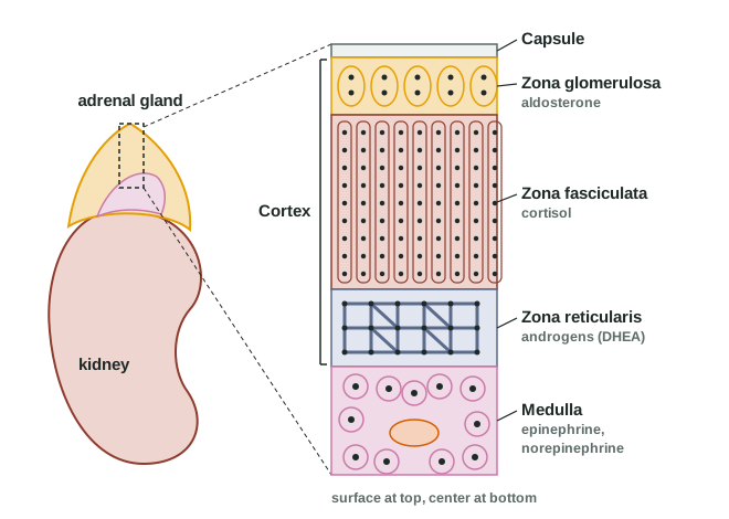
The adrenal cortex and its three zones
Under the capsule, the cortex has three layers, or zones, named for how their cells are arranged:
- Zona glomerulosa (glomerul- = little ball): a thin outer layer of cells in rounded clusters. It makes mineralocorticoids, mainly aldosterone.
- Zona fasciculata (fascicul- = little bundle): the thickest layer, cells lined up in straight columns. It makes glucocorticoids, mainly cortisol.
- Zona reticularis (reticul- = little net): the innermost layer, cells in a branching network. It makes adrenal androgens, mainly DHEA, and a little cortisol.
A memory hook, from the outside in: salt, sugar, sex. The deeper you go, the sweeter it gets.
Every hormone of the cortex is a steroid, built from cholesterol. That tells you how they behave. Steroids are lipid-soluble, so the cortex does not store them in vesicles: it makes them as they are needed and they diffuse straight out. In the blood, most of each hormone rides on carrier proteins; about 90 percent of cortisol is bound. At the target, the hormone crosses the plasma membrane and binds an intracellular receptor protein, and the complex switches genes on or off. New proteins take time to build, so these hormones act over hours and their effects last.
Aldosterone: sodium in, potassium out
Eat a large banana-and-potato meal and your plasma potassium creeps up. Within minutes, the zona glomerulosa releases more aldosterone (named for the aldehyde group on its structure). It is the main mineralocorticoid, a steroid of the cortex that acts on minerals, meaning the ions sodium and potassium.
Three signals raise aldosterone release:
- A rise in plasma potassium, acting directly on the glomerulosa cells. Even a small rise works.
- Angiotensin II, the end product of the renin–angiotensin–aldosterone system (next section), which rises when blood volume or blood pressure falls.
- ACTH, which has a smaller, short-lived effect.
Its main target is the last stretch of the kidney's tubes, where the final adjustments to the urine are made. There, aldosterone makes the cells build more sodium channels and more sodium–potassium pumps. The result:
- More sodium is reabsorbed from the urine into the blood.
- More potassium is secreted into the urine.
- Water follows the sodium by osmosis, as long as ADH has made the tubes permeable to water. Blood volume rises.
Sweat glands, the glands that make saliva, and the colon respond the same way, keeping sodium and losing potassium. Because aldosterone works by building new proteins, its effect takes hours to build and outlasts the hormone itself.
Both main triggers are negative feedback. As potassium falls back, the glomerulosa cells ease off. As volume and pressure rise, angiotensin II falls, and aldosterone with it.
The renin–angiotensin–aldosterone system
Aldosterone's volume trigger comes from a chain of blood-borne steps that starts in the kidney, the renin–angiotensin–aldosterone system (RAAS). Here is the short version; it returns in full with long-term blood pressure control.
- When blood pressure in the kidney's small arteries falls, when sympathetic nerves fire (through beta-1 receptor proteins), or when less salt flows past sensing cells in the kidney, specialized kidney cells release renin, an enzyme, into the blood.
- Renin cuts angiotensinogen, an inactive protein the liver releases into the plasma all the time, into angiotensin I.
- Angiotensin-converting enzyme (ACE), on the inner lining of blood vessels and most plentiful in the lungs, converts angiotensin I into angiotensin II, the active hormone.
- Angiotensin II makes the zona glomerulosa release aldosterone, constricts small arteries (vasoconstriction), and acts on the hypothalamus to raise ADH release and thirst.
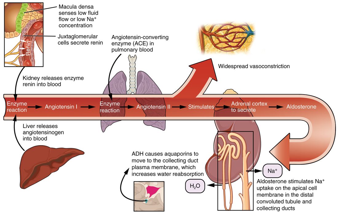
No organ in the chain has to sit near another, because every product travels in the blood (Figure 2). Drugs that block ACE, or block angiotensin II's receptor protein, lower aldosterone and are among the most used treatments for raised blood pressure. A person taking one tends to keep more potassium, which follows directly from what aldosterone does.
Cortisol and the pituitary–adrenal axis
How cortisol release is controlled
Cortisol is the main glucocorticoid (gluc/o = glucose, cortic- = cortex, -oid = like): a steroid of the cortex named for its effect on blood glucose. Its release is set by the hypothalamic–pituitary–adrenal axis, the pattern you met with the pituitary (Figure 3):
- Neurons in the hypothalamus release corticotropin-releasing hormone (CRH) into the hypophyseal portal system.
- CRH makes the anterior pituitary release ACTH (adrenocorticotropic hormone).
- ACTH binds receptor proteins on zona fasciculata cells, which make and release cortisol within minutes. Over weeks, ACTH also keeps the zona fasciculata and zona reticularis large; without it they shrink.
- Cortisol inhibits CRH release by the hypothalamus and ACTH release by the pituitary: long-loop negative feedback.
Two inputs override the set point. First, a daily rhythm: cortisol is lowest around midnight, climbs through the early morning and peaks within an hour of waking. A cortisol blood test is therefore timed. Second, stress: physical stress such as injury, surgery, infection, severe illness or low blood glucose, and psychological stress, raise CRH release through the hypothalamus, and cortisol can rise severalfold within an hour.
What cortisol does
Cortisol acts on almost every tissue. Its effects fall into four groups.
- Fuel: raises blood glucose. In the liver, cortisol switches on gluconeogenesis. In muscle, it breaks down protein, releasing amino acids the liver turns into glucose. In fat, it helps release fatty acids and glycerol. It also makes muscle and fat respond less to insulin. The net effect: blood glucose rises, and less of it is taken up by muscle and fat, so more is left for the brain during a long fast or a long stress.
- Inflammation and immunity: damps both. Cortisol reduces the release of prostaglandins and other local signals of inflammation, lowers the number of some white blood cells in the blood, and makes capillaries less leaky. That is why synthetic glucocorticoid drugs such as prednisone and hydrocortisone treat allergies, arthritis and inflammatory lung disease, and stop the immune system from attacking a transplanted organ.
- Blood vessels: permissive. Cortisol keeps the receptor proteins and signaling machinery that let vascular smooth muscle respond to norepinephrine and epinephrine. Without cortisol, vessels respond weakly to sympathetic signals and blood pressure falls, especially under stress.
- Other tissues. It slows bone building, so long-term excess thins bone (osteoporosis). It affects mood, sleep and memory, and at high levels it reduces the release of the other anterior pituitary axes.
Cortisol can bind the same receptor protein as aldosterone, and it circulates at far higher levels. In the kidney's aldosterone-sensitive cells, an enzyme converts cortisol to an inactive form, so aldosterone's receptor proteins answer only to aldosterone.
Adrenal androgens
The zona reticularis makes adrenal androgens, mainly dehydroepiandrosterone (DHEA) and its sulfate form. These are weak androgens: they act mostly after other tissues, such as skin and fat, convert them into stronger ones. Their release is driven by ACTH, but they do not feed back on it.
In adult men, the testes make far more androgen than the adrenal glands, so adrenal androgens add little. In women, they are a main source of androgen: they help drive the growth of pubic and underarm hair at puberty and contribute to sex drive. Their output rises from about age 6 to 8 (before puberty itself), peaks in the twenties and falls steadily with age.
The adrenal medulla, revisited
You met the medulla as the sympathetic division's hormone gland: preganglionic fibers release acetylcholine onto nicotinic receptor proteins of chromaffin cells, which release about 80 percent epinephrine and 20 percent norepinephrine into the blood within seconds. Two points connect it to the cortex and to fuel.
- Blood flows from cortex to medulla. Much of the medulla's blood has already passed through the cortex and arrives rich in cortisol. Cortisol keeps up the enzyme that converts norepinephrine into epinephrine. When cortisol is low, the medulla makes less epinephrine.
- Epinephrine is a fuel hormone too. It switches on glycogenolysis in the liver, cuts insulin release and raises glucagon release, and releases fatty acids from fat. Blood glucose rises within minutes.
Its circulatory effects are the ones you learned with the autonomic receptor proteins. Circulating epinephrine speeds and strengthens the heart through beta-1. In skeletal muscle, nerve-released norepinephrine constricts small arteries through alpha-1, while low levels of circulating epinephrine dilate them through beta-2.
| Aldosterone | Cortisol | Epinephrine | |
|---|---|---|---|
| Source | Zona glomerulosa of the cortex | Zona fasciculata of the cortex | Chromaffin cells of the medulla |
| Class | Steroid (mineralocorticoid) | Steroid (glucocorticoid) | Amine (catecholamine) |
| Solubility and mechanism | Lipid-soluble; intracellular receptor protein, new proteins made | Lipid-soluble; intracellular receptor protein, new proteins made | Water-soluble; membrane receptor proteins (alpha and beta), second messengers |
| Main stimulus | High plasma potassium; angiotensin II | ACTH (stress, daily rhythm) | Sympathetic preganglionic fibers (acetylcholine) |
| Speed and duration | Hours; lasts a day or more | Hours; lasts hours to days | Seconds; lasts a few minutes |
| Main effects | Kidney keeps sodium and water, loses potassium | Raises blood glucose; damps inflammation; permissive for vessel responses | Faster, stronger heart; glucose and fatty acids released; airways widen |
| Too little | Low sodium, high potassium, low blood volume | Weakness, low blood glucose, low blood pressure | Usually no disease, because sympathetic nerves cover |
The stress response
A hiker breaks her ankle on a remote trail. In the first seconds her heart pounds and her glucose climbs; hours later, still waiting for rescue, her body is breaking down muscle protein to make glucose. The same hormones act on different time scales. In the 1930s, Hans Selye described this pattern as the general adaptation syndrome, in three stages:
- Alarm reaction (seconds to minutes): the fight-or-flight response. Sympathetic nerves and epinephrine from the medulla raise heart rate, blood pressure, breathing and blood glucose, and shift blood toward skeletal muscle.
- Stage of resistance (hours to weeks): the hormones of the cortex carry the load. The hypothalamic–pituitary–adrenal axis keeps cortisol high, which supplies glucose from protein and fat. Angiotensin II, aldosterone and ADH hold on to salt and water.
- Stage of exhaustion (weeks to months): if the stress never ends, the lasting hormone exposure itself does damage: muscle wasting, high blood glucose, raised blood pressure, weaker immune defenses and poor healing, some of the same harms seen with cortisol excess.
Adrenal disorders
Too little: Addison disease
Addison disease is primary adrenal insufficiency: the cortex itself is destroyed. In high-income countries the usual cause is the immune system attacking the cortex; worldwide, infections are also common. All three zones fail, so both cortisol and aldosterone are low.
- From low cortisol: tiredness, weakness, weight loss, low blood glucose, and low blood pressure that worsens on standing.
- From low aldosterone: the kidneys lose sodium and water and keep potassium. Plasma sodium falls, plasma potassium rises, and people crave salt.
- Darker skin, especially in skin creases, scars and the gums. With cortisol low, feedback no longer holds back the pituitary, and ACTH rises very high. ACTH is cut from a larger precursor protein that also yields a hormone that stimulates melanocytes, and ACTH itself stimulates them at high levels.
An infection or injury can tip a person with Addison disease into an adrenal crisis: collapse, very low blood pressure and low glucose. It is treated with intravenous hydrocortisone, salt and fluid.
When the pituitary fails instead (secondary adrenal insufficiency), ACTH is low. Cortisol falls, but the skin does not darken, and aldosterone is mostly spared, because angiotensin II and potassium, not ACTH, are its main drivers.
Too much: Cushing syndrome
Cushing syndrome is the effect of long-term excess glucocorticoid. By far the most common cause is glucocorticoid medication, taken for lung disease, arthritis or after an organ transplant. Among causes inside the body, the most common is a pituitary tumor releasing ACTH (called Cushing disease); others are an adrenal tumor making cortisol on its own, or a tumor elsewhere releasing ACTH.
Each sign follows from something cortisol does:
- Fat moves to the trunk, face and upper back, giving a round face and a fat pad between the shoulders, while the arms and legs thin.
- Protein breakdown thins the skin, weakens capillaries and muscles: easy bruising, wide purple stretch marks on the abdomen, weak thigh and shoulder muscles.
- High blood glucose, from gluconeogenesis and insulin resistance, often to the point of diabetes.
- Raised blood pressure, partly because excess cortisol overwhelms the kidney enzyme and acts like aldosterone.
- Thin bones, frequent infections, poor wound healing, and mood changes.
| Addison disease | Cushing syndrome | |
|---|---|---|
| Problem | Too little cortex hormone | Too much glucocorticoid |
| Most common cause | Immune attack on the cortex | Glucocorticoid medication |
| Cortisol | Low | High |
| Aldosterone | Low | Usually normal |
| ACTH | High | Low with medication or an adrenal tumor; high with a pituitary or other ACTH-releasing tumor |
| Blood pressure | Low | High |
| Blood glucose | Low | High |
| Body weight and shape | Weight loss | Weight gain on the trunk and face; thin limbs |
| Skin | Darker | Thin, bruises easily, purple stretch marks |
Stopping steroid drugs
A person who takes a glucocorticoid drug for more than a few weeks has feedback working against them. The drug suppresses CRH and ACTH. Without ACTH, the zona fasciculata shrinks and stops making cortisol. If the drug is stopped suddenly, there is no cortisol from either source, and the axis takes weeks to months to recover. The person can go into adrenal crisis. That is why these drugs are tapered, reduced step by step, giving the pituitary and cortex time to restart.
Summary
Each adrenal gland is two glands. The cortex makes steroids from cholesterol, in three zones: the zona glomerulosa makes aldosterone, the zona fasciculata makes cortisol, and the zona reticularis makes adrenal androgens such as DHEA. The medulla, part of the sympathetic division, releases epinephrine. Aldosterone, released in response to high potassium and angiotensin II, makes the kidney keep sodium and water and excrete potassium. The renin–angiotensin–aldosterone system links a fall in pressure or volume to aldosterone: renin from the kidney converts angiotensinogen to angiotensin I, and ACE converts that to angiotensin II. Cortisol, driven by CRH and ACTH and held in check by negative feedback, raises blood glucose, damps inflammation and keeps vessels responsive to sympathetic signals. In the stress response, epinephrine acts in seconds and cortisol over hours to weeks. Addison disease (too little cortex hormone) brings low glucose, low blood pressure, salt loss and darker skin; Cushing syndrome (too much glucocorticoid, usually from medication) brings central fat, thin skin, high glucose and raised blood pressure.