Tubular reabsorption and secretion decide what your urine contains. Your glomeruli filter about 180 liters of plasma a day, and the renal tubule hands almost all of it back. This page follows that work segment by segment: the two routes across the tubule wall, the proximal tubule that returns most of the filtrate, the transport maximum that lets glucose spill into urine in diabetes, the salt pumps of the nephron loop, the fine adjustments hormones make in the distal tubule and collecting duct, the substances the tubule secretes, and the split between water you must reabsorb and water your body can choose to keep.
From 180 liters to 1.5
A healthy adult's glomerular filtration rate (GFR) is about 125 mL/min. Over a day that adds up to about 180 L of glomerular filtrate, more than three times the water in your whole body. Yet you pass only 1 to 2 L of urine. The difference is returned to the blood as the fluid flows along the tubule.
Worked example 1: how much filtered water comes back
Problem. A man filters 180 L a day and passes 1.5 L of urine. What percentage of the filtered water does his renal tubule return to the blood?
- Find the water returned. 180 L − 1.5 L = 178.5 L.
- Divide by the water filtered. 178.5 ÷ 180 = 0.992.
- Convert to a percentage. 0.992 × 100 = 99.2%.
Answer. About 99%. Less than 1% of what he filters leaves as urine.
Worked example 2: the sodium the tubule handles
Problem. His plasma sodium is 140 mmol/L, and sodium is filtered freely, so the filtrate carries the same concentration. His urine contains 150 mmol of sodium a day. How much sodium does he filter, and what percentage does he reabsorb?
- Filtered load = GFR × plasma concentration = 180 L/day × 140 mmol/L = 25,200 mmol/day. That is about 1.5 kg of table salt.
- Reabsorbed = filtered − excreted = 25,200 − 150 = 25,050 mmol/day.
- Percentage. 25,050 ÷ 25,200 × 100 = 99.4%.
Answer. He filters 25,200 mmol a day and reabsorbs 99.4% of it. A change of just 1% in reabsorption would change his sodium excretion by 252 mmol a day, more than he eats.
That last step is why the tubule, not the glomerulus, fine-tunes your urine: small changes in how much it reabsorbs make large changes in what you excrete.
Filter, reabsorb, secrete
Three processes shape urine, and you can keep track of any substance with one line of bookkeeping:
- Filtration moves plasma, minus cells and most proteins, from the glomerulus into the capsule. You met it in the last topic.
- Tubular reabsorption (re- = back, ab- = away, sorb- = suck in) moves a substance from the fluid inside the tubule back into the blood of the peritubular capillaries.
- Tubular secretion moves a substance the other way: from the peritubular capillaries into the tubule, adding it to what was filtered.
This is mass balance applied to the kidney. For any substance:
amount excreted = amount filtered − amount reabsorbed + amount secreted
Glucose is filtered and then fully reabsorbed, so none is excreted. Potassium is filtered, mostly reabsorbed, and then partly secreted again near the end. Some drugs are filtered and also heavily secreted, so more leaves in urine than was filtered.
Two routes across the tubule wall
The tubule wall is a single layer of epithelial cells. Its apical surface faces the lumen, the fluid inside the tubule. Its basolateral membrane (baso- = base, later- = side) faces the interstitial fluid and, beyond it, the peritubular capillaries. A reabsorbed substance can cross in one of two ways:
- The transcellular route (trans- = across, cell- = cell) goes through a cell: in across the apical membrane, through the cytoplasm, and out across the basolateral membrane. Each membrane needs its own channel or carrier.
- The paracellular route (para- = beside) goes between cells, through the tight junctions that join neighboring cells near their apical ends. No membrane is crossed, so no carrier is needed, and movement is always passive, down a gradient.
How much the paracellular route carries depends on how tight the tight junctions are. In the proximal tubule they are leaky tight junctions: water and small ions such as chloride, calcium and potassium slip between cells. Further along, the junctions get tighter, and the collecting duct's are tight enough that almost everything must go through cells, where hormones can control it.
| Transcellular route | Paracellular route | |
|---|---|---|
| Path | Through the cell: apical membrane, cytoplasm, basolateral membrane | Between cells, through the tight junctions |
| Membranes crossed | Two | None |
| Needs a carrier or channel | Yes, one on each membrane | No |
| Can move a substance against its gradient | Yes, with active transport | No, passive only |
| Can hormones regulate it directly | Yes, by adding or removing transport proteins | Only indirectly |
| Example | Glucose in the proximal tubule | Chloride and calcium in the proximal tubule |
The engine: the sodium–potassium pump
Almost every transport step in the tubule starts with the same protein. The sodium–potassium pump sits only on the basolateral membrane. Using ATP, it moves 3 Na+ out of the cell into the interstitial fluid and 2 K+ in. Three things follow:
- Sodium inside the tubule cells stays low, about 10 to 20 mmol/L, while the filtrate holds about 140 mmol/L.
- The inside of the cell is negative. So sodium in the lumen has a steep electrochemical gradient into the cell.
- Carriers on the apical membrane use that downhill sodium flow to pull other substances into the cell with it (symport) or push substances out in exchange (antiport). That is secondary active transport: the pump spends the ATP, and the carriers spend the gradient.
Stop the pump and the gradient runs down within minutes. Reabsorption of sodium, glucose, amino acids and much else then stops too, even though none of the apical carriers uses ATP directly.
Reabsorption in the proximal tubule
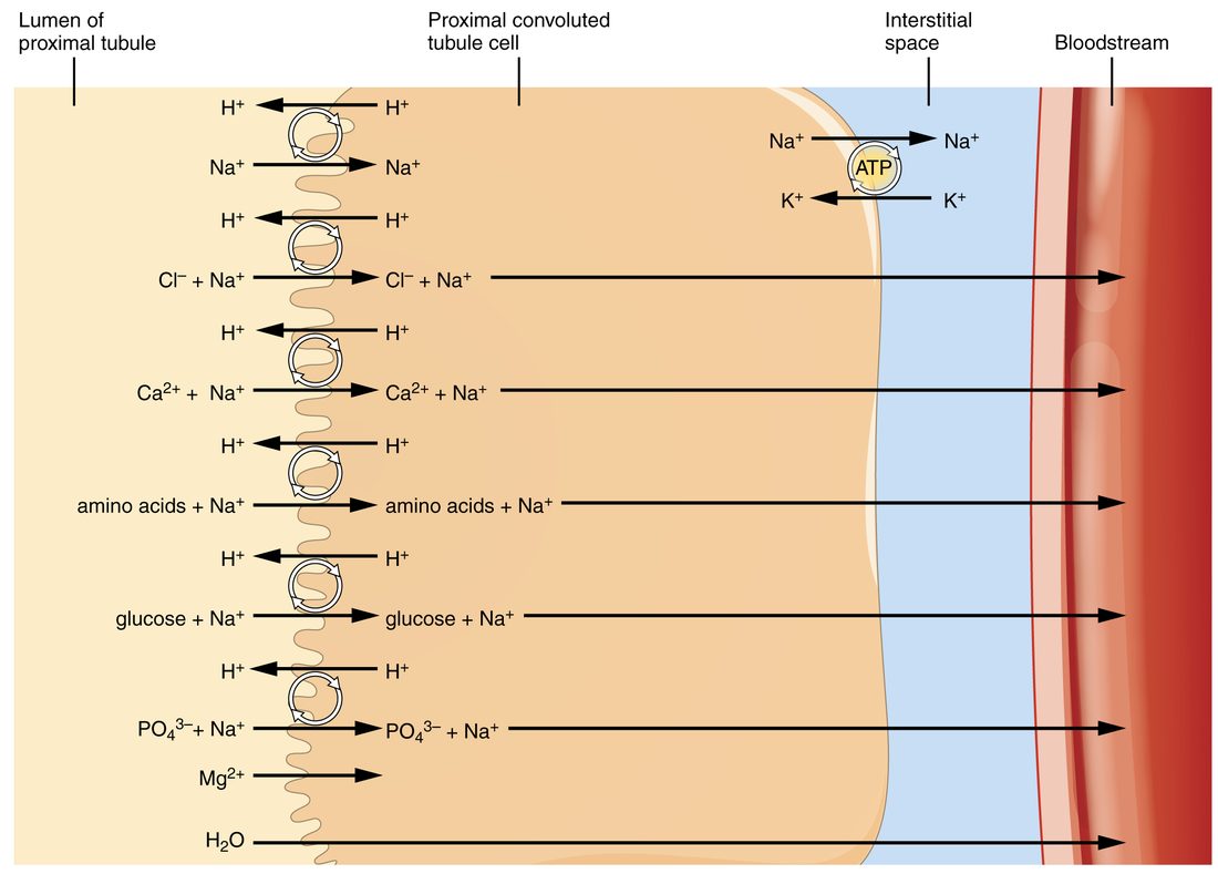
Reabsorption in the proximal tubule is the bulk step. The proximal convoluted tubule (PCT) returns about two thirds of the filtered sodium and water, and nearly all the nutrients. Its cells are built for volume: a dense brush border of microvilli on the apical surface multiplies the membrane area, and the cells are packed with mitochondria that make ATP for the pumps (Figure 1).
What the proximal tubule returns
- Sodium, about 65%. It enters mostly in exchange for H+ (the sodium–hydrogen exchanger, an antiporter) and with nutrients (symporters).
- Glucose and amino acids, essentially 100% when their plasma levels are normal. Each rides in with sodium on its own symporter, then leaves the cell across the basolateral membrane by facilitated diffusion.
- Bicarbonate, about 80%. The H+ that the exchanger sends into the lumen combines with filtered bicarbonate; the details belong to acid–base balance, later in the course.
- Water, about 65%. Removing solute leaves the lumen slightly more dilute than the cells and interstitial fluid, and water follows by osmosis, through aquaporins and through the leaky tight junctions.
- Chloride, potassium, calcium and urea, passively. As water leaves, these become more concentrated in the lumen than in the blood, and they diffuse out, much of it by the paracellular route. About 65% of filtered potassium and calcium and about half the urea come back here.
- Phosphate, about 80%, on a sodium–phosphate symporter. Parathyroid hormone (PTH) removes these carriers from the membrane, so more phosphate is lost in urine.
- Small proteins and peptides that slipped through the filter are taken up by endocytosis and broken down to amino acids inside the cells.
Because solute and water leave together, the fluid at the end of the proximal tubule has the same osmolarity as plasma, about 300 mOsm/L, even though its volume has fallen by two thirds.
Into the peritubular capillaries
Everything reabsorbed first lands in the interstitial fluid. From there it moves into the peritubular capillaries, because the capillary pressures favor uptake:
- Hydrostatic pressure is low. Blood reaching them has already passed through two sets of resistance vessels, the afferent and efferent arterioles.
- Colloid osmotic pressure is high. About a fifth of the plasma was filtered off in the glomerulus, but the proteins stayed behind, so the blood leaving through the efferent arteriole is richer in protein.
Transport maximum and renal threshold
A person with uncontrolled diabetes mellitus and a blood glucose of 400 mg/dL has glucose in the urine. A healthy person at 90 mg/dL has none. The tubule has not changed; the load has.
Glucose leaves the lumen only on carriers, and there is a fixed number of them. When every carrier is busy, adding more glucose cannot speed reabsorption: this is carrier saturation. For the kidney, the highest rate at which the tubules can reabsorb a substance is its transport maximum (Tm). For glucose, both kidneys together manage about 375 mg/min.
The filtered load is how much reaches the tubule each minute: GFR × plasma concentration.
Worked example 3: normal glucose
Problem. GFR is 125 mL/min and plasma glucose is 100 mg/dL. Is any glucose excreted?
- Convert the concentration. 100 mg/dL = 100 mg per 100 mL = 1 mg/mL.
- Filtered load = 125 mL/min × 1 mg/mL = 125 mg/min.
- Compare with Tm. 125 mg/min is well below 375 mg/min, so the carriers can take all of it.
Answer. All 125 mg/min is reabsorbed and none is excreted.
Worked example 4: high glucose
Problem. The same person's plasma glucose rises to 500 mg/dL. How much glucose is excreted each minute?
- Convert. 500 mg/dL = 5 mg/mL.
- Filtered load = 125 × 5 = 625 mg/min.
- Reabsorbed. The carriers are saturated, so reabsorption is capped at Tm: 375 mg/min.
- Excreted = filtered − reabsorbed = 625 − 375 = 250 mg/min.
Answer. 250 mg of glucose leaves in the urine every minute, about 360 g a day if it lasted.
Figure 2 plots all three amounts against plasma glucose.
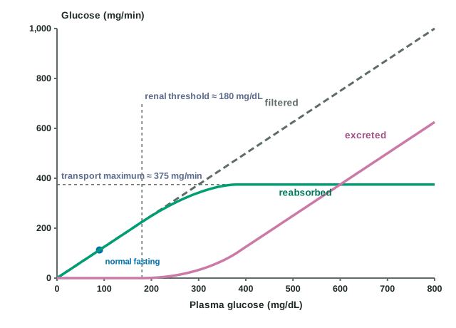
The renal threshold is the plasma concentration at which a substance first appears in urine. For glucose it is about 180 mg/dL. You might expect it to be Tm ÷ GFR = 375 ÷ 125 = 3 mg/mL, which is 300 mg/dL. It is lower because nephrons differ: some have fewer carriers for the glucose they filter and saturate early, so glucose starts to leak through them before the kidney as a whole reaches its maximum. That early leak is the curved "splay" on the graph between about 180 and 350 mg/dL.
Glucose in the urine is glucosuria (also called glycosuria; glyc- = sugar, -uria = urine condition). The glucose left in the tubule holds water with it, which is why untreated diabetes mellitus causes polyuria and thirst.
Transport in the nephron loop
Transport in the nephron loop follows one rule: the two limbs let different things through.
- The descending limb is thin and full of aquaporins, but has few salt carriers. As it dips into the medulla, the fluid around it gets saltier, and water leaves the tubule by osmosis. About 15% of filtered water is reabsorbed here.
- The ascending limb has almost no aquaporins, so water cannot follow salt out of it. Its thin lower part lets NaCl diffuse out. Its thick ascending limb pumps salt out actively.
The Na–K–2Cl cotransporter
The key carrier of the thick ascending limb is the Na–K–2Cl cotransporter (NKCC2), a symporter on the apical membrane. Each cycle moves 1 Na+, 1 K+ and 2 Cl− into the cell together, powered by the sodium gradient the basolateral pump makes. Then:
- Sodium leaves the cell on the basolateral sodium–potassium pump, and chloride leaves through chloride channels. Both reach the interstitial fluid.
- Most of the potassium leaks straight back into the lumen through apical potassium channels. That recycling keeps NKCC2 supplied with potassium, and it leaves the lumen positively charged.
- The positive lumen pushes cations out between the cells: calcium, magnesium and more sodium leave by the paracellular route.
- No water follows, because this segment is nearly impermeable to it.
The loop reabsorbs about 25% of the filtered sodium, most of it here. Because it removes salt but not water, the fluid leaving the thick ascending limb is more dilute than plasma, about 100 mOsm/L. That is why it is called the diluting segment. The salt it pumps out stays in the medulla and makes the fluid around the loop saltier; the next topic shows how that lets you make concentrated urine.
| Descending limb | Thick ascending limb | |
|---|---|---|
| Permeability to water | High (aquaporins) | Very low |
| Salt transport | Little | Active, through NKCC2 |
| What leaves the tubule | Water | Na+, Cl−, K+, Ca2+, Mg2+ |
| Tubular fluid along the segment | Grows more concentrated | Grows more dilute |
| Share of filtered load reabsorbed | About 15% of water | About 25% of sodium |
Transport in the distal tubule and collecting duct
By the time fluid reaches the distal convoluted tubule (DCT), about 90% of the filtered sodium and 80% of the water are gone. What is left gets adjusted to match your intake, mostly under hormonal control. This is transport in the distal tubule and collecting duct.
The distal convoluted tubule
- A sodium–chloride symporter on the apical membrane reabsorbs about 5% of filtered sodium.
- The DCT has little water permeability, so the fluid gets more dilute still.
- Calcium is reabsorbed through channels, and PTH increases it. That is how PTH keeps calcium in your blood while it sends phosphate out (Figure 3).
Principal cells
The last part of the distal tubule and the collecting duct contain two kinds of cell. Principal cells are the more common. They handle sodium, potassium and water:
- Sodium enters them through apical sodium channels, down the gradient the basolateral pump makes. Potassium leaves them into the lumen through apical potassium channels. So the more sodium they reabsorb, the more potassium they secrete.
- Aldosterone acts here. It makes principal cells build more sodium channels, potassium channels and sodium–potassium pumps. Sodium reabsorption and potassium secretion both rise, and water follows the sodium when water can cross.
- ADH acts here too. It makes principal cells put aquaporins into their apical membrane, so water can leave the duct. Without ADH, these cells are nearly waterproof.
- ANP reduces sodium reabsorption by the collecting duct, so more sodium, and the water with it, stays in the urine.
Intercalated cells
Intercalated cells (inter- = between, calated = inserted) are scattered among the principal cells. They secrete acid (H+) or bicarbonate into the urine, depending on your blood pH; you will see how in acid–base balance. When your body is short of potassium, some of them also reabsorb potassium.
| Principal cells | Intercalated cells | |
|---|---|---|
| How common | Most of the cells lining the duct | A minority, scattered among the principal cells |
| Main job | Sodium, potassium and water balance | Acid–base balance |
| Key apical proteins | Sodium channels, potassium channels, aquaporins (with ADH) | Proton pumps or bicarbonate carriers |
| Hormones that act on them | Aldosterone, ADH, ANP | Mainly responds to blood pH (aldosterone plays a smaller part) |
| Effect on potassium | Secrete it | Can reabsorb it when the body is short |
Tubular secretion
Filtration is limited: only about a fifth of the plasma reaching a glomerulus is filtered, and anything bound to plasma proteins stays in the blood. Tubular secretion reaches the other four fifths. Carriers in the tubule cells take substances from the interstitial fluid around the peritubular capillaries and move them into the lumen.
- Potassium is secreted by principal cells. Nearly all the potassium in your urine was secreted there; the filtered potassium was mostly reabsorbed earlier. This lets you excrete more potassium after a potassium-rich meal.
- H+ is secreted in the proximal tubule and by intercalated cells, which is how the kidneys remove acid.
- Organic acids and bases are secreted in the proximal tubule by broad carriers that recognize many different molecules. They clear drugs such as penicillin, waste products such as uric acid, and toxins. Penicillin is secreted so efficiently that early doctors, when it was scarce, gave a second drug that competes for the same carrier, to keep penicillin in the blood longer.
Because secretion adds to filtration, a heavily secreted substance can be excreted faster than it is filtered, and some are cleared from almost all the plasma that passes through the kidney.
Obligatory and facultative water reabsorption
Some water reabsorption happens whatever your body needs; the rest is adjustable.
- Obligatory water reabsorption (obligare = to bind) is the water that follows solute out of the proximal tubule and the descending limb. These segments are always permeable to water, so this water comes back whether you are dehydrated or overhydrated: roughly 80 to 85% of what you filter.
- Facultative water reabsorption (facultas = ability, option) happens in the last part of the distal tubule and the collecting duct, and only when ADH puts aquaporins into principal cells. It covers the remaining 15 to 20%, and it is what sets the volume of your urine.
Some textbooks round the split to about 90% obligatory and 10% facultative. The exact figure matters less than the idea: most water returns whatever you drink, and a smaller, adjustable share sets your urine volume.
Worked example 5: what ADH controls
Problem. A woman filters 180 L a day. About 82% is reabsorbed obligatorily. How much water reaches the distal parts of her nephrons, and what is the range of urine volumes ADH can produce, if her collecting ducts can reabsorb anywhere from none to all but 0.5 L of it? (Real kidneys reabsorb some water here even with no ADH, so the upper end is not reached in practice.)
- Obligatory reabsorption. 0.82 × 180 L = 147.6 L, about 148 L.
- Water left. 180 − 148 = 32 L a day reach the distal tubule and collecting duct.
- High ADH. Almost all of it is reabsorbed: urine volume about 0.5 L.
- No ADH. Little of it is reabsorbed: urine volume rises toward tens of liters.
Answer. About 32 L a day are open to adjustment. With plenty of ADH she passes about half a liter; without ADH, many liters. People who lack ADH (diabetes insipidus) can pass more than 10 L a day.
Segment by segment
Figure 3 and the table below put the segments side by side.
| Proximal tubule | Nephron loop | Distal tubule | Collecting duct | |
|---|---|---|---|---|
| Share of filtered sodium reabsorbed | About 65% | About 25% (thick ascending limb) | About 5% | 2 to 5%, adjustable |
| Share of filtered water reabsorbed | About 65% | About 15% (descending limb) | Very little | From a few percent to nearly all of the remaining 15 to 20%, set by ADH |
| Main apical carriers | Sodium–hydrogen exchanger; sodium symporters for glucose, amino acids and phosphate | NKCC2 | Sodium–chloride symporter | Sodium channels; aquaporins when ADH is present |
| Tight junctions | Leaky | Moderately tight | Tight | Tightest |
| Hormones acting | Angiotensin II (more sodium reabsorbed); PTH (less phosphate reabsorbed) | None of the main hormones is its chief control | PTH (more calcium reabsorbed) | Aldosterone, ADH, ANP |
| Secretes | H+, organic acids and bases, drugs | Little | Little | K+ (principal cells), H+ (intercalated cells) |
| Fluid leaving it | About 300 mOsm/L, same as plasma | About 100 mOsm/L, dilute | About 100 mOsm/L or less | 50 to 1,200 mOsm/L, depending on ADH |
Read the table from left to right and a pattern appears: each segment handles less volume than the one before, but under tighter control. The proximal tubule moves the most and is hardly regulated; the collecting duct moves the least and decides the final urine.