The nephron is the microscopic unit that makes urine, and each of your kidneys holds about a million of them. This page is a structure-and-function tour of one nephron, with a diagram of every part: the renal corpuscle, where blood is filtered; the renal tubule, a long, folded tube whose cells change the filtered fluid; the collecting duct that it drains into; the two kinds of nephrons; the unusual chain of blood vessels that serves each one; and the juxtaglomerular apparatus, where the tubule reports back to its own blood supply. The next topics explain what each part does in detail.
The nephron: the unit that makes urine
In the last topic you followed urine from the renal papilla out of the body. Now zoom in on where it comes from. Each pyramid and the cortex above it are packed with microscopic tubes, each about as wide as a hair, coiled and looped and bundled with capillaries.
A nephron (nephr- = kidney) is one of those units: a ball of capillaries with a hollow cup around it, and a single tube leading from the cup. Each kidney has roughly a million nephrons, though the number varies widely between people, from about 200,000 to over 2 million. You are born with all you will ever have. Nephrons lost to injury or disease are not replaced, and after about age 40 you lose some every year.
Every nephron does the same three things to blood plasma, one after another:
- It filters. Pressure pushes plasma fluid out of the capillary ball into the cup. Blood cells and most proteins stay behind.
- It takes back. As the fluid flows along the tube, the cells lining it move most of the water and nearly all the useful solutes back into the blood.
- It adds. The same cells move some substances, such as extra potassium, hydrogen ions and many drugs, from the blood into the fluid.
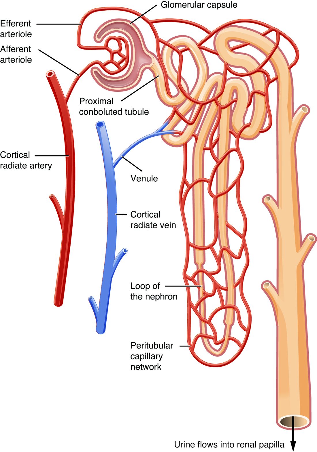
What is left at the end is urine. Each of these three processes has its own topic next. This one is about the structure that does them (Figure 1).
A nephron has two main parts: the renal corpuscle, which filters, and the renal tubule, which changes the filtered fluid.
The renal corpuscle
The renal corpuscle (corpus = body, -cle = small) is the filtering end of the nephron, a round structure about 0.2 mm across, just big enough to see as a dot with a magnifying glass. Every renal corpuscle sits in the cortex. It has two parts (Figure 3):
- The glomerulus (glomus = ball of yarn, -ulus = small; plural glomeruli) is a tuft of about 30 to 50 looping capillaries. They are fenestrated capillaries, the type with pores through their endothelial cells that you met with blood vessels, so they leak fluid far faster than ordinary capillaries.
- The glomerular capsule, also called Bowman's capsule, is the cup around the glomerulus: the blind, swollen start of the tubule, pushed in by the capillary tuft like a fist pushed into a soft balloon. It has two layers with a space between them:
- The parietal layer (pariet- = wall) is the outer wall of the cup, a simple squamous epithelium. It only holds fluid in.
- The visceral layer clings to the capillaries. It is made of podocytes (pod- = foot, -cyte = cell), cells with long branches that wrap around each capillary. Their smallest branches interlock like the fingers of two clasped hands, leaving narrow gaps between them. You will meet those gaps as part of the filter in the next topic.
- The capsular space lies between the two layers. Fluid filtered out of the glomerulus collects here, then flows into the tubule.
A renal corpuscle has two poles. At the vascular pole, blood vessels enter and leave the glomerulus. At the tubular pole, on the opposite side, the capsular space opens into the tubule.
The renal tubule
The renal tubule is the tube that carries fluid away from the capsule. It is about 3 cm long if you straighten it, and each part has cells built for its job. Follow the fluid through it (Figure 1):
- Proximal convoluted tubule (PCT; proximal = nearest, convoluted = coiled). The first, longest and most coiled part, in the cortex near its own corpuscle. Its cuboidal cells carry a dense brush border of microvilli on the side facing the fluid, and they are packed with mitochondria. Microvilli multiply the surface for transport, and mitochondria supply the ATP that the pumps use. This is where most of the filtered water and solutes are taken back: roughly two thirds of the water and salt, and almost all the glucose and amino acids.
- Nephron loop, also called the loop of Henle, after the anatomist who described it. The tubule straightens and dives toward the medulla, turns in a hairpin and climbs back to the cortex.
- The descending limb runs down. Most of it is a thin segment of simple squamous cells.
- The ascending limb runs back up. It starts thin in long loops, then becomes the thick ascending limb, lined with cuboidal cells full of mitochondria, which pump salt out of the fluid.
- Distal convoluted tubule (DCT; distal = farther away). Back in the cortex, the ascending limb passes right beside its own renal corpuscle and continues as the DCT, a shorter coiled segment of cuboidal cells with few microvilli. Here, and in the collecting duct, hormones such as aldosterone fine-tune how much salt the tubule takes back.
- Collecting duct. A short connecting segment joins each DCT to a collecting duct. Each collecting duct receives fluid from many nephrons and runs straight down through the medulla toward the papilla. Collecting ducts merge into larger ducts that open on the tip of the papilla, where the fluid, now urine, drips into a minor calyx. The collecting duct is where ADH acts.
Strictly speaking, the collecting duct is not part of any one nephron, because many nephrons share it, and it grows from a different part of the embryo. This course counts it as the last part of the renal tubule, as most A&P courses do, because the fluid in it is still being changed.
| Proximal convoluted tubule | Nephron loop | Distal convoluted tubule | Collecting duct | |
|---|---|---|---|---|
| Where | Cortex | Dips into the medulla | Cortex | Cortex, then through the medulla to the papilla |
| Cells | Cuboidal | Thin parts squamous; thick ascending limb cuboidal | Cuboidal | Cuboidal, taller near the papilla |
| Microvilli | Dense brush border | Few | Few | Few |
| Mitochondria | Very many | Few in thin parts; many in the thick ascending limb | Many | Moderate |
| Main job | Takes back most water and solutes | Sets up the conditions for concentrating urine | Fine-tunes salt under hormone control | Final adjustment of water and salt; ADH acts here |
Cortical and juxtamedullary nephrons
Not all nephrons are the same shape. Where the renal corpuscle sits decides how long the loop is (Figure 1):
- Cortical nephrons, about 85 percent of the total, have their corpuscles in the outer part of the cortex. Their loops are short and dip only a little way into the medulla.
- Juxtamedullary nephrons (juxta- = next to), about 15 percent, have their corpuscles deep in the cortex, next to the medulla. Their loops are long and plunge deep into the medulla, some almost to the tip of the papilla.
The long loops of juxtamedullary nephrons are what let the kidney make urine more concentrated than blood. Desert animals show this clearly: the kangaroo rat, which can live without drinking, has very long loops and can make urine several times as concentrated as yours.
| Cortical nephron | Juxtamedullary nephron | |
|---|---|---|
| Share of nephrons | About 85% | About 15% |
| Where the renal corpuscle sits | Outer cortex | Inner cortex, next to the medulla |
| Nephron loop | Short, dips just into the outer medulla | Long, reaches deep into the medulla |
| Capillaries around the tubule | Peritubular capillaries | Peritubular capillaries plus the vasa recta |
| Main contribution | Most of the filtering and taking back | Building the conditions for concentrated urine |
The blood vessels of the nephron
In the last topic you followed arteries out to the cortical radiate arteries. From there, the blood vessels of the nephron are arranged in a way found almost nowhere else in the body: two capillary beds, one after the other, with an arteriole between them (Figure 2).
- An afferent arteriole (af- = toward, fer- = carry) branches off a cortical radiate artery and carries blood into the glomerulus.
- Blood runs through the glomerular capillaries, the first capillary bed. Here, fluid is filtered out.
- An efferent arteriole (ef- = away) carries blood out of the glomerulus. It is an arteriole, not a venule, and in most nephrons it is narrower than the afferent arteriole. (In juxtamedullary nephrons it is about as wide or wider, but it still resists flow.)
- The efferent arteriole feeds the second capillary bed. For most nephrons, this is the peritubular capillaries (peri- = around), a low-pressure mesh wrapped around the tubules in the cortex. The water and solutes the tubule takes back enter these capillaries. You met them in capillary exchange as one of the few beds that take fluid in all along their length.
- For juxtamedullary nephrons, the efferent arteriole also gives rise to the vasa recta (vasa = vessels, recta = straight), long, thin, hairpin-shaped capillaries that run down into the medulla alongside the long loops and back up again. Blood moves through them slowly.
- Both beds drain into venules and then the cortical radiate veins.
Why does this layout matter? Because there is an arteriole on each side of the glomerulus, the kidney can set the pressure in the glomerulus by narrowing or widening either one. Narrowing the efferent arteriole backs pressure up into the glomerulus, which keeps its capillary pressure unusually high, far higher than in the capillaries of your skin or muscles. That high pressure drives filtration. By the time blood reaches the peritubular capillaries, it has passed through two resistances, its pressure is low, and fluid moves into the blood instead. You will use this layout in the next topic to predict how narrowing each arteriole changes filtration.
| Afferent arteriole | Efferent arteriole | |
|---|---|---|
| Direction | Into the glomerulus | Out of the glomerulus |
| Comes from | A cortical radiate artery | The glomerular capillaries |
| Leads to | The glomerulus | Peritubular capillaries and, in juxtamedullary nephrons, the vasa recta |
| Width | Wider in most nephrons | Narrower in most nephrons |
| Special cells in its wall | Juxtaglomerular cells, which release renin | None |
The juxtaglomerular apparatus
After its trip down and up the loop, every nephron's tubule comes back to touch its own renal corpuscle, right at the vascular pole, between the afferent and efferent arterioles. That meeting point is the juxtaglomerular apparatus (JGA; juxta- = next to): a small cluster of specialized cells where the tubule can signal to the arterioles that feed it (Figure 3). It has three parts:
- The macula densa (macula = spot, densa = dense) is a patch of tall, tightly crowded cells in the tubule wall, at the end of the thick ascending limb where it meets the arterioles. These cells sense how much sodium chloride is in the fluid flowing past them.
- Juxtaglomerular cells, also called granular cells, are modified smooth muscle cells in the wall of the afferent arteriole. Their granules hold renin. They release it when the pressure stretching the arteriole falls, when sympathetic nerves stimulate them through beta-1 receptor proteins, and when the macula densa signals that little sodium chloride is reaching it. You met these three triggers when you studied the renin–angiotensin–aldosterone system; now you can put names to the cells.
- Mesangial cells (mes- = middle, angi- = vessel). A few lie outside the glomerulus, in the angle between the arterioles and the macula densa, and seem to pass signals between them. Many more sit inside the glomerulus, between its capillary loops. These support the capillaries, can contract to change the filtering surface, and engulf debris trapped by the filter.
Figure 4 shows the same cells in the context of the whole renin pathway.
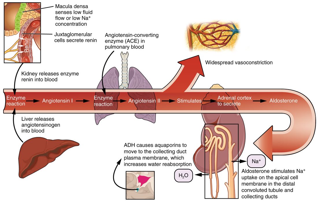
The JGA works in two directions. Through renin, it helps set blood pressure and blood volume for the whole body. Through signals from the macula densa to the afferent arteriole, it adjusts how much its own nephron filters. That second job is part of how the kidney keeps its filtering steady, and it is the subject of the next topic.
Filtrate through the nephron
Put the parts together and follow one drop of fluid from blood to urine:
- Blood arrives in the afferent arteriole and enters the glomerulus.
- Pressure pushes plasma fluid out of the capillaries, past the podocytes, into the capsular space.
- The fluid flows into the proximal convoluted tubule, which takes back most of it.
- What remains runs down the descending limb of the nephron loop and up the ascending limb.
- It passes the macula densa and enters the distal convoluted tubule.
- It drains into a collecting duct, which carries it through the medulla to the tip of the renal papilla, and from there into a minor calyx as urine.
Meanwhile, the blood that was not filtered leaves in the efferent arteriole and flows through the peritubular capillaries or the vasa recta, collecting everything the tubule returns, and drains back into the veins.