The nephron
1Why this matters
Ana, 34, donates a kidney to her brother. Her surgeon tells her that her remaining kidney will grow a little and filter more, but it will not make a single new nephron: she was born with all she will ever have. Within a year, her one kidney does about 70 percent of the work her two used to do, because each of its million or so nephrons works harder.
2What this builds on
3Quick check before you start
1. Which type of capillary has pores through its endothelial cells and is found where large amounts of fluid are filtered?
- Continuous capillary
- Fenestrated capillary
- Sinusoid
Show the answer
Fenestrated capillaries (fenestra = window) have pores through their endothelial cells on an intact basement membrane. They are found in the kidneys, the intestinal lining and endocrine glands.
- Continuous capillary:
- Correct: Fenestrated capillary:
- Sinusoid:
2. In the kidney, which artery arches along the boundary between the cortex and the medulla?
- Arcuate artery
- Segmental artery
- Interlobar artery
Show the answer
Arcuate arteries (arcu- = bow) arch over the bases of the pyramids, where cortex meets medulla, and give off the cortical radiate arteries.
- Correct: Arcuate artery:
- Segmental artery:
- Interlobar artery:
3. What does renin do?
- It converts angiotensin I into angiotensin II
- It makes the adrenal cortex release aldosterone
- It cuts angiotensinogen into angiotensin I
Show the answer
Renin is an enzyme released by the kidney. It cuts angiotensinogen, made by the liver, into angiotensin I. ACE then converts angiotensin I into angiotensin II, which releases aldosterone.
- It converts angiotensin I into angiotensin II:
- It makes the adrenal cortex release aldosterone:
- Correct: It cuts angiotensinogen into angiotensin I:
4Anatomy
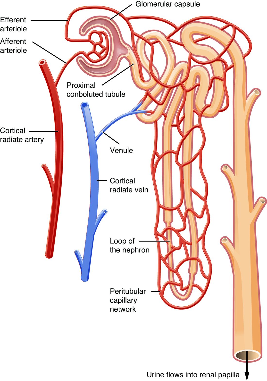
With labels hidden, select a box to reveal its label.
5How it works, step by step
- Blood enters the glomerulus through the afferent arteriole and can leave only through the efferent arteriole, which in most nephrons is narrower and resists the outflow.Pressure inside the glomerular capillaries stays unusually high.
- High pressure pushes on leaky, fenestrated capillary walls wrapped in podocytes.Plasma fluid, without blood cells and most proteins, is pushed into the capsular space.
- The fluid flows into the proximal convoluted tubule, whose cells have a brush border of microvilli and many mitochondria.Most of the water and nearly all the useful solutes are taken back into the peritubular capillaries.
- The remaining fluid runs down and up the nephron loop and passes the macula densa, which senses its sodium chloride.The macula densa signals the juxtaglomerular cells and the afferent arteriole of its own nephron.
- The fluid continues through the distal convoluted tubule into a collecting duct shared with other nephrons.The collecting duct carries it through the medulla to the renal papilla, where it drips into a minor calyx as urine.
6Core concepts
7A common mistake
The wrong idea: Blood leaving the glomerulus flows into a venule, as it does after any other capillary bed.
What actually happens: Blood leaves the glomerulus through the efferent arteriole, an arteriole, and then flows through a second capillary bed, the peritubular capillaries (and in juxtamedullary nephrons, the vasa recta), before reaching a venule. Having an arteriole on both sides is what lets the kidney keep glomerular pressure high and adjust it from either side.
8Check yourself
Anything you miss goes into your review queue.
1. Name the pinned structure.
- Glomerulus
- Glomerular capsule
- Proximal convoluted tubule
- Afferent arteriole
Show the answer
The glomerular capsule (Bowman's capsule) is the hollow cup around the glomerulus, the swollen start of the tubule. Filtered fluid collects in the space inside it.
- Glomerulus: The glomerulus is the tuft of capillaries inside the cup, not the cup itself.
- Correct: Glomerular capsule: Correct. The cup wrapped around the capillary tuft is the glomerular capsule.
- Proximal convoluted tubule: The proximal convoluted tubule is the coiled tube that leaves the capsule at the tubular pole.
- Afferent arteriole: The afferent arteriole is the blood vessel that carries blood into the capillary tuft.
2. In the inset, name the pinned group of crowded cells in the tubule wall.
- Juxtaglomerular cells
- Podocytes
- Macula densa
- Mesangial cells
Show the answer
The macula densa (dense spot) is a patch of tall, crowded cells in the wall of the tubule where it touches its own arterioles. They sense how much sodium chloride is in the fluid flowing past.
- Juxtaglomerular cells: Juxtaglomerular cells sit in the wall of the afferent arteriole, not the tubule, and release renin.
- Podocytes: Podocytes wrap the glomerular capillaries inside the capsule.
- Correct: Macula densa: Correct. These tubule cells are the macula densa.
- Mesangial cells: Mesangial cells lie between the arterioles and inside the glomerulus, not in the tubule wall.
3. Order the parts of the nephron that filtered fluid flows through, starting where it is first collected.
- Glomerular capsule
- Proximal convoluted tubule
- Descending limb of the nephron loop
- Ascending limb of the nephron loop
- Distal convoluted tubule
- Collecting duct
Show the answer
Fluid filtered from the glomerulus collects in the capsular space of the glomerular capsule, flows into the proximal convoluted tubule, down the descending limb and up the ascending limb of the nephron loop, through the distal convoluted tubule, and into a collecting duct.
- Correct order: 1. Glomerular capsule 2. Proximal convoluted tubule 3. Descending limb of the nephron loop 4. Ascending limb of the nephron loop 5. Distal convoluted tubule 6. Collecting duct
4. Fill the gap: afferent arteriole → glomerulus → ____ → peritubular capillaries.
- Venule
- Efferent arteriole
- Vasa recta
- Cortical radiate vein
Show the answer
Blood leaves the glomerulus through the efferent arteriole, which then feeds the second capillary bed, the peritubular capillaries.
- Venule: A venule would drain a capillary bed toward the veins. Here blood still has a second capillary bed to cross, so the vessel between them is an arteriole.
- Correct: Efferent arteriole: Correct. The efferent arteriole links the two capillary beds.
- Vasa recta: The vasa recta are a second capillary bed like the peritubular capillaries, fed by the efferent arteriole; they do not come between the glomerulus and the peritubular capillaries.
- Cortical radiate vein: Cortical radiate veins are much larger vessels that collect blood after both capillary beds.
5. Under the microscope, one part of the tubule has cuboidal cells with a dense brush border of microvilli and very many mitochondria. What does this structure suggest about its job?
- It stores urine between emptyings
- It lets water through passively, without spending any ATP at all
- It releases renin into the blood
- It moves large amounts of material back into the blood
Show the answer
This is the proximal convoluted tubule. Microvilli multiply the surface area for transport, and mitochondria supply the ATP that its pumps use. It takes back most of the filtered water and solutes.
- It stores urine between emptyings: Urine is stored in the bladder, lined by transitional epithelium. No tubule segment stores urine.
- It lets water through passively, without spending any ATP at all: Many mitochondria mean heavy ATP use. A segment built only to let water through would not need them.
- It releases renin into the blood: Renin comes from juxtaglomerular cells in the afferent arteriole wall, not from tubule cells.
- Correct: It moves large amounts of material back into the blood: Correct. A large surface and plenty of ATP point to heavy transport, which is the proximal tubule's job.
6. Why is blood pressure in the glomerular capillaries much higher than in capillaries elsewhere, such as in your skin?
- The glomerulus is close to the heart
- The glomerular capillaries have thicker walls than other capillaries
- An arteriole on the way out holds pressure back in the glomerulus
- The podocytes pump blood through the capillaries
Show the answer
Most capillary beds drain into venules, which offer little resistance. The glomerulus drains into the efferent arteriole, a resistance vessel, so pressure backs up in the glomerular capillaries.
- The glomerulus is close to the heart: The kidneys are no closer to the heart than many other organs. The layout of their vessels, not distance, sets the pressure.
- The glomerular capillaries have thicker walls than other capillaries: Glomerular capillaries are thin, leaky fenestrated capillaries. Wall thickness does not raise the pressure inside.
- Correct: An arteriole on the way out holds pressure back in the glomerulus: Correct. Resistance downstream, in the efferent arteriole, keeps glomerular pressure high.
- The podocytes pump blood through the capillaries: Podocytes wrap the capillaries; they do not pump. The heart supplies the pressure.
7. A tiny clot completely blocks the afferent arteriole of one nephron. Predict the change in each variable for that nephron, compared with before the clot.
| Variable | Change |
|---|---|
| Blood flow into its glomerulus | — |
| Fluid entering its capsular space | — |
| Blood flow in the peritubular capillaries fed by its efferent arteriole | — |
| Blood flow into the glomerulus of a neighboring nephron | — |
Show the answer
The two capillary beds of a nephron are in series: afferent arteriole, glomerulus, efferent arteriole, peritubular capillaries. Blocking the first vessel starves everything downstream in that nephron but leaves its neighbors untouched.
- Blood flow into its glomerulus: down. The afferent arteriole is the only way blood enters the glomerulus, so blocking it stops the flow.
- Fluid entering its capsular space: down. With no blood and no pressure in the glomerulus, nothing is pushed into the capsule.
- Blood flow in the peritubular capillaries fed by its efferent arteriole: down. The efferent arteriole receives its blood from the glomerulus, so the second capillary bed downstream loses its supply too.
- Blood flow into the glomerulus of a neighboring nephron: no change. Each nephron has its own afferent arteriole from the cortical radiate artery, so a clot in one does not block the others.
9Summary
A nephron is the unit that makes urine; each kidney has about a million, and none are replaced once lost. Its renal corpuscle, in the cortex, is a glomerulus of fenestrated capillaries inside the glomerular (Bowman's) capsule, whose visceral layer is made of podocytes and whose capsular space collects the filtered fluid. The renal tubule runs from the capsule: the proximal convoluted tubule, with its brush border and many mitochondria, takes back most of the fluid; the nephron loop, with descending and ascending limbs, dips into the medulla; the distal convoluted tubule returns to touch its own corpuscle; and a collecting duct, shared by many nephrons, carries the fluid to the papilla. Cortical nephrons (about 85%) have short loops; juxtamedullary nephrons (about 15%) have long loops and the vasa recta. Blood flows through an afferent arteriole, the glomerulus, an efferent arteriole and then the peritubular capillaries or vasa recta. The juxtaglomerular apparatus, where the tubule meets its arterioles, joins the macula densa, the renin-releasing juxtaglomerular cells and mesangial cells.