Structure of blood vessels
1Why this matters
Ms. Delgado, 52, has worked 12-hour shifts on her feet as a nurse for 25 years. Her lower legs ache by evening, and swollen, twisting blue veins now bulge under the skin of her calves. The tiny one-way valves inside those veins have stopped sealing, so blood falls back down her legs every time she stands, pools, and stretches the thin vein walls a little more each year.
2What this builds on
3Quick check before you start
1. Which tissue lines the inside of every blood vessel?
- Simple squamous epithelium (endothelium)
- Stratified squamous epithelium
- Dense regular connective tissue
Show the answer
Blood vessels are lined by endothelium, a simple squamous epithelium: one layer of flat cells, thin enough for substances to cross easily.
- Correct: Simple squamous epithelium (endothelium):
- Stratified squamous epithelium:
- Dense regular connective tissue:
2. Which fibers let a tissue stretch and then spring back to its original shape?
- Collagen fibers
- Elastic fibers
- Reticular fibers
Show the answer
Elastic fibers, made of elastin, stretch and recoil. Collagen fibers resist stretching, and reticular fibers form fine supporting meshes.
- Collagen fibers:
- Correct: Elastic fibers:
- Reticular fibers:
3. When the smooth muscle in a vessel wall contracts, the vessel:
- Widens (vasodilation)
- Narrows (vasoconstriction)
- Gets longer
Show the answer
The smooth muscle wraps around the vessel in rings, so its contraction squeezes the vessel narrower: vasoconstriction.
- Widens (vasodilation):
- Correct: Narrows (vasoconstriction):
- Gets longer:
4Anatomy
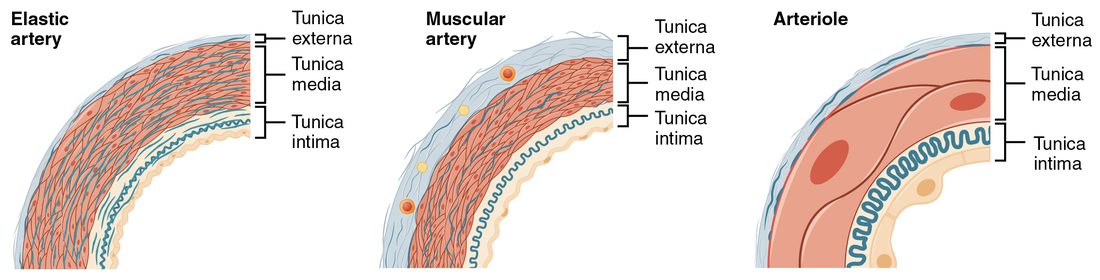
With labels hidden, select a box to reveal its label.
5How it works, step by step
- The left ventricle ejects blood into the aorta faster than it can flow on into smaller arteries.The elastic fibers of the aortic wall stretch and store energy.
- The aortic valve closes and the stretched elastic fibers recoil.Blood keeps flowing forward during diastole, and aortic pressure falls only to about 80 mm Hg.
- Blood reaches the arterioles, whose smooth muscle holds them partly contracted (vascular tone).Their radius sets the resistance at the entrance to each capillary bed, and so how much blood each bed receives.
- Blood passes through capillaries whose wall is a single layer of endothelium.Gases, nutrients and wastes cross the thin wall; continuous, fenestrated and sinusoidal capillaries let through progressively larger substances.
- Blood enters thin-walled, compliant veins at low pressure.Veins hold most of the blood volume, and the skeletal muscle pump, the respiratory pump and venoconstriction push it back to the heart through one-way valves.
6Core concepts
7A common mistake
The wrong idea: Veins carry blood back to the heart at high pressure, just like arteries in reverse, and their valves are pumps.
What actually happens: By the time blood reaches the veins, most of its pressure has been spent in the arterioles and capillaries: venules are at about 15 mm Hg and the right atrium near 0. Venous valves are passive one-way flaps that only stop backflow. What actually moves blood up your legs is the squeeze of your skeletal muscles and the pressure changes of breathing, with the valves making sure the movement goes only one way.
8Check yourself
Anything you miss goes into your review queue.
1. Which layer of an artery wall contains most of its smooth muscle?
- Tunica intima
- Tunica externa
- Tunica media
- The basement membrane
Show the answer
The tunica media is the middle layer: rings of smooth muscle mixed with elastic fibers. Its contraction narrows the vessel.
- Tunica intima: The tunica intima is the endothelial lining with a thin layer of connective tissue. It has no muscle layer.
- Tunica externa: The tunica externa is mostly collagen. It anchors the vessel and resists overstretching.
- Correct: Tunica media: Correct. The media is the muscle layer.
- The basement membrane: The basement membrane is the thin sheet the endothelium sits on, part of the tunica intima.
2. Compare the three walls in the figure. Which vessel stretches most with each ventricular ejection and recoils during diastole, keeping blood moving between beats?
- The arteriole
- The muscular artery
- All three stretch about equally
- The elastic artery
Show the answer
The elastic artery's tunica media is packed with layers of elastic fibers. They stretch as blood surges in during ejection and recoil when the aortic valve closes, pushing blood onward during diastole.
- The arteriole: The arteriole's wall is mostly a few smooth muscle cells. It adjusts resistance rather than storing stretch.
- The muscular artery: The muscular artery's media is mostly smooth muscle with fewer elastic fibers, so it stretches much less.
- All three stretch about equally: The walls differ in elastic content, and so in how much they stretch and recoil.
- Correct: The elastic artery: Correct. Elastic arteries, such as the aorta, act as the pressure reservoir between beats.
3. Why are arterioles, rather than large arteries, the main site of resistance to blood flow?
- They carry blood at the highest pressure in the body
- They have the thickest walls of any vessel
- Their radius is small and tightly controlled, and resistance depends on radius⁴
- They lack an endothelial lining, leaving blood in direct contact with their smooth muscle
Show the answer
Resistance depends on 1 ÷ radius to the fourth power, so small vessels resist flow strongly and small changes in their radius matter a great deal. Arterioles also have the most smooth muscle relative to their size, so they can change radius widely.
- They carry blood at the highest pressure in the body: Pressure is highest in the large arteries; it drops as blood passes through the arterioles.
- They have the thickest walls of any vessel: Large arteries have thicker walls. Arterioles have thick walls only relative to their small lumen.
- Correct: Their radius is small and tightly controlled, and resistance depends on radius⁴: Correct. Small radius plus strong control of radius makes arterioles the resistance vessels.
- They lack an endothelial lining, leaving blood in direct contact with their smooth muscle: Every vessel, arterioles included, is lined by endothelium.
4. An artery and a vein of similar size lie side by side on a slide. Which features identify the vein? Select all that apply.
- A thin tunica media
- A tunica externa thicker than its media
- A wide lumen, often collapsed into an oval
- A prominent wavy internal elastic membrane
- A thick ring of smooth muscle holding it round
Show the answer
Veins carry blood at low pressure, and their walls reflect it: a thin media, a relatively thick externa, and a wide lumen that collapses when empty.
- Correct: A thin tunica media: Identifies the vein. Low pressure needs little muscle.
- Correct: A tunica externa thicker than its media: Identifies the vein. Its thickest layer is the collagen-rich externa.
- Correct: A wide lumen, often collapsed into an oval: Identifies the vein. The thin wall collapses without blood pressure holding it open.
- A prominent wavy internal elastic membrane: Identifies the artery. The internal elastic membrane is prominent in arteries.
- A thick ring of smooth muscle holding it round: Identifies the artery. A thick muscular media keeps the artery round even when empty.
5. A student describes the skeletal muscle pump. One step is wrong. Find it.
- The calf muscle contracts and squeezes the veins running through it.
- Pressure in the squeezed segment of vein rises.
- The valve above the segment opens and the valve below it closes, each pushed by the pressure difference across it.
- Blood moves up toward the heart.
- When the muscle relaxes, the valve above opens wider and blood falls back into the segment.
Show the answer
When the muscle relaxes, pressure in the segment falls. Blood above starts to flow back, fills the cusps of the valve above and closes it, so blood cannot fall back. The segment refills from below.
- The calf muscle contracts and squeezes the veins running through it.: This step is right. Many limb veins run between and through skeletal muscles.
- Pressure in the squeezed segment of vein rises.: This step is right. Squeezing a closed segment raises its pressure.
- The valve above the segment opens and the valve below it closes, each pushed by the pressure difference across it.: This step is right. Higher pressure in the squeezed segment pushes the valve above open and the valve below shut.
- Blood moves up toward the heart.: This step is right. The closed lower valve leaves only one way out.
- Correct: When the muscle relaxes, the valve above opens wider and blood falls back into the segment.: This is the error. The valve above closes on relaxation; that is what stops blood from falling back.
6. According to current evidence in humans, which structures mainly control how much blood enters a capillary bed?
- The arterioles feeding the bed
- The venous valves downstream
- The pericytes of the venules
- The capillaries' own endothelial pores
Show the answer
Arteriolar smooth muscle sets the resistance at the entrance to each bed and responds to nerves and to local chemicals. Discrete precapillary sphincters have been found in only some tissues and species, and their role in humans is unsettled. For your exam, many tests still expect the answer "precapillary sphincters".
- Correct: The arterioles feeding the bed: Correct. Arterioles are the resistance vessels that set flow into each bed.
- The venous valves downstream: Venous valves only stop backflow in veins. They do not control flow into capillaries.
- The pericytes of the venules: Venule pericytes support the wall and affect its leakiness; they do not meter flow into the bed.
- The capillaries' own endothelial pores: Pores affect what crosses the wall, not how much blood flows through the bed.
7. A blood donor gives about 500 mL of blood. Within minutes, sympathetic nerves fire more strongly to her veins. How does that help keep blood flowing to her heart and brain?
- It dilates the veins so they can hold the donated volume
- It closes every venous valve in the body at once and holds the remaining blood in place there
- It constricts the veins, shrinking the venous reservoir and moving stored blood toward the heart
- It makes the capillaries fenestrated so fluid enters faster
Show the answer
About 60 to 65 percent of blood sits in the compliant systemic veins. Sympathetic venoconstriction reduces their capacity, raises venous pressure and pushes that stored blood toward the heart, partly making up for the volume lost.
- It dilates the veins so they can hold the donated volume: Dilating the veins would let more blood pool in them, which is the opposite of what she needs.
- It closes every venous valve in the body at once and holds the remaining blood in place there: Valves are passive. Nerves do not open or close them, and blocking flow would stop venous return.
- Correct: It constricts the veins, shrinking the venous reservoir and moving stored blood toward the heart: Correct. The veins act as a blood reservoir the body can draw on.
- It makes the capillaries fenestrated so fluid enters faster: Capillary type is a fixed structural feature of each tissue; nerves do not change it within minutes.
9Summary
Vessel walls have three tunics: the intima (endothelium on a basement membrane, with an internal elastic membrane in arteries), the media (smooth muscle and elastic fibers) and the externa (collagen, carrying the vasa vasorum and nervi vasorum). Elastic arteries near the heart stretch and recoil to keep pressure up between beats; muscular arteries distribute blood to organs; arterioles are the resistance vessels, and their vascular tone sets flow into each capillary bed. Capillaries are one endothelial layer, continuous, fenestrated or sinusoidal; metarterioles, thoroughfare channels and arteriovenous anastomoses can shunt blood past them. Venules and veins have thin walls, wide lumens and valves; as compliant capacitance vessels they hold about 60 to 65 percent of the blood. Venous return depends on the skeletal muscle pump, the respiratory pump and venoconstriction, and failed valves lead to varicose veins.