Liver, gallbladder and pancreas
1Why this matters
Mrs. Alvarez, 48, has had gallstones for years without trouble. This week her eyes turned yellow, her urine went the color of cola and her stools turned pale gray. One stone has slipped out of her gallbladder and jammed in the duct that carries bile to her intestine. To see why one small stone changes her eyes, urine and stools at once, you need to follow bile from the liver cell that makes it to the duodenum.
2What this builds on
3Quick check before you start
1. Where does blood from the stomach and intestines go first?
- Straight to the inferior vena cava
- To the liver, through the hepatic portal vein
- To the kidneys, before it returns to the heart
Show the answer
Veins from the stomach, intestines, spleen and pancreas join into the hepatic portal vein, which carries their blood to the liver's sinusoids before it returns to the heart.
- Straight to the inferior vena cava:
- Correct: To the liver, through the hepatic portal vein:
- To the kidneys, before it returns to the heart:
2. What makes a gland exocrine rather than endocrine?
- It releases its product into a duct that leads to a surface
- It releases a hormone into the blood
- It is made of only one cell
Show the answer
An exocrine gland releases its product into a duct that carries it to a surface, such as the lining of the gut. An endocrine gland has no duct and releases hormones toward capillaries.
- Correct: It releases its product into a duct that leads to a surface:
- It releases a hormone into the blood:
- It is made of only one cell:
3. Which hormone do duodenal cells release when fat and protein arrive?
- Gastrin
- Secretin
- Cholecystokinin
Show the answer
Fat and protein in the duodenum trigger cholecystokinin (CCK). Acid triggers secretin; gastrin comes from the stomach.
- Gastrin:
- Secretin:
- Correct: Cholecystokinin:
4Anatomy
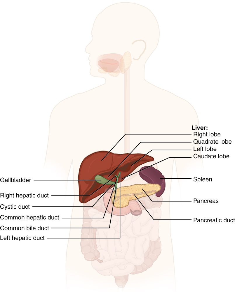
With labels hidden, select a box to reveal its label.
5How it works, step by step
- Fatty, acidic chyme enters the duodenum.Enteroendocrine cells in the duodenal wall release cholecystokinin (in response to fat and protein) and secretin (in response to acid) into the blood.
- Cholecystokinin reaches the gallbladder and the hepatopancreatic sphincter.The gallbladder contracts and the sphincter relaxes, so concentrated bile flows down the common bile duct into the duodenum.
- Cholecystokinin and secretin reach the pancreas.Acinar cells release enzymes and duct cells release bicarbonate, and pancreatic juice flows into the duodenum through the same opening.
- Bicarbonate neutralizes the acid chyme.Duodenal pH rises toward neutral, where pancreatic enzymes work, and enteropeptidase on the brush border converts trypsinogen to trypsin, which activates the other protein-digesting enzymes.
- Bile salts coat the fat and keep churned droplets small (emulsification).The fat's surface area multiplies, so pancreatic lipase can digest it far faster.
6Core concepts
7A common mistake
The wrong idea: Bile digests fat.
What actually happens: Bile has no digestive enzymes. Its bile salts emulsify fat: they coat it and keep it in tiny droplets, which multiplies the surface that pancreatic lipase can reach. Lipase does the chemical digestion. Without bile, fat digestion is slow and much fat is lost in the feces; without lipase, fat is emulsified but barely digested at all.
8Check yourself
Anything you miss goes into your review queue.
1. Name the pinned structures.
- Bile canaliculi
- Sinusoids
- Central veins
- Portal venules
Show the answer
The channels running between the plates of hepatocytes, from the edge of the lobule to the central vein, are the hepatic sinusoids: wide, leaky capillaries where portal and arterial blood mix.
- Bile canaliculi: Bile canaliculi are tiny grooves between neighboring hepatocytes, far too small to show as channels here, and they carry bile, not blood.
- Correct: Sinusoids: Correct. These are the hepatic sinusoids.
- Central veins: There is one central vein per lobule, running up its middle. The pinned channels run between the plates toward it.
- Portal venules: Portal venules are the blue vessels at the corners of the lobule, in the portal triads. They feed the sinusoids.
2. A gallstone lodges in the common bile duct, above the point where it meets the pancreatic duct, and blocks it completely. Predict the change in each variable over the next few days.
| Variable | Change |
|---|---|
| Bile reaching the duodenum | — |
| Conjugated bilirubin in the blood | — |
| Darkness of the urine | — |
| Brown color of the feces | — |
| Fat in the feces | — |
| Pancreatic juice reaching the duodenum | — |
Show the answer
A blocked common bile duct stops bile from reaching the gut. Conjugated bilirubin backs up into the blood and urine, the feces lose their brown pigment, and less fat is digested and absorbed for lack of bile salts. Pancreatic juice still flows because its duct is below the blockage.
- Bile reaching the duodenum: down. The only route for bile from both the liver and the gallbladder to the duodenum is through the common bile duct.
- Conjugated bilirubin in the blood: up. Hepatocytes keep conjugating bilirubin, but bile cannot drain, so conjugated bilirubin backs up into the blood.
- Darkness of the urine: up. Conjugated bilirubin is water-soluble, so the extra in the blood spills into the urine and darkens it.
- Brown color of the feces: down. With no bilirubin reaching the intestine, bacteria make no stercobilin, so the feces turn pale.
- Fat in the feces: up. Without bile salts, fat is not emulsified, so lipase has less surface to work on. Bile salts also carry the digested fat to the lining, so much of what is digested is not absorbed either.
- Pancreatic juice reaching the duodenum: no change. The stone sits above the junction with the pancreatic duct, so pancreatic juice still drains through the ampulla.
3. Bile contains no enzymes. How, then, does it speed up the digestion of fat?
- Its bicarbonate splits the bonds that hold fatty acids to glycerol
- Its bilirubin activates pancreatic lipase
- Its bile salts break fat into many small droplets
- Its cholesterol dissolves the fat into the water
Show the answer
Bile salts have a hydrophobic face that sticks into fat and a hydrophilic face that stays in the water. They coat fat droplets and keep them small once churning breaks them up. Many small droplets have far more surface than a few large ones, and lipase works at that surface.
- Its bicarbonate splits the bonds that hold fatty acids to glycerol: Bicarbonate neutralizes acid. It does not split the bonds in triglycerides; lipase does that.
- Its bilirubin activates pancreatic lipase: Bilirubin is a waste pigment from old red blood cells, carried out in bile. It does not activate lipase.
- Correct: Its bile salts break fat into many small droplets: Correct. Emulsification multiplies the surface area lipase can reach.
- Its cholesterol dissolves the fat into the water: Cholesterol does not dissolve in water, and bile has to carry it with bile salts and phospholipids. It does not dissolve fat.
4. A 34-year-old woman had the last part of her ileum removed because of Crohn disease. Months later she has pale, greasy stools after fatty meals. Her liver, gallbladder and pancreas are normal. What best explains her symptoms?
- Her gallbladder can no longer concentrate bile
- Her pancreas no longer releases lipase in response to fat
- Her bile salts are lost instead of being recycled
- Her liver has stopped conjugating bilirubin
Show the answer
About 95% of bile salts are reabsorbed in the last part of the ileum and returned to the liver in the hepatic portal vein: the enterohepatic circulation. Without that segment they are lost in the feces. The liver makes new ones from cholesterol, but not fast enough, so too few bile salts reach the gut to emulsify a fatty meal.
- Her gallbladder can no longer concentrate bile: Her gallbladder is normal and still concentrates bile. The problem is how much bile salt there is to concentrate.
- Her pancreas no longer releases lipase in response to fat: Her pancreas is normal. Lipase is still released; it simply has too little emulsified fat surface to work on.
- Correct: Her bile salts are lost instead of being recycled: Correct. Loss of the terminal ileum breaks the enterohepatic circulation, so bile salts are lost in the feces.
- Her liver has stopped conjugating bilirubin: Her liver is normal. Failure to conjugate bilirubin would cause jaundice, not fatty stools.
5. An experiment infuses acid into a volunteer's duodenum. Which change in pancreatic secretion follows?
- A small volume of juice rich in enzymes, driven by cholecystokinin
- A large volume of juice rich in bicarbonate, driven by secretin
- No change in pancreatic secretion
- Juice rich in trypsin, activated inside the pancreatic duct
Show the answer
Acid lowers duodenal pH, which releases secretin. Secretin acts mainly on the duct cells, which pour out a large volume of bicarbonate-rich fluid. The bicarbonate neutralizes the acid, removing the stimulus: negative feedback.
- A small volume of juice rich in enzymes, driven by cholecystokinin: Cholecystokinin is released by fat and protein, not acid, and it drives the enzyme-rich secretion of the acinar cells.
- Correct: A large volume of juice rich in bicarbonate, driven by secretin: Correct. Acid releases secretin, which drives bicarbonate secretion.
- No change in pancreatic secretion: The pancreas does respond: acid is the main stimulus for secretin, which drives bicarbonate secretion.
- Juice rich in trypsin, activated inside the pancreatic duct: Trypsin is activated in the duodenum by enteropeptidase, never normally in the duct.
6. A 52-year-old man with gallstones has sudden, severe pain in his upper middle abdomen that goes through to his back. His blood lipase is many times normal. Which event most likely started his illness?
- Bile salts leaked from the liver into the blood and dissolved the pancreas
- A stone blocked the cystic duct and stopped bile from reaching the duodenum after his meals
- Stomach acid flowed back up the pancreatic duct and burned the pancreas
- A stone at the ampulla blocked the pancreatic duct, so trypsin was activated in the gland
Show the answer
This is acute pancreatitis. A gallstone stuck at the ampulla blocks the pancreatic duct. Enzymes are trapped, trypsin is activated inside the gland, and it activates the other enzymes, which digest pancreatic tissue. Lipase leaks from the damaged gland into the blood.
- Bile salts leaked from the liver into the blood and dissolved the pancreas: Bile salts emulsify fat; they do not digest tissue, and they do not cause pancreatitis by entering the blood.
- A stone blocked the cystic duct and stopped bile from reaching the duodenum after his meals: A cystic duct stone causes gallbladder pain under the right ribs, and bile from the liver still reaches the duodenum. It does not block the pancreatic duct or raise lipase.
- Stomach acid flowed back up the pancreatic duct and burned the pancreas: Stomach acid is neutralized in the duodenum and does not travel up the pancreatic duct.
- Correct: A stone at the ampulla blocked the pancreatic duct, so trypsin was activated in the gland: Correct. Gallstones at the ampulla are one of the two commonest causes of acute pancreatitis.
7. Blood and bile both move through a liver lobule. How do their directions compare?
- Both flow outward from the central vein to the portal triads
- Both flow inward from the portal triads to the central vein
- Blood flows inward to the central vein; bile flows outward to the portal triads
- Blood flows outward to the portal triads; bile flows inward and drains into the central vein
Show the answer
Blood enters at the portal triads, crosses the sinusoids and leaves by the central vein. Bile is secreted into canaliculi between hepatocytes and flows the other way, out to the bile ducts in the portal triads.
- Both flow outward from the central vein to the portal triads: The central vein collects blood; it is where blood leaves, not where it enters, and bile never enters it.
- Both flow inward from the portal triads to the central vein: Blood flows inward, but bile flows outward to the bile ducts.
- Correct: Blood flows inward to the central vein; bile flows outward to the portal triads: Correct. The two flows run in opposite directions and never mix.
- Blood flows outward to the portal triads; bile flows inward and drains into the central vein: This reverses both flows.
8. Why does the pancreas release its protein-digesting enzymes as trypsinogen and chymotrypsinogen rather than as active trypsin and chymotrypsin?
- Active enzymes inside the gland would digest the acinar cells and ducts
- The active forms are too large to pass through the pancreatic duct
- The precursors digest protein faster in the acid of the stomach
- The active forms would be destroyed by bile salts
Show the answer
Protein-digesting enzymes do not tell the gland's own proteins from food proteins. Kept as inactive precursors, they cannot digest the pancreas; they are switched on only in the duodenum, where enteropeptidase starts the cascade. When this fails, as in pancreatitis, the gland digests itself.
- Correct: Active enzymes inside the gland would digest the acinar cells and ducts: Correct. Zymogens protect the tissue that makes them.
- The active forms are too large to pass through the pancreatic duct: Size is not the issue; activation cuts off a small piece and makes the enzyme slightly smaller, not larger.
- The precursors digest protein faster in the acid of the stomach: Pancreatic juice never reaches the stomach, and the precursors do not digest protein anywhere.
- The active forms would be destroyed by bile salts: Bile salts do not destroy trypsin or chymotrypsin; both work alongside bile in the duodenum.
9Summary
The liver is built of lobules: plates of hepatocytes between leaky sinusoids, with a central vein in the middle and portal triads (portal vein branch, hepatic artery branch, bile duct) at the corners. Blood flows inward to the central vein; bile flows outward in canaliculi. Hepatocytes store and release glucose, make plasma proteins and clotting factors, process drugs and toxins, excrete bilirubin, store iron and vitamins, and make bile. Bile salts emulsify fat and are recycled through the enterohepatic circulation. The gallbladder stores and concentrates bile and empties it when cholecystokinin contracts it and relaxes the hepatopancreatic sphincter. Pancreatic juice brings bicarbonate (driven by secretin) and enzymes (driven by CCK); its protein-digesting enzymes travel as zymogens that enteropeptidase and trypsin activate in the duodenum. When trypsin is activated inside the gland, the result is pancreatitis. Hepatitis is liver inflammation; cirrhosis is the scarring that follows years of injury.